
Respiratory causes of palpitations
Palpitations from respiratory causes typically involve hypoxia, hypercapnia, acid-base changes, or mechanical effects on the heart. Key mechanisms include hypoxic tachycardia, sympathetic activation from CO2 retention, and direct arrhythmia triggers in conditions like PE, cor pulmonale, or obstructive sleep apnea. Pulmonary hypertension can cause right heart strain with ectopic beats, and severe bronchospasm triggers catecholamine surges.
Searching sources...
Reading File
Now I'll run parallel searches across the library and PubMed.
Finding Sources
Finding Sources
Finding Sources
Excellent - I found dedicated palpitations sections in Harrison's, Braunwald's, and Goldman-Cecil. Let me read those in parallel.
Reading File
Reading File
Reading File
Good content from Harrison's and Braunwald's. Now let me search specifically for respiratory causes - PE, OSA, pulmonary hypertension, hypoxia, hypercapnia, COPD.
Finding Sources
Finding Sources
Finding Sources
Finding Sources
Finding Sources
Finding Sources
I found a dedicated section in Murray & Nadel's on arrhythmias in OSA. Let me read that, plus read from Braunwald's assessment section, and also get the full palpitations assessment from Braunwald's block 7.
Reading File
Reading File
I now have rich content from authoritative sources. Let me fetch the OSA arrhythmia figure and also run a quick PubMed search on recent evidence.
Loading Image
Searching PubMed
respiratory causes palpitations arrhythmia hypoxia
I have comprehensive material from authoritative sources. Now I can compile a thorough answer.
Respiratory Causes of Palpitations
Palpitations are defined as an unpleasant awareness of the heartbeat - "thumping," "pounding," or "fluttering" - and can arise from a rapid rate, irregular rhythm, or increased contractile force. While cardiac and psychiatric causes dominate (43% and 31% respectively in large series), several respiratory conditions are well-recognised triggers. - Harrison's Principles of Internal Medicine 22E
1. Pulmonary Embolism (PE)
PE is among the cardiovascular causes of palpitations listed in Harrison's, and can produce them through multiple mechanisms:
- Acute right heart strain: sudden increase in right ventricular afterload stretches the right atrium and ventricle, triggering atrial fibrillation, flutter, or sinus tachycardia
- Hypoxia-driven sympathetic activation: reduced V/Q matching lowers PaO2, stimulating the adrenal medulla and raising circulating catecholamines
- Reflex tachycardia: baroreceptor activation from the drop in cardiac output
Massive PE is listed as an anatomic cause of syncope through obstruction to blood flow, with preceding palpitations as a less severe warning symptom. - Braunwald's Heart Disease
2. Obstructive Sleep Apnea (OSA)
OSA produces the broadest and most evidence-based spectrum of arrhythmias among respiratory conditions. The Sleep Heart Health Study (n = 6,441) found that every arrhythmia type was more frequent in severe OSA (AHI ≥30) compared to no OSA:
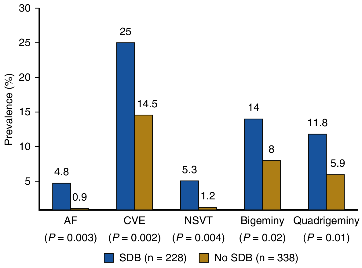
From Murray & Nadel's Textbook of Respiratory Medicine - Figure 119.2. Blue = severe OSA (SDB), Gold = no OSA. AF = atrial fibrillation; CVE = complex ventricular ectopy; NSVT = non-sustained ventricular tachycardia.
Key findings:
- Atrial fibrillation: 4.8% vs 0.9% (OR 4.0, P = 0.003) - 4x more common
- Complex ventricular ectopy: 25% vs 14.5% (OR 1.7, P = 0.002)
- NSVT: 5.3% vs 1.2% (OR 3.4, P = 0.004)
- Conduction delay arrhythmias were also significantly more common
Mechanisms driving these arrhythmias include:
- Cyclic hypoxia and hypercapnia during apnoeic episodes
- Negative intrathoracic pressure swings stretching atrial walls
- Increased sympathetic tone (loss of normal nocturnal parasympathetic dominance)
- Systemic and pulmonary hypertension over time
- Right and left ventricular remodelling
Central sleep apnea (Cheyne-Stokes pattern, e.g. in heart failure) was independently associated with new-onset atrial fibrillation (OR 3.0, 95% CI 1.4-6.4). - Murray & Nadel's Textbook of Respiratory Medicine
3. Hypoxia (Any Cause)
Regardless of aetiology (pneumonia, ARDS, altitude, respiratory failure), hypoxia drives palpitations through:
- Sinus tachycardia: the most common response, mediated via peripheral chemoreceptors (carotid bodies) → sympathoadrenal activation
- Catecholamine surge: adrenaline and noradrenaline enhance myocardial automaticity and reduce the fibrillation threshold
- Metabolic acidosis: accompanying lactic acidosis further alters membrane potentials
At high altitude, the constellation of headache, fatigue, dizziness, nausea, palpitations, and insomnia is attributable to acute hypoxia and respiratory alkalosis. - Costanzo Physiology 7th Edition
4. Hypercapnia (CO2 Retention)
In chronic obstructive respiratory conditions (COPD with type 2 respiratory failure, obesity-hypoventilation syndrome):
- Hypercapnia causes sympathetic nervous system activation through central chemoreceptors
- Acidosis (respiratory acidosis, pH <7.35) directly alters cardiac ion channel function - particularly fast sodium channels - predisposing to ectopic beats and atrial flutter
- CO2 is also a direct vasodilator - the resulting hyperdynamic circulation produces a bounding pulse that patients feel as palpitations even without a true arrhythmia
5. Cor Pulmonale / Pulmonary Arterial Hypertension
Chronic right ventricular pressure overload leads to:
- Right atrial dilatation, a substrate for atrial flutter and fibrillation
- Right ventricular hypertrophy with increased wall stress - susceptible to ventricular ectopics
- Goodman & Gilman's notes PAH treatments specifically address the arrhythmia-palpitations link in pulmonary vascular disease
6. Hyperventilation Syndrome / Panic with Respiratory Component
Kaplan & Sadock's highlights hyperventilation as producing palpitations through:
- Respiratory alkalosis (low PaCO2 → high pH) → hypocalcaemia (reduced ionised Ca2+) → increased myocardial excitability
- Symptoms: onset at rest, sighing respirations, apprehension, palpitations, depersonalisation
- Catecholamine release during acute hyperventilation further drives sinus tachycardia
This is important because hyperventilation is both a cause and a consequence of anxiety/panic - creating a self-reinforcing loop.
7. Acute Asthma / Severe Bronchospasm
- Endogenous catecholamine surge from respiratory distress - adrenaline from the adrenal medulla drives sinus tachycardia and can cause PVCs
- Exogenous bronchodilators: beta-2 agonists (salbutamol, terbutaline) have beta-1 cross-reactivity at high doses, directly causing palpitations and tachycardia; theophylline and aminophylline are specifically listed in Harrison's as causing palpitations
- Hypoxia from V/Q mismatch adds to arrhythmia risk
Summary Table
| Respiratory Condition | Primary Arrhythmia / Palpitation Mechanism | Key Arrhythmias |
|---|---|---|
| Pulmonary embolism | RV strain, hypoxia, reflex tachycardia | Sinus tachycardia, AF, flutter |
| Obstructive sleep apnea | Cyclic hypoxia, sympathetic activation, atrial stretch | AF (4x increased), CVE, NSVT |
| Hypoxia (any cause) | Chemoreceptor → catecholamine surge | Sinus tachycardia, PVCs |
| Hypercapnia / COPD | Sympathetic activation, respiratory acidosis | Sinus tachycardia, atrial flutter |
| Cor pulmonale / PAH | RA/RV dilatation and hypertrophy | AF, flutter, PVCs |
| Hyperventilation | Respiratory alkalosis → hypocalcaemia | Sinus tachycardia, PVCs |
| Acute asthma | Endogenous + exogenous catecholamines | Sinus tachycardia, PVCs |
Clinical Pearl
Positional palpitations suggest a structural process - either within the heart (atrial myxoma) or adjacent to the heart (mediastinal mass, e.g. from a respiratory tumour or lymphoma compressing cardiac structures). A mediastinal mass can directly mechanically irritate the heart or great vessels. - Harrison's Principles of Internal Medicine 22E
When evaluating palpitations in any patient with a respiratory history, key tests are a 12-lead ECG (for right heart strain pattern, P-pulmonale, RBBB), ABG (hypoxia/hypercapnia), echo (RV/RA dilatation), and ambulatory Holter monitoring. In suspected OSA, an overnight polysomnography is indicated given the strong and independent arrhythmia risk described above.
This is a shared conversation. Sign in to Orris to start your own chat.