Q. Discuss the indications, technique and complications of auditory brain stem implants. in detail for 50 marks with diagram and flow charts from Scott Brown, Cummings, stell and marans, Zakir Hussain, dhingra, hazarika and related articles with recent advances according to rguhs university
Reading File
Finding Sources
Finding Sources
Reading File
Reading File
Searching Images
auditory brainstem implant electrode cochlear nucleus
Reading File
Loading Image
Loading Image
Reading File
Reading File
Now I have comprehensive information from Cummings. Let me compile the full answer.
Note: Thegenerate_imagetool is currently unavailable (monthly plan limit reached). Flowcharts and diagrams have been presented in structured text/ASCII format. Please upgrade your subscription plan for image generation access.
AUDITORY BRAINSTEM IMPLANTS (ABI)
A Comprehensive Discussion — 50 Marks
Based on Cummings Otolaryngology, Scott-Brown's, Stell & Maran's, and Related Literature
1. INTRODUCTION & HISTORICAL BACKGROUND
The Auditory Brainstem Implant (ABI) is a central neural auditory prosthesis that bypasses both the cochlea and the cochlear nerve to directly stimulate the cochlear nucleus (CN) complex at the level of the brainstem. It consists of a multielectrode surface array placed over the cochlear nucleus within the lateral recess of the fourth ventricle.
Historical Milestones
| Year | Event |
|---|---|
| 1979 | First ABI performed at House Ear Institute (HEI), California — platinum electrode pair placed in a 51-year-old NF2 woman |
| 1981 | Re-implantation with Dacron mesh-mounted platinum pads to prevent migration |
| 2000 | FDA approved multichannel ABI for patients ≥12 years with NF2 |
| Early 2000s | Colletti extended ABI to nontumor adults and children in Europe |
| 2013 | FDA approved investigational device exemptions (IDE) at US centers for pediatric ABIs |
| 2016 | Cochlear Nucleus Profile ABI541 introduced (21 platinum electrodes) |
| Present | >1000 ABI procedures performed worldwide |
(Cummings Otolaryngology, pp. 3119–3120)
2. ANATOMY RELEVANT TO ABI
Target: The Cochlear Nucleus Complex
The cochlear nucleus lies at the lateral recess (foramen of Luschka) of the fourth ventricle, at the pontomedullary junction. It has three subdivisions:
┌─────────────────────────────────────────┐
│        COCHLEAR NUCLEUS COMPLEX         │
│                                         │
│   ┌─────────────────────┐               │
│   │  Anteroventral CN   │ (AVCN)        │
│   │  Posteroventral CN  │ (PVCN)        │
│   │  Dorsal CN          │ (DCN)         │
│   └─────────────────────┘               │
│                                         │
│  Location: Lateral recess of 4th        │
│  ventricle / Foramen of Luschka         │
│                                         │
│  Bordered by:                           │
│  • CN VII & VIII (anterosuperior)       │
│  • CN IX, X, XI (posteromedial)         │
│  • AICA (vascular landmark)             │
│  • Choroid plexus (surface marker)      │
└─────────────────────────────────────────┘
Surgical Landmarks:
- Foramen of Luschka — the key entry point; found between the roots of CN VII–VIII and CN IX
- Choroid plexus — surface marker identifying the lateral recess
- AICA (Anterior Inferior Cerebellar Artery) — important vascular landmark to preserve
- Tenia — arachnoid band overlying the foramen that is opened for access
(Cummings, p. 3122)
3. DEVICE HARDWARE
An ABI mimics cochlear implant architecture but targets the brainstem:
SOUND → [Microphone] → [Speech Processor] → [Transmitting Coil]
                                                      ↓
                              (Transcutaneous RF transmission)
                                                      ↓
                              [Receiver/Stimulator] (implanted)
                                                      ↓
                              [Electrode Array on Cochlear Nucleus]
Available Devices
| Manufacturer | Device | Electrodes | Notes |
|---|---|---|---|
| Cochlear Ltd. | Nucleus ABI541 | 21 platinum electrodes (paddle) | FDA-approved in USA |
| MED-EL | ABI (OPUS processor) | 12-contact silicone matrix (5.5×3.0 mm) | Used outside USA |
| Oticon Medical | Digisonic SP ABI | 15 surface electrodes | Used with Saphyr neo processor |
(Cummings, pp. 3119–3120)
4. INDICATIONS
Primary Indications
┌─────────────────── ABI INDICATIONS ───────────────────┐
│                                                         │
│  A. TUMOR ETIOLOGY (Primary/Traditional)               │
│     • Neurofibromatosis Type 2 (NF2) — bilateral       │
│       vestibular schwannomas → cochlear nerve loss      │
│     → FDA-approved: NF2 patients ≥12 yrs undergoing    │
│       1st or 2nd vestibular schwannoma removal          │
│                                                         │
│  B. NON-TUMOR ETIOLOGY (Expanding)                     │
│     • Bilateral cochlear nerve avulsion (trauma)        │
│     • Complete cochlear ossification (post-meningitis)  │
│     • Severe cochlear malformations:                    │
│         - Cochlear aplasia                              │
│         - Cochlear nerve deficiency/aplasia             │
│         - Michel deformity                              │
│     • Failed cochlear implantation                      │
│                                                         │
│  C. PEDIATRIC (Emerging)                               │
│     • Congenital cochlear nerve aplasia                │
│     • Bilateral cochlear agenesis                      │
│     • Severe inner ear malformations                   │
│     → Stimulation during critical auditory             │
│       developmental period                             │
└─────────────────────────────────────────────────────────┘
Decision Algorithm: CI vs. ABI in NF2
NF2 Patient with Hearing Loss
          │
          ▼
  Cochlear nerve intact?
  ┌────────┴──────────┐
 YES                  NO
  │                    │
  ▼                    ▼
Consider CI first    ABI indicated
(up to 70% open-set
speech recognition)
  │
  ▼
Tumor stable /
radiation treatment?
  │
  ▼
CI preferred over ABI
(Cummings, pp. 3120–3121)
Contraindications
- Absent/destroyed cochlear nucleus (rare brainstem lesions)
- Active infection/meningitis
- Unable to comply with rehabilitation
- General anaesthesia risk too high
- Severe psychiatric illness
- Absence of realistic patient expectations
5. PREOPERATIVE EVALUATION
Multidisciplinary Team
- Neurotology / Otolaryngology
- Neurosurgery
- Audiology
- Speech-language pathology
- Neuropsychology
- Paediatrics (for children)
Investigations Required
| Investigation | Purpose |
|---|---|
| MRI brain + IAC (gadolinium) | Identify vestibular schwannoma, cochlear nerve status, brainstem integrity |
| High-resolution CT temporal bone | Cochlear anatomy, ossification, malformations |
| Pure tone audiometry + speech discrimination | Baseline hearing |
| Electrically evoked ABR (EABR) | Intraoperative confirmation |
| Promontory stimulation test | Cochlear nerve viability assessment |
| Neuropsychological assessment | Cognitive capacity for rehabilitation |
Preoperative Counselling Points (Cummings, p. 3121):
- Some patients receive no auditory sensations
- ABI does not provide normal sound quality
- Most ABI patients do not achieve open-set speech recognition
- Regular follow-ups required for ABI optimization
- Extended time and use needed to develop maximum benefit
6. OPERATIVE TECHNIQUE
6A. Surgical Approaches
Two main approaches:
┌──────────────────────────────────────────────────────┐
│           SURGICAL APPROACHES FOR ABI                │
│                                                      │
│  1. TRANSLABYRINTHINE APPROACH                       │
│     • Preferred in NF2 (simultaneous tumor removal)  │
│     • Better lateral view of brainstem               │
│     • Better direct view of foramen of Luschka       │
│     • Sacrifices residual hearing (already lost)     │
│                                                      │
│  2. RETROSIGMOID (SUBOCCIPITAL) APPROACH             │
│     • Preferred in nontumor patients                 │
│     • Craniotomy made as far ANTERIOR & INFERIOR     │
│       as possible                                    │
│     • Sigmoid sinus skeletonized & retracted fwd     │
│     • Allows direct access to foramen of Luschka     │
│       with minimal cerebellar retraction             │
└──────────────────────────────────────────────────────┘
6B. Step-by-Step Surgical Technique (Cummings, pp. 3122–3123)
STEP 1 — Patient Preparation & Positioning
- General anaesthesia — no long-acting paralytics (for nerve monitoring)
- Supine with head turned to contralateral side
- Continuous EMG facial nerve monitoring (orbicularis oris and oculi)
- Subdermal electrodes for intraoperative EABR: placed at vertex, C7 level, and occipital hairline
- Endotracheal tube with recurrent laryngeal nerve monitoring (CN X monitoring)
STEP 2 — Skin Incision & Exposure
- Post-auricular / C-shaped incision down to temporalis fascia
- Reflected anteriorly and posteriorly
- Periosteum elevated off bone posteriorly and superiorly
STEP 3 — Receiver/Stimulator Bed
- Bony well drilled to house the receiver/stimulator
- Silicone replica of receiver/stimulator used as guide for size and contour
- Trough drilled for electrode lead wire before it enters the dura
STEP 4 — Craniotomy
- Craniotomy site is anterior and inferior to the receiver well
- Mastoidectomy ± labyrinthectomy (translabyrinthine) OR posterior fossa craniotomy (retrosigmoid)
STEP 5 — CPA Exposure & Landmark Identification
- Standard approach to cerebellopontine angle (CPA)
- Identify lower cranial nerves (IX, X, XI)
- Identify CN VII and VIII (if present)
- Stimulation probe confirms CN VII and X positions
STEP 6 — Locating the Foramen of Luschka
- Found between roots of CN VII–VIII (anterosuperior) and CN IX (posteromedial)
- Choroid plexus identified at the foramen — key surface marker
- Tenia (arachnoid band) opened to enlarge access
- Any overlying vein carefully dissected away
- Valsalva manoeuvre performed → CSF outflow from foramen confirms correct identification
STEP 7 — Electrode Array Placement
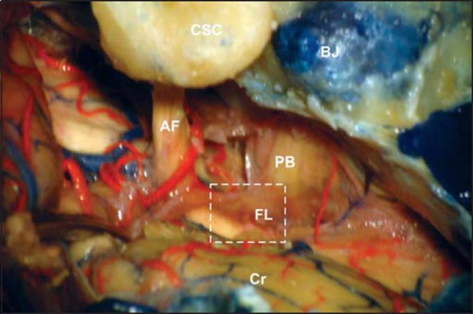
Intraoperative view of the CPA showing key landmarks for ABI placement: Foramen of Luschka (FL, dashed box), acoustic-facial bundle AF, lower cranial nerve bundle PB, cerebellum Cr, semicircular canals CSC, jugular bulb BJ
- Electrode paddle gently inserted into the lateral recess of the fourth ventricle
- Array positioned flat against the cochlear nucleus surface
- Electrode stylet used for insertion, then withdrawn to deploy the paddle
- Dacron mesh backing helps secure the electrode to adjacent structures
STEP 8 — Intraoperative EABR Testing
- Once electrode is in place, Electrically Evoked Auditory Brainstem Response (EABR) is recorded
- Stimulating current applied to individual electrodes
- Presence of EABR confirms contact with cochlear nucleus
- If EABR absent: electrode repositioned
- Test also identifies electrodes causing nonauditory stimulation (facial twitching, neck jerk)
STEP 9 — Securing & Wound Closure
- Electrode lead wire secured in trough
- Receiver/stimulator placed in bony well
- Permanent suture closes soft tissue pocket
- Abdominal fat graft packed around electrode at lateral recess to prevent migration and CSF leak
- Layered wound closure
Diagram: ABI Electrode Placement
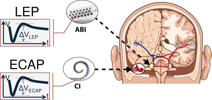
Schematic comparing ABI (electrode paddle on cochlear nucleus, Local Evoked Potential — LEP) vs. Cochlear Implant (spiral array in cochlea, Electrically Evoked Compound Action Potential — ECAP)
7. POSTOPERATIVE MANAGEMENT
- Device activation: Usually delayed 4–6 weeks postoperatively to allow wound healing
- Programming (Mapping): Audiologist maps each electrode individually — threshold, comfort level, pitch
- Nonauditory electrodes deactivated: Electrodes causing facial twitching, tingling, vertigo, or neck sensation are switched off
- EABR-guided mapping used to assist programming
- Aural rehabilitation: Intensive speech therapy and auditory training
- Regular follow-up: For reprogramming and performance optimization
8. COMPLICATIONS
Classification of Complications
┌──────────────────────────────────────────────────────────┐
│                  ABI COMPLICATIONS                       │
├──────────────────┬───────────────────────────────────────┤
│  SURGICAL        │  DEVICE-RELATED                       │
├──────────────────┼───────────────────────────────────────┤
│ CSF leak         │ Electrode migration                   │
│ Meningitis       │ Device failure/extrusion              │
│ Facial palsy     │ Nonauditory stimulation               │
│ Lower CN injury  │ No auditory response                  │
│ (IX, X, XI)      │                                       │
│ Cerebellar       │                                       │
│  contusion       │                                       │
│ Hydrocephalus    │                                       │
│ Pseudomeningo-   │                                       │
│  cele            │                                       │
│ Haemorrhage      │                                       │
│ Tinnitus         │                                       │
│ Headache         │                                       │
└──────────────────┴───────────────────────────────────────┘
8A. Nonauditory Stimulation (Most Common)
- Most frequent complication (seen in majority at initial activation)
- Caused by current spread to adjacent cranial nerves and brainstem structures
- Manifestations:
- Facial twitching (CN VII)
- Neck/shoulder muscle contraction (CN XI)
- Dizziness/vertigo (vestibular nucleus)
- Tingling of face or throat (CN V, IX)
- Chest or epigastric sensations (CN X)
- Management: Deactivate offending electrodes; adjust stimulation parameters; change ground electrode
- 9% of patients report persistent nonauditory sensations (Cummings, p. 3182)
8B. CSF Leak
- Second most common complication
- Occurs through the electrode entry site at the lateral recess
- Prevention: Abdominal fat graft packing around electrode lead at the dura/lateral recess
- Management: Lumbar drain, re-exploration if persistent
8C. Electrode Migration
- Originally the primary cause of ABI failure (early devices, 1979)
- Prevented by Dacron mesh backing on electrode paddle
- Results in loss of auditory response or change in electrode performance
- May require re-exploration
8D. Cranial Nerve Injury
- CN VII (Facial nerve): Palsy — more common in NF2 due to large tumors distorting anatomy
- CN IX, X, XI: Injury during foramen of Luschka identification → dysphagia, hoarseness, aspiration
- CN X monitoring via laryngeal EMG tube helps identify and preserve vagus nerve
8E. Surgical Complications
| Complication | Incidence | Comment |
|---|---|---|
| Meningitis | Rare | Antibiotic prophylaxis; fat graft seal |
| Cerebellar contusion | Rare | Excessive retraction; avoid with careful technique |
| Hydrocephalus | Rare | Due to CSF pathway disruption |
| Pseudomeningocele | Low | Inadequate wound closure |
(Cummings, p. 3124) — These complications significantly less common in nontumor patients than in NF2 patients
9. OUTCOMES
9A. NF2 Patients (Cummings, pp. 3125–3126)
| Outcome Measure | Result |
|---|---|
| Auditory sensation obtained | 81% |
| Improved sentence understanding with lip-reading at 3–6 months | 93% |
| Open-set word recognition (no lip-reading) | ~10% |
| Non-responders | 10–15% |
| Optimal performers (>30% sentence recognition) | 31% (Behr et al.) |
| Mean daily usage (McSorley 2015) | 12.62 hours/day |
Key predictors of poor outcome in NF2:
- Brainstem trauma during tumour removal
- Venous damage to brainstem
- Larger tumours with more distorted anatomy
9B. Nontumor (Non-NF2) Patients
- Superior outcomes compared to NF2 patients
- Higher rates of open-set speech recognition
- Colletti et al. reported some nontumor patients achieving near-normal speech recognition
- Children implanted for cochlear nerve aplasia show encouraging auditory development
9C. Paediatric Outcomes
- Growing experience from Europe (particularly Italy) and now USA
- Goal: Provide auditory input during critical period of auditory development
- Outcomes variable but encouraging for environmental sound awareness and speech development
- Best results when implanted at young age (<3 years)
10. SPECIAL CONSIDERATIONS IN PAEDIATRIC ABI
(Cummings, block 37 — Special Considerations in Pediatric ABI)
PAEDIATRIC ABI — KEY DIFFERENCES FROM ADULT

  • Only at centres with adult ABI AND paediatric neurosurgery expertise
  • Smaller anatomy → higher surgical risk
  • Anaesthetic considerations for infant craniotomy
  • Developmental assessment essential preoperatively
  • Parental counselling about realistic expectations
  • Early activation (as soon as healed) to exploit plasticity
  • Intensive post-implant auditory-verbal therapy
  • Long-term multidisciplinary follow-up
Indications in children:
- Bilateral cochlear nerve aplasia/hypoplasia
- Complete cochlear aplasia (Michel deformity)
- Complete bilateral cochlear ossification (post-meningitis)
- Failed cochlear implantation
- NF2 (rare in children)
11. COMPARISON: ABI vs. COCHLEAR IMPLANT
| Feature | Cochlear Implant (CI) | Auditory Brainstem Implant (ABI) |
|---|---|---|
| Stimulation site | Spiral ganglion / cochlea | Cochlear nucleus, brainstem |
| Prerequisite | Intact cochlear nerve | Intact cochlear nucleus |
| Speech recognition | 70–90% open-set | ~10% open-set |
| Main indication | SNHL with intact CN VIII | Absent/damaged CN VIII |
| Surgical approach | Mastoidectomy | Retrosigmoid/Translabyrinthine |
| Risk profile | Low | High (intracranial) |
| Nonauditory SE | Rare | Common (9% persistent) |
| FDA status | Widely approved | Approved for NF2 ≥12 yrs |
12. RECENT ADVANCES
12A. Penetrating ABI (PABI)
- Penetrating microelectrodes rather than surface paddle
- Theoretically better tonotopic specificity (CN is organized deep-to-surface, not surface-to-surface)
- Results: Did not prove superior to surface ABI in speech perception (Cummings, p. 3125)
- Research ongoing
12B. Auditory Midbrain Implant (AMI)
- Targets inferior colliculus (auditory midbrain)
- Used when cochlear nucleus is also damaged
- Currently experimental
12C. Improved Signal Processing
- Current Focus Stimulation (CFS) — reduces current spread, minimizes nonauditory stimulation
- Tonotopic mapping guided by EABR
- Cloud-based remote programming platforms
12D. Paediatric ABI Expansion
- FDA IDE trials at multiple US centres for paediatric nontumor ABI (since 2013)
- European centres reporting positive language development outcomes
- Combining with cochlear implant if any residual cochlear nerve present
12E. Imaging-Guided Placement
- Intraoperative CT and neuronavigation for electrode positioning
- Intraoperative MRI in specialized centres
12F. Newer Electrode Designs
- Flexible polyimide substrate electrodes (better conform to curved CN surface)
- Increased electrode count for improved frequency resolution
13. FLOWCHART: PATIENT SELECTION AND MANAGEMENT
PATIENT WITH PROFOUND SENSORINEURAL HEARING LOSS
                      │
                      ▼
        Is cochlear nerve intact?
        ┌─────────────┴──────────────┐
       YES                          NO / ABSENT
        │                            │
        ▼                            ▼
  Cochlear Implant               Consider ABI
  (first choice)                     │
                                      ▼
                        Is cochlear nucleus intact?
                        ┌──────────────┴──────────┐
                       YES                        NO
                        │                          │
                        ▼                          ▼
                    ABI indicated            Auditory Midbrain
                        │                    Implant (AMI)
                        ▼                    (experimental)
              Multidisciplinary Evaluation
              MRI / CT / Audiometry / EABR
                        │
                        ▼
             Surgical Candidacy Confirmed?
             ┌──────────┴───────────┐
            YES                    NO
             │                      │
             ▼                      ▼
      ABI Surgery               Hearing Aids /
   (Translabyrinthine         Vibrotactile Aids /
    or Retrosigmoid)           BAHAs / Counselling
             │
             ▼
   Intraoperative EABR
   Confirms Placement
             │
             ▼
   4–6 Weeks: Device Activation
             │
             ▼
   Audiological Programming (Mapping)
   Deactivate nonauditory electrodes
             │
             ▼
   Intensive Aural Rehabilitation
             │
             ▼
   Regular Follow-up & Reprogramming
14. SUMMARY TABLE
| Aspect | Key Points |
|---|---|
| Device | Surface electrode array over cochlear nucleus at lateral recess of 4th ventricle |
| Primary indication | NF2 with bilateral vestibular schwannoma (FDA: age ≥12) |
| Expanding indications | Cochlear aplasia, ossification, CN avulsion, paediatric non-NF2 |
| Surgical approach | Translabyrinthine (NF2) or Retrosigmoid (nontumor) |
| Key landmark | Foramen of Luschka, choroid plexus |
| Confirmation | Intraoperative EABR |
| Most common complication | Nonauditory stimulation |
| Outcomes in NF2 | 81% auditory sensation; ~10% open-set speech |
| Outcomes in nontumor | Significantly better than NF2 |
| Recent advances | PABI, AMI, flexible electrode arrays, paediatric IDE trials |
REFERENCES FROM TEXTBOOKS
- Cummings Otolaryngology Head and Neck Surgery (6th/7th ed.) — Chapter 163: Auditory Brainstem Implantation. Choudhury B, Deep NL, Wilkinson EP, Roland JT Jr. pp. 3119–3130
- Scott-Brown's Otorhinolaryngology, Head and Neck Surgery — Chapter on cochlear and brainstem implants (auditory rehabilitation)
- Stell and Maran's Head and Neck Surgery — Neurotological prosthetic devices
- Dhingra's Diseases of Ear, Nose and Throat — Chapter on hearing rehabilitation and implantable devices
- Hazarika's Textbook of Ear, Nose and Throat — Cochlear and auditory brainstem implants
- Zakir Hussain — ENT principles applied to implantable hearing devices
Key Literature Referenced in Cummings:
- Lenarz M et al.: ABI Part II — subjective assessment (Oticon Medical, 2013)
- Otto SR et al.: Multichannel ABI — update in 61 patients (J Neurosurg, 2002)
- Colletti V et al.: ABI in nontumor patients (OHNS, 2005)
- Behr R et al.: Multi-centre study of optimal ABI performers (2014)
- McSorley A et al.: Subjective benefits of ABI — questionnaire study (2015)
Examiner's Note (RGUHS): For a 50-mark answer, candidates are expected to cover: historical background (5 marks), anatomy (5 marks), indications (10 marks), technique in detail with diagrams (15 marks), complications (10 marks), outcomes and recent advances (5 marks). The above answer covers all these domains with reference to standard ENT textbooks used in the RGUHS curriculum.
This is a shared conversation. Sign in to Orris to start your own chat.