
Show some of the standard medical books
Finding Sources
Finding Sources
Finding Sources
Finding Sources
Finding Sources
Finding Sources
Finding Sources
Here is a full listing of the medical textbooks available in the library, organized by specialty:
Anatomy
- Color Atlas of Ultrasound Anatomy, 3e
- Color Atlas of Human Anatomy Vol. 1, 2, 3
- General Anatomy and Musculoskeletal System (THIEME Atlas)
- Gray's Anatomy for Students
- Imaging Anatomy: Bones, Joints, Vessels and Nerves
- Netter's Atlas of Human Anatomy
Anesthesiology
- Barash, Cullen & Stoelting's Clinical Anesthesia, 9e
- Miller's Anesthesia, 2-Volume Set, 10e
- Morgan and Mikhail's Clinical Anesthesiology, 7e
Biochemistry
- Basic Medical Biochemistry - A Clinical Approach, 6e
- Biochemistry, 8th ed (Lippincott Illustrated Reviews)
- Harper's Illustrated Biochemistry, 32nd Ed
Cardiology
- Braunwald's Heart Disease, 2-Volume Set
- Fuster and Hurst's The Heart, 15th Edition
- Textbook of Clinical Echocardiography
Community Medicine
- Park's Textbook of Preventive and Social Medicine
Dermatology
- Andrews' Diseases of the Skin Clinical Atlas, 2e
- Andrews' Diseases of the Skin (Clinical Dermatology)
- Dermatology, 2-Volume Set, 5e
- Fitzpatrick's Dermatology, Volume 1 & 2
Embryology
- Langman's Medical Embryology
- The Developing Human: Clinically Oriented Embryology
Emergency Medicine
- Rosen's Emergency Medicine, Concepts and Clinical Practice
- Roberts and Hedges' Clinical Procedures in Emergency Medicine
- Tintinalli's Emergency Medicine: A Comprehensive Study
ENT (Otolaryngology)
- Cummings Otolaryngology Head and Neck Surgery
- K.J. Lee's Essential Otolaryngology
- Scott-Brown's Otorhinolaryngology Head & Neck Surgery (Vol 1 & 2)
- Shambaugh Surgery of the Ear
Family Medicine
- Pfenninger and Fowler's Procedures for Primary Care, 3e
- Swanson's Family Medicine Review
- Textbook of Family Medicine, 9e
Forensic Medicine
- Brogdon's Forensic Radiology
- DiMaio's Forensic Pathology, 3rd Edition
- Forensic Anthropology: A Comprehensive Introduction, 2e
- P.C. Dikshit Textbook of Forensic Medicine and Toxicology
- Parikh's Textbook of Medical Jurisprudence
- The Essentials of Forensic Medicine and Toxicology, 36th ed (2026)
Gastroenterology
- Clinical Gastrointestinal Endoscopy, 3e
- Sleisenger & Fordtran's Gastrointestinal and Liver Disease
- Yamada's Textbook of Gastroenterology, 3-Volume Set, 7e
Gastro Surgery
- Maingot's Abdominal Operations
General Surgery
- Bailey and Love's Short Practice of Surgery, 28th Edition
- Current Surgical Therapy, 14e
- Fischer's Mastery of Surgery, 8e
- Mulholland & Greenfield's Surgery, 7e
- Pye's Surgical Handicraft, 22nd Edition
- S. Das: A Manual on Clinical Surgery, 13th Edition
- Schwartz's Principles of Surgery, 2-Volume Set, 11th Edition
- Sabiston Textbook of Surgery, Biological Basis of Modern Surgical Practice
Genetics
- Emery's Elements of Medical Genetics and Genomics
- Thompson & Thompson Genetics and Genomics in Medicine, 9e
Histology
- Histology: A Text and Atlas (with Correlated Cell and Molecular Biology)
- Junqueira's Basic Histology: Text and Atlas, 17e
Immunology
- Cellular and Molecular Immunology
- Janeway's Immunobiology, 10e
- Roitt's Essential Immunology
Internal Medicine
- Firestein & Kelley's Textbook of Rheumatology, 2-Volume Set
- Frameworks for Internal Medicine
- Goldman-Cecil Medicine, 2-Volume Set (International Edition)
- Harrison's Principles of Internal Medicine, 22e (2025)
- Rheumatology, 2-Volume Set (2022)
- Symptom to Diagnosis: An Evidence-Based Guide, 4e
- The Washington Manual of Medical Therapeutics
Laboratory Medicine
- Henry's Clinical Diagnosis and Management by Laboratory Methods
- Quick Compendium of Clinical Pathology, 5e
- Tietz Textbook of Laboratory Medicine, 7e
Microbiology
- Jawetz, Melnick & Adelberg's Medical Microbiology, 28e
- Medical Microbiology, 9e
- Sherris & Ryan's Medical Microbiology, 8e
Nephrology
- Brenner and Rector's The Kidney, 2-Volume Set
- Comprehensive Clinical Nephrology, 7e
- National Kidney Foundation Primer on Kidney Diseases, 8e
Neurology
- Adams and Victor's Principles of Neurology, 12e
- Bradley and Daroff's Neurology in Clinical Practice
- Localization in Clinical Neurology, 8e
- Plum and Posner's Diagnosis and Treatment of Stupor and Coma
Neuroscience
- Eric Kandel's Principles of Neural Science, 6e
- Neuroanatomy through Clinical Cases, 3e
- Neuroscience: Exploring the Brain, 5e
Obstetrics & Gynecology
- Berek & Novak's Gynecology
- Creasy & Resnik's Maternal-Fetal Medicine: Principles and Practice
Ophthalmology
- Kanski's Clinical Ophthalmology: A Systematic Approach, 10e
- The Wills Eye Manual, Office and Emergency Room Diagnosis
Orthopedics
- Campbell's Operative Orthopaedics, 15th Ed (2026)
- Miller's Review of Orthopaedics, 9e
- Rockwood and Green's Fractures in Adults, 10th ed (2025)
Pathology
- Robbins & Kumar Basic Pathology (Robbins Pathology)
- Robbins, Cotran & Kumar Pathologic Basis of Disease
Pediatrics
- Red Book 2021: Report of the Committee on Infectious Diseases
- The Harriet Lane Handbook, 23e (Johns Hopkins)
Pharmacology
- Goodman & Gilman's Pharmacological Basis of Therapeutics
- Katzung's Basic and Clinical Pharmacology, 16e
- Lippincott Illustrated Reviews: Pharmacology
Physiology
- Costanzo Physiology, 7e
- Ganong's Review of Medical Physiology, 26e
- Guyton and Hall Textbook of Medical Physiology
- Medical Physiology
Psychiatry
- Kaplan & Sadock's Comprehensive Textbook of Psychiatry
- Kaplan and Sadock's Synopsis of Psychiatry
- Stahl's Essential Psychopharmacology
- The Clozapine Handbook
- The Maudsley Deprescribing Guidelines
- The Maudsley Prescribing Guidelines in Psychiatry, 15e
Pulmonology
- Fishman's Pulmonary Diseases and Disorders, 2-Volume Set
- Murray & Nadel's Textbook of Respiratory Medicine, 2-Volume Set
Radiology
- Grainger & Allison's Diagnostic Radiology
Urology
- Campbell Walsh Wein Urology, 3-Volume Set
- Hinman's Atlas of Urologic Surgery
- Smith and Tanagho's General Urology, 19e
Total: 126 textbooks spanning 35+ medical specialties. The library includes both foundational texts (Guyton, Robbins, Gray's) and the latest editions of major clinical references (Harrison's 22e 2025, Campbell's Orthopaedics 15e 2026, etc.). You can ask me questions on any of these topics and I'll search the relevant books for you.
Open any one standard book
I'll open Gray's Anatomy for Students - one of the most iconic anatomy textbooks. Let me pull up its table of contents.
Finding Sources
Finding Sources
Finding Sources
Finding Sources
Finding Sources
Finding Sources
Reading File
Reading File
Loading Image
Here is Gray's Anatomy for Students (ISBN: 9780323934237) - opened to its introductory chapter, pages 17 onwards:
📖 Gray's Anatomy for Students
Chapter 1 - Introduction to Anatomy
What is Anatomy?
Anatomy includes structures that can be seen grossly (without magnification) and microscopically (with magnification). The term by itself typically refers to gross or macroscopic anatomy - the study of structures visible to the naked eye. Microscopic anatomy (histology) uses a microscope to study cells and tissues.
"Anatomy forms the basis for the practice of medicine. Anatomy leads the physician toward an understanding of a patient's disease, whether carrying out a physical examination or using the most advanced imaging techniques."
Observation and visualization are the primary techniques for learning anatomy - not mere memorization. Understanding the context of structures (how they relate to each other and function) is what separates true anatomical knowledge from a list of names.
How Can Gross Anatomy Be Studied?
The word "anatomy" derives from the Greek temnein - meaning "to cut" - reflecting its historical roots in cadaver dissection. Today, dissection is augmented by prosected material, plastic models, computer modules, and virtual/augmented reality experiences.
Two main approaches exist:
| Approach | Method | Strength | Weakness |
|---|---|---|---|
| Regional | Study each body region (thorax, abdomen, limb, etc.) as a complete unit | Works well with cadaver dissection | Harder to follow a system throughout the whole body |
| Systemic | Follow one body system (cardiovascular, nervous, etc.) throughout the entire body | Great for understanding whole-system continuity | Can lose regional context |
Important Anatomical Terms
The Anatomical Position: Body erect, face forward, arms at sides with palms facing forward. All directional terms are referenced to this standard position.
Anatomical Planes:
- Median/Sagittal plane - divides body into left and right
- Coronal/Frontal plane - divides body into anterior and posterior
- Transverse/Axial plane - divides body into superior and inferior
Terms of Location:
- Medial / Lateral
- Superior / Inferior
- Anterior / Posterior
- Proximal / Distal
- Superficial - closer to the body surface
- Deep - further from the body surface
Diagnostic Imaging Techniques
In 1895, Wilhelm Roentgen used X-rays from a cathode ray tube to produce the first radiographic exposure - of his wife's hand.
Plain Radiography (X-rays)
X-rays are photons (electromagnetic radiation) generated by a cathode ray tube. As X-rays pass through the body, tissues attenuate (reduce) them differently:
- Air - attenuates X-rays very little (appears black on film)
- Fat - attenuates more than air, less than water
- Soft tissue/water - intermediate attenuation (appears grey)
- Bone - attenuates X-rays the most (appears white/bright)
This is how the classic black-and-white X-ray image is formed.
Fig. 1.2 - Cathode Ray Tube for the Production of X-rays (from the textbook):
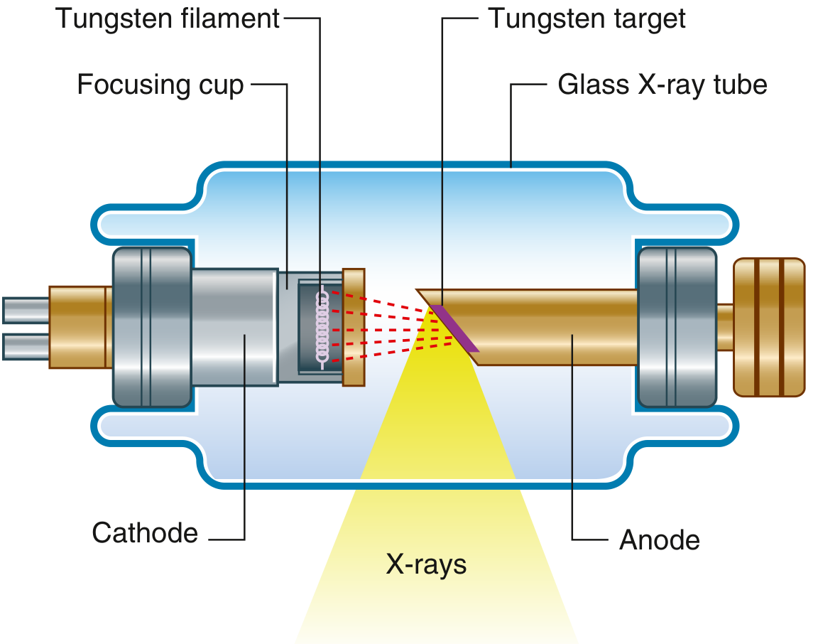
The diagram above shows the cathode (negative electrode with tungsten filament and focusing cup) firing electrons (red dashed lines) at the angled tungsten target (anode), producing the X-ray beam (yellow cone) that exits the glass tube.
- Gray's Anatomy for Students, pp. 17-20
This is just the opening of Block 1 (pages 17-288). The book goes on to cover the full body systematically - Back, Thorax, Abdomen, Pelvis, Lower Limb, Upper Limb, Head & Neck - with rich diagrams, clinical correlations ("In the Clinic" boxes), and imaging anatomy throughout. Would you like me to open any specific chapter or topic from this book?
Search Gray's for clinical applications in a specific area
Which area would you like to explore?
Asking for Preferences
Which anatomical area should I search for clinical applications?
This is a shared conversation. Sign in to Orris to start your own chat.