
Chronic liver disease, etiology,clinical features,diagnosis,treatment
Reading File
Now let me search the medical library for comprehensive coverage of chronic liver disease.
Finding Sources
Finding Sources
Searching PubMed
"chronic liver disease" AND management
Good results. Now let me read the most comprehensive sources in parallel - Harrison's and Sleisenger for etiology/clinical features, and the surgical source for management.
Reading File
Reading File
Reading File
Good start. The Bailey & Love excerpt is relevant. Let me now read the main chronic liver disease section from Bailey & Love and Harrison's block 51 in parallel, plus search for cirrhosis/complications.
Reading File
Reading File
Finding Sources
I see the Harrison's block 51 has neuropathy content. Let me get the dedicated Harrison's cirrhosis section and the Goldman-Cecil section on CLD which will have comprehensive etiology, features, diagnosis and treatment.
Reading File
Reading File
Excellent content coming in. Let me now get the Harrison's section on cirrhosis etiology/diagnosis/treatment and more from Goldman-Cecil on CLD treatment.
Reading File
Reading File
I now have rich content. Let me get the specific Harrison's cirrhosis section and the Goldman-Cecil treatment section to complete the picture.
Finding Sources
Reading File
Excellent - I have comprehensive treatment information now. Let me get the Goldman-Cecil etiology/clinical section and one more read for the Harrison's cirrhosis etiology section.
Finding Sources
Reading File
Let me read the Goldman-Cecil cirrhosis section to get etiology and clinical features, and also the Harrison's cirrhosis main section.
Reading File
Reading File
I now have excellent, comprehensive content from multiple authoritative textbooks. Let me also get the Goldman-Cecil clinical features section and fetch the cirrhosis diagram.
Reading File
Loading Image
I now have comprehensive information from multiple authoritative sources. Let me compile the full, detailed answer.
Chronic Liver Disease (CLD) / Cirrhosis
Definition
Chronic liver disease refers to a spectrum of progressive hepatic injury lasting more than 6 months, with cirrhosis representing its end stage. Cirrhosis is a diffuse process characterized by fibrosis and conversion of normal hepatic architecture to structurally abnormal regenerative nodules. These nodules lack normal lobular organization and are surrounded by fibrous bands. The process is considered irreversible once established, though early fibrosis can regress with treatment of the underlying cause. Clinically, cirrhosis is classified as compensated or decompensated - decompensation being defined by the presence of ascites, variceal bleeding, hepatic encephalopathy, or jaundice.
- Goldman-Cecil Medicine, Chapter 139
- Bailey & Love's Short Practice of Surgery, Chapter 69
Etiology
Major Causes (responsible for the vast majority of cases)
| Cause | Notes |
|---|---|
| Alcohol-related liver disease | Leading cause in Western/industrialized countries |
| Nonalcoholic steatohepatitis (NASH/MASLD) | Increasingly prevalent; closely linked to obesity, T2DM, metabolic syndrome |
| Chronic Hepatitis B | Primary cause in China and Southeast Asia; indefinite antiviral therapy indicated |
| Chronic Hepatitis C | Was the most common cause in the USA until direct-acting antivirals became available |
Other Causes (<2% of all cases each)
-
Autoimmune liver diseases: Primary biliary cholangitis (PBC), primary sclerosing cholangitis (PSC), autoimmune hepatitis
-
Metabolic disorders: Hemochromatosis, Wilson disease, alpha-1 antitrypsin (A1AT) deficiency, glycogen storage diseases, porphyria
-
Biliary obstruction: Biliary atresia, cystic fibrosis, mechanical obstruction
-
Vascular: Budd-Chiari syndrome, veno-occlusive disease, right-sided heart failure (cardiac cirrhosis)
-
Drugs and toxins: Methotrexate, amiodarone, isoniazid, industrial toxins
-
Cryptogenic cirrhosis: Many now thought to be unrecognized NASH
-
Goldman-Cecil Medicine, Table 139-1
-
Harrison's Principles of Internal Medicine 22E, Chapter 355
Pathobiology
The key pathogenic event is activation of hepatic stellate cells (Ito cells) in the space of Disse. Normally quiescent vitamin A-storing cells, they are activated by injury and begin to:
- Lose retinoid stores
- Proliferate
- Secrete excess extracellular matrix (collagen types I and III)
- Transform into myofibroblasts causing progressive fibrosis
Collagen deposition in the space of Disse leads to defenestration of sinusoidal endothelial cells ("capillarization"), reducing sinusoidal diameter and impairing hepatocyte-plasma exchange. Portal hypertension results from both a fixed component (fibrous compression of sinusoids) and a functional/dynamic component (intrahepatic vasoconstriction due to nitric oxide deficiency).
Clinical Features
Symptoms
- Lethargy and weakness - universal, independent of aetiology, often precede jaundice
- Anorexia, weight loss, muscle wasting
- Abdominal distension (ascites)
- Haematemesis or melaena (variceal bleeding)
- Confusion, altered behaviour (hepatic encephalopathy)
- Pruritus (especially in cholestatic disease)
Signs
General:
- Jaundice (scleral icterus)
- Fetor hepaticus (sweet musty breath)
- Parotid enlargement, Dupuytren's contracture (alcoholic CLD)
Skin (stigmata of chronic liver disease):
- Spider naevi (cutaneous vascular abnormalities that blanch on pressure)
- Palmar erythema
- White nails (leukonychia)
- Caput medusae (dilated abdominal veins)
- Bruising/purpura (coagulopathy)
Endocrine:
- Hypogonadism (testicular atrophy)
- Gynaecomastia
- Loss of body hair
Neurological:
- Flapping tremor (asterixis) - the most useful clinical sign of hepatic encephalopathy
- Mental derangement: memory impairment, confusion, personality changes, altered sleep patterns, slow slurred speech
Abdominal:
- Hepatomegaly (early) or small shrunken liver (late)
- Splenomegaly (portal hypertension)
- Ascites (shifting dullness, fluid thrill)
Cardiovascular (hyperdynamic circulation):
-
High cardiac output
-
Large pulse volume
-
Low blood pressure
-
Flushed warm extremities
-
Bailey & Love's Short Practice of Surgery, Chapter 69
-
Goldman-Cecil Medicine, Chapter 139
Complications
The two main mechanisms driving complications are portal hypertension and liver insufficiency:
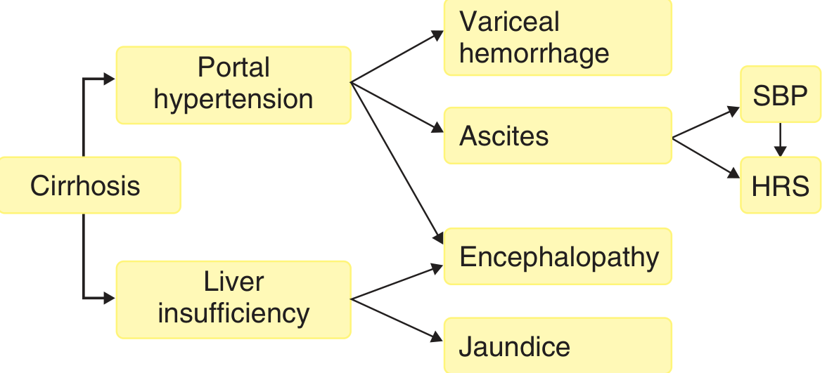
| Complication | Mechanism |
|---|---|
| Gastroesophageal variceal hemorrhage | Portal hypertension - portosystemic collaterals |
| Ascites | Sinusoidal portal hypertension + splanchnic vasodilation + reduced oncotic pressure |
| Spontaneous bacterial peritonitis (SBP) | Bacterial translocation into ascitic fluid (PMN >250/μL) |
| Hepatic encephalopathy | Portosystemic shunting + liver insufficiency (ammonia and other toxins) |
| Hepatorenal syndrome (HRS) | Renal vasoconstriction secondary to splanchnic vasodilation in advanced portal hypertension |
| Hepatopulmonary syndrome | Intrapulmonary vascular dilation → hypoxemia (PaO2 <80 mmHg) |
| Portopulmonary hypertension | Mean PA pressure >25 mmHg on right heart catheterization |
| Coagulopathy | Reduced hepatic synthesis of clotting factors + thrombocytopenia (hypersplenism) |
| Hepatocellular carcinoma (HCC) | Major long-term complication; risk highest in viral cirrhosis |
| Bone disease | Osteopenia, osteoporosis (malabsorption of fat-soluble vitamins) |
| Peripheral neuropathy | Axonal sensorimotor neuropathy, especially with alcoholic/viral CLD |
| Malnutrition | Reduced synthetic function, reduced absorption |
- Harrison's Principles of Internal Medicine 22E, Table 355-2
- Goldman-Cecil Medicine, Chapter 139
Diagnosis
Liver Blood Tests (LFTs)
| Test | Normal Range | Significance |
|---|---|---|
| Bilirubin | 5-17 μmol/L | Impaired conjugation/excretion in CLD; elevated in decompensation |
| ALT/AST | 5-40 IU/L | Hepatocellular damage; AST:ALT >2 suggests alcohol |
| ALP | 30-140 IU/L | Elevated in cholestatic disease; also from bone sources |
| GGT | 10-48 IU/L | Useful marker of alcohol intake |
| Albumin | 35-50 g/L | Reduced in chronic hepatic insufficiency (synthetic function) |
| Prothrombin time (PT) | 12-16 s | Prolonged with impaired synthesis of clotting factors |
The standard monitoring of liver function in CLD is serial measurement of bilirubin, albumin, and PT - Bailey & Love's, Chapter 69
Scoring Systems
- Child-Turcotte-Pugh (CTP) score: Uses bilirubin, albumin, PT, ascites, encephalopathy. Grades A (compensated) to C (decompensated)
- MELD score (Model for End-stage Liver Disease): Uses serum creatinine, bilirubin, INR. Used for transplant organ allocation; score >15 generally qualifies for listing
Specific Investigations
Imaging:
- Ultrasound (US): First-line; assesses liver echogenicity, nodularity, portal vein diameter (>13 mm = portal hypertension), splenomegaly, ascites. Also used for HCC screening (every 6 months in cirrhosis)
- CT/MRI: Better characterization of hepatic architecture, HCC staging, vascular anatomy
- Fibroscan (transient elastography): Non-invasive assessment of liver stiffness/fibrosis
Endoscopy:
- Upper GI endoscopy to grade oesophageal/gastric varices; screen every 2-3 years if no varices, 1-2 years if small varices
Liver Biopsy:
- The gold standard for confirming diagnosis, grading fibrosis (Metavir F0-F4), assessing aetiology, and guiding prognosis
- Contraindicated with coagulopathy (INR >1.5, platelets <60,000)
- Characteristic findings: PAS+/diastase-resistant globules in A1AT deficiency; Mallory hyaline in alcoholic hepatitis
Ascitic Fluid Analysis:
- Serum-Ascites Albumin Gradient (SAAG): >1.1 g/dL = portal hypertension (cirrhosis, cardiac ascites); <1.1 g/dL = non-portal hypertension (malignancy, TB)
- Ascitic PMN >250/μL = SBP (diagnostic)
Cause-Specific Tests:
- Viral serology: HBsAg, HBeAg, HBV DNA; anti-HCV, HCV RNA
- Autoimmune: ANA, anti-smooth muscle Ab (AIH), AMA (PBC), pANCA (PSC)
- Ferritin, transferrin saturation (hemochromatosis); ceruloplasmin, 24h urinary copper, slit-lamp exam for KF rings (Wilson disease)
- Alpha-1 antitrypsin level and phenotype; serum protein electrophoresis
Treatment
General Principles
Treatment of CLD/cirrhosis is directed at:
- Treating the underlying cause to halt/reverse fibrosis
- Avoiding factors that worsen liver disease
- Lowering portal pressure to prevent decompensation
- Screening for and treating complications
- Liver transplantation for end-stage disease
Treating the Underlying Cause
| Cause | Specific Treatment |
|---|---|
| HBV cirrhosis | Indefinite nucleos(t)ide analogue therapy (tenofovir disoproxil fumarate [TDF], tenofovir alafenamide [TAF], or entecavir); PegIFN in compensated cirrhosis only |
| HCV cirrhosis | Direct-acting antivirals (DAAs) - high SVR rates; fibrosis regression documented after cure |
| Alcoholic CLD | Absolute alcohol cessation; corticosteroids (prednisolone) or pentoxifylline in severe alcoholic hepatitis (Maddrey's DF >32); nutritional support |
| NASH/MASLD | Weight reduction (>7-10% body weight), exercise, treatment of metabolic syndrome; resmetirom (THR-β agonist) recently approved |
| Autoimmune hepatitis | Prednisolone ± azathioprine |
| PBC | Ursodeoxycholic acid (UDCA); obeticholic acid if inadequate response |
| PSC | UDCA (controversial); treat dominant strictures endoscopically; transplantation |
| Hemochromatosis | Phlebotomy (weekly venesection until ferritin <50 μg/L); deferasirox/desferrioxamine if not tolerating venesection |
| Wilson disease | D-penicillamine or trientine (copper chelation); zinc (maintenance) |
| A1AT deficiency | Liver transplantation is the only effective treatment |
Portal Pressure Reduction (Prevention of Decompensation)
- Non-selective beta-blockers (NSBBs): Carvedilol 3.125-12.5 mg/day (preferred - also vasodilatory), or propranolol/nadolol. Reduce portal pressure by splanchnic vasoconstriction and decreasing portal inflow. Prevent first variceal hemorrhage and reduce decompensation and death in compensated cirrhosis with significant portal hypertension
- Endoscopic variceal ligation (EVL/banding): Alternative if NSBBs are contraindicated; does not reduce portal pressure but obliterates varices. Combined EVL + NSBBs for secondary prophylaxis
Management of Specific Complications
Ascites:
- Sodium restriction (<88 mmol/day)
- Diuretics: Spironolactone 100 mg/day (first-line, anti-aldosterone), add furosemide 40 mg/day if needed; titrate in 100:40 ratio
- Therapeutic paracentesis with albumin infusion (8 g/L of ascites drained) for tense/refractory ascites
- TIPS (Transjugular Intrahepatic Portosystemic Shunt) for refractory ascites
Spontaneous Bacterial Peritonitis (SBP):
- Empiric IV antibiotics: Cefotaxime 2 g IV q8h (or ceftriaxone) for 5 days
- IV albumin 1.5 g/kg on day 1 and 1 g/kg on day 3 reduces HRS risk
- Long-term prophylaxis: Norfloxacin 400 mg/day or ciprofloxacin 500 mg/day
Variceal Hemorrhage (Acute):
- Vasoactive drugs: Terlipressin (preferred), somatostatin, or octreotide started immediately
- Antibiotic prophylaxis: Ceftriaxone 1 g/day or norfloxacin
- Urgent endoscopy with EVL (within 12 hours)
- TIPS if endoscopic therapy fails
Hepatic Encephalopathy:
- Identify and treat precipitants (infection, GI bleed, constipation, electrolyte disturbance, excessive diuresis, sedatives)
- Lactulose (titrate to 2-3 soft stools/day) - acidifies colon, reduces ammonia absorption
- Rifaximin 550 mg BD - non-absorbable antibiotic, reduces gut bacterial ammonia production; added to lactulose for recurrent episodes
- Protein restriction is no longer recommended (adequate dietary protein 1.2-1.5 g/kg/day)
Hepatorenal Syndrome (HRS):
- HRS type 1 (acute): Terlipressin + albumin (first-line); noradrenaline as alternative
- Discontinue nephrotoxic drugs, NSAIDs, diuretics
- Bridge to transplantation
HCC Surveillance:
- Ultrasound ± AFP every 6 months in all cirrhotic patients
Liver Transplantation
The only definitive cure for end-stage CLD. Indicated for:
- Decompensated cirrhosis (CTP class B/C; MELD >15)
- Acute liver failure
- HCC within Milan criteria (single nodule ≤5 cm; ≤3 nodules each ≤3 cm)
- Metabolic liver disease causing systemic complications
Absolute contraindications: Extrahepatic malignancy, uncontrolled systemic sepsis, severe cardiopulmonary disease, active alcohol/substance use.
Current 1-year survival >90%. In the US in 2022, 9,528 patients received liver allografts.
- Harrison's Principles of Internal Medicine 22E, Chapter 355-356
- Goldman-Cecil Medicine, Chapter 139
- Bailey & Love's Short Practice of Surgery, Chapter 69
Prognosis
| CTP Class | Points | 1-year survival | 2-year survival |
|---|---|---|---|
| A (compensated) | 5-6 | 100% | 85% |
| B | 7-9 | 80% | 60% |
| C (decompensated) | 10-15 | 45% | 35% |
MELD score >25 carries approximately 3-month mortality of 50% without transplantation.
Recent evidence note: A 2025 meta-analysis (PMID 40204206) reports high and growing global prevalence of steatotic liver disease (MASLD/MAFLD), reinforcing it as a rapidly emerging major cause of CLD worldwide alongside traditional causes.
This is a shared conversation. Sign in to Orris to start your own chat.