
Deep tendon reflexes slow in hypothyroidism mechanisms
- Reduced Na+/K+-ATPase activity slowing muscle membrane repolarization
- Decreased myosin ATPase activity impairing muscle fiber relaxation
- Altered muscle fiber composition (shift toward slow Type I fibers)
- Reduced actin-myosin cross-bridge cycling speed
- Impaired calcium reuptake by the sarcoplasmic reticulum
hypothyroidism deep tendon reflex relaxation mechanism
deep tendon reflex arc mechanism diagram
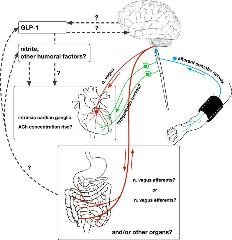
This pathophysiology diagram illustrates the 'remote preconditioning reflex arc,' a complex neural and humoral pathway involved in remote ischemic preconditioning (RIPre). The diagram depicts a stimulus applied to an arm via a blood pressure cuff, which triggers afferent somatic nerves that project to the brain. Efferent signals are shown descending from the central nervous system via the vagus nerve (n. vagus) and sympathetic nerves to the heart and other visceral organs, specifically the intestine. Key cardiovascular features include the involvement of intrinsic cardiac ganglia and a potential rise in acetylcholine (ACh) concentration in the myocardium. The diagram highlights several scientific uncertainties using question marks, specifically regarding the role of sympathetic nerves, whether abdominal vagal fibers are afferent or efferent, and the origin of humoral factors like GLP-1 and nitrite. Humoral pathways are shown as dashed lines connecting systemic factors to the brain and heart, indicating a parallel mechanism of cardioprotection alongside the primary neural reflex arc.
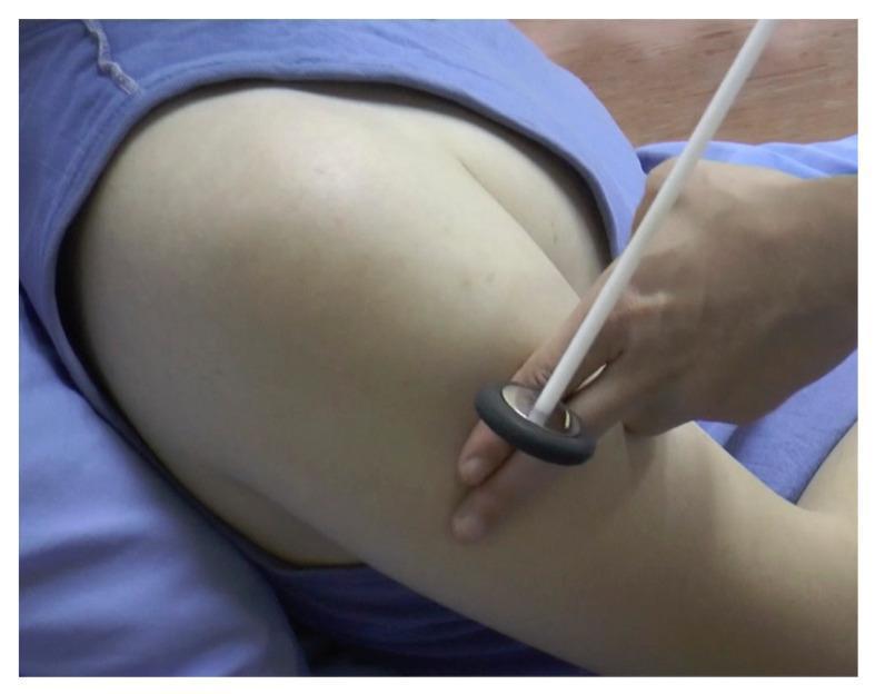
This clinical procedural photograph demonstrates the technique for eliciting a deep tendon reflex in the upper extremity, specifically targeting the deltoid tendon. A healthcare provider is shown using a Babinski-style reflex hammer, characterized by a slender, light-colored handle and a circular, disk-shaped head with a dark rubberized peripheral ring. The examiner's hand holds the handle between the thumb and fingers to provide a controlled, swinging motion. The patient is positioned supine, with the shoulder and upper arm exposed. The reflex hammer's head is shown at the point of impact on the lateral aspect of the upper arm, near the insertion of the deltoid muscle. This procedure is fundamental in a neurological examination to assess the integrity of the C5 and C6 spinal nerve roots and the axillary nerve. The image serves as an educational tool for medical students and clinicians to visualize proper tool handling and anatomical targeting for upper limb reflex testing.
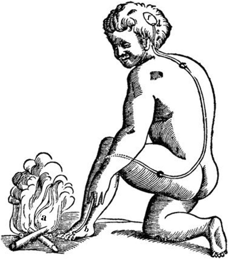
This historical anatomical illustration depicts René Descartes' early 17th-century mechanistic theory of the reflex arc. The diagram shows a human figure kneeling by a fire, illustrating the physiological response to a noxious stimulus. Key elements are labeled to represent a functional pathway: (a) indicates the external stimulus (fire); (b) represents the peripheral sensory site on the skin of the foot; (c) shows a 'fine thread' or nerve fiber extending from the foot, up the leg and back, to the head. The path includes nodes representing the spinal column or ganglia. Within the brain, (d) and (e) represent mechanical valves that govern the release of 'animal spirits' from a central cavity (f). This anatomical diagram serves as a foundational concept in the history of neuroscience, demonstrating an early understanding of the stimulus-response pathway, where sensory input travels to the central nervous system to trigger a motor reflex. It is a critical visual resource for teaching the evolution of neurophysiology and the history of medicine.
Slow ("Hung-Up") Deep Tendon Reflexes in Hypothyroidism
1. Reduced Myosin ATPase Activity (Slowed Contraction Phase)
- Thyroid hormone upregulates fast MHC isoforms (MHC-I, MHC-II) and increases myosin ATPase activity.
- In hypothyroidism, myosin ATPase activity is decreased. Since the speed of muscle contraction is proportional to the rate of ATP hydrolysis by myosin cross-bridges, both the contraction phase and the relaxation phase are prolonged.
"The speed of the contractile process is related to the quantity of myosin adenosine triphosphate (ATPase), which is increased in hyperthyroid muscle and decreased in hypothyroid muscle."
- Adams and Victor's Principles of Neurology, 12th Ed.
2. Impaired Calcium Reuptake by the Sarcoplasmic Reticulum (Key Mechanism of Slowed Relaxation)
- Muscle relaxation requires rapid re-uptake of cytosolic Ca²⁺ back into the sarcoplasmic reticulum (SR) by the SERCA pump (sarcoplasmic/endoplasmic reticulum Ca²⁺-ATPase).
- Thyroid hormone stimulates SERCA pump expression and activity. In hypothyroidism, SERCA activity is reduced, so Ca²⁺ removal from the cytoplasm is slow.
- As long as cytoplasmic [Ca²⁺] remains elevated, actin-myosin cross-bridges remain engaged and the muscle cannot fully relax.
"The speed of relaxation depends on the rate of release and reaccumulation of calcium in the endoplasmic reticulum. This is slowed in hypothyroidism and increased in hyperthyroidism."
- Adams and Victor's Principles of Neurology, 12th Ed.
"The basis of this disorder [pseudomyotonia] appears to be slowness in the reaccumulation of calcium ions in the endoplasmic reticulum and in the disengagement of actin and myosin filaments."
- Adams and Victor's Principles of Neurology, 12th Ed.
3. Slowed Actin-Myosin Cross-Bridge Cycling and Disengagement
4. Shift in Muscle Fiber Composition
- There is a shift toward slow-twitch Type I fibers, which have inherently slower contraction-relaxation kinetics.
- Biopsy findings in hypothyroid myopathy show altered proportions of fiber types (increase in small fibers, either type 1 or 2) and slight distension of the sarcoplasmic reticulum - Adams & Victor's.
- This compounding of slow-fiber physiology on top of impaired Ca²⁺ handling amplifies the clinical finding.
5. Reduced Na⁺/K⁺-ATPase Activity (Membrane-Level Contribution)
- Alters muscle membrane resting potential
- Slows membrane repolarization after action potentials
- Contributes to the overall sluggishness of the neuromuscular response, though this is secondary to the Ca²⁺ mechanism.
Clinical Correlates
| Feature | Mechanism |
|---|---|
| Prolonged relaxation of Achilles reflex | Impaired SERCA Ca²⁺ reuptake |
| Slowed contraction phase | Reduced myosin ATPase |
| Pseudomyotonia (no true myotonic EMG discharges) | Ca²⁺/cross-bridge mechanism, not ionic channel defect |
| Myoedema (local mounding on percussion) | Abnormal muscle membrane excitability + slow Ca²⁺ clearance |
| Elevated CK (up to 10x normal) | Muscle fiber injury from metabolic slowing |
| Corrects with thyroxine replacement | All mechanisms are reversible |
"Features of slow muscle contraction and relaxation occur in 25% of patients [with hypothyroidism]; the relaxation phase of muscle stretch reflexes is characteristically prolonged and best observed at the ankle or biceps brachii reflexes."
- Harrison's Principles of Internal Medicine, 22nd Ed.