

Pathology practical peripheral blood smear interpretation 2 mbbs
CLL chronic lymphocytic leukemia peripheral blood smear smudge cells lymphocytosis
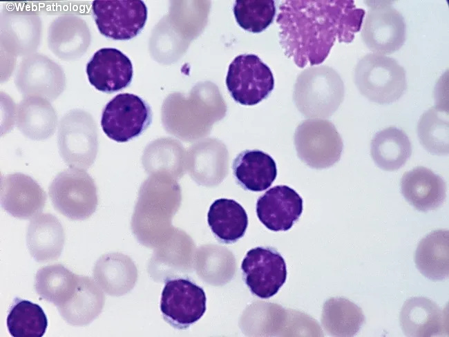
Peripheral blood smear from a patient with suspected chronic lymphocytic leukemia (CLL) shows a predominance of small, mature-appearing lymphocytes with scant cytoplasm and clumped nuclear chromatin, accompanied by fragile cells that readily disrupt during smear preparation, producing numerous smudge or basket cells visible on the slide. The image depicts a single smudge cell in the upper right, with numerous small darkly staining lymphocytes occupying the field. The smear was prepared with Wright-Giemsa stain and examined under light microscopy at high magnification (approximately 1000x total magnification with oil immersion). Clinically, CLL is defined by absolute mature lymphocytosis of ≥5.0 × 10^9/L sustained for ≥3 months, often asymptomatic and detected incidentally on complete blood count. Morphology alone is insufficient for diagnosis; immunophenotyping showing co-expression of CD5, CD19, CD23 with weak surface immunoglobulin and light chain restriction supports CLL diagnosis. Smudge cells result from fragile leukemic cells and reflect underlying cytoskeletal fragility, though they are not specific. The image illustrates typical features: monomorphic lymphocytes, scant cytoplasm, condensed chromatin, and absent prominent nucleoli. This finding has diagnostic significance, guiding further workup, including flow cytometry, cytogenetics, and molecular studies, to distinguish CLL from other chronic lymphoproliferative disorders such as small lymphocytic lymphoma (SLL), mantle cell lymphoma, prolymphocytic leukemia, and reactive lymphocytosis.
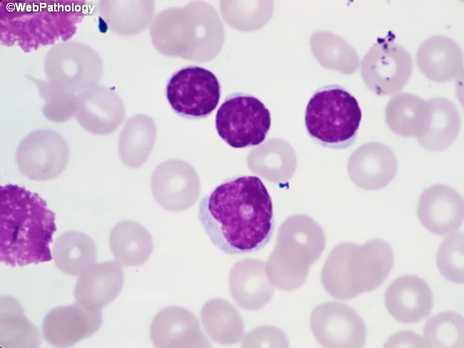
Imaging Modality: Brightfield light microscopy of a Wright-Giemsa stained peripheral blood smear. The primary subject is leukocytes from circulating blood, with focus on small mature-appearing lymphocytes and rare prolymphocytes. At center-right, several lymphocytes exhibit condensed chromatin and scant cytoplasm, consistent with chronic lymphocytic leukemia (CLL) morphology. Along the left edge, characteristic smudge cells are visible, reflecting fragile lymphocytes commonly seen in CLL. A prolymphocyte located just below center presents with slightly irregular nuclear contours, more dispersed chromatin, a prominent nucleolus, and modestly increased cytoplasm. In CLL, prolymphocytes usually comprise less than 2% of neoplastic cells; when 10–15%, the term atypical CLL is used and carries implications of aberrant immunophenotype, cytogenetic abnormalities, cytopenias, refractoriness to therapy, and worse prognosis. If prolymphocytes predominate, consideration should be given to B-cell prolymphocytic leukemia. This image demonstrates key diagnostic features including lymphoid morphology, prolymphocytic variant, and smear artifacts. Clinically, these findings correlate with lymphocytosis and potential anemia or thrombocytopenia in affected patients. Definitive characterization requires ancillary testing such as flow cytometry, immunophenotyping, and cytogenetics. The morphology supports a differential diagnosis that includes CLL with prolymphocytic transformation, atypical CLL, and B-PLL, guiding prognosis and treatment planning.
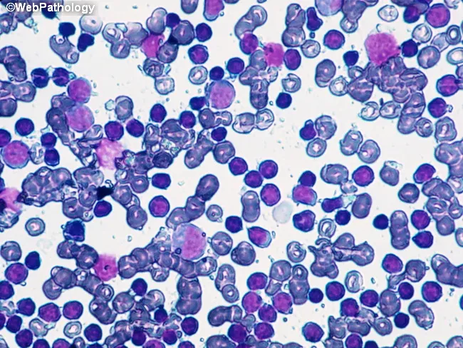
Imaging modality: Brightfield light microscopy of a Wright-Giemsa stained peripheral blood smear. The slide shows a predominant population of small to medium-sized lymphoid cells with scant pale cytoplasm and dense, clumped chromatin. Numerous cells exhibit mature-appearing B lymphocyte morphology. A fine background of erythrocytes and occasional smudge cells is present, with no overt blasts or granulocytic abnormalities. The overall pattern is lymphocytosis with a monomorphic lymphocytic population, compatible with monoclonal B-cell lymphocytosis (MBL) or the chronic lymphocytic leukemia (CLL) spectrum. Immunophenotypic confirmation (CD5+, CD23+, CD19+, surface Ig) is typically required for definitive classification, but is outside the scope of this image. The cluster appears relatively uniform, suggesting clonality rather than reactive lymphocytosis. Genetic associations commonly reported with CLL/MBL include 13q14 deletion and trisomy 12, though such findings require molecular testing. Clinically, CLL is defined by an absolute lymphocyte count ≥5.0 x 10^9/L in peripheral blood for at least 3 months; values below this threshold meet criteria for MBL. The image illustrates morphologic correlates of indolent clonal B-cell expansions and underscores the need to integrate flow cytometry and cytogenetics for diagnosis, prognosis, and management decisions. This image is educational for hematology, pathology, and cytology reference libraries.
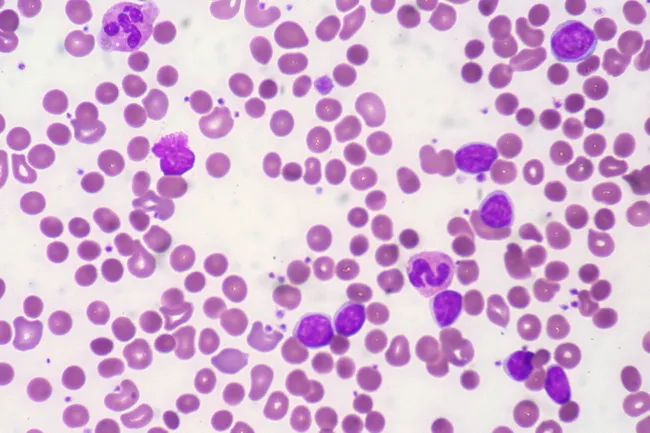
Peripheral blood smear from a patient with known chronic lymphocytic leukemia (CLL) showing autoimmune hemolytic anemia (AIHA). Brightfield microscopy of Wright-Giemsa stained smear at 1000x (oil immersion) reveals two dominant cell populations: numerous small mature lymphocytes typical of CLL and red blood cells showing spherocytic deformation. A smudge cell is evident just left of center, a classical clue for CLL. Red cells display reduced central pallor consistent with spherocytes, indicating intravascular or extravascular hemolysis driven by autoantibodies. The background shows mild polychromasia and rare nucleated RBCs, consistent with a robust reticulocyte response; the corrected reticulocyte count in this case is markedly elevated (>7%). Platelets are not significantly abnormal. These features collectively support autoimmune destruction of erythrocytes in the setting of CLL. Diagnostic significance lies in recognizing AIHA as a common autoimmune complication in CLL, with a clinical picture of sudden Hb decline and compensatory erythropoiesis. Differential considerations include drug-induced hemolysis, other causes of hemolytic anemia, or marrow infiltration by CLL. Clinical correlation includes therapy implications (steroids, rituximab) and close monitoring of hemolysis markers. This image is a valuable teaching resource for hematology morphology, AIHA in CLL, and differential diagnosis in anemia.
Peripheral Blood Smear Interpretation - Case H7
DIAGNOSIS: Chronic Lymphocytic Leukemia (CLL)
Step 1: Analyse the CBC Parameters
| Parameter | Patient Value | Normal Range | Interpretation |
|---|---|---|---|
| Haemoglobin | 9.3 g/dL | M: 14-17 | Anaemia (moderate) |
| Total RBC | 3 million/mm³ | M: 4.5-5.5 | Reduced |
| Total WBC | 4,00,000/mm³ | 4,000-11,000 | Markedly elevated (leukocytosis) |
| Neutrophils | 4% | 40-80% | Relative neutropenia |
| Prolymphocytes | 5% | - | Abnormal cells present |
| Lymphocytes | 90% | 20-40% | Marked lymphocytosis |
| PCV | 28.5% | M: 40-50% | Low |
| MCV | 95.6 fL | 82-92 | Slightly high (normocytic-macrocytic) |
| MCHC | 32.6 g/dL | 32-35 | Normal |
| RDW | 13% | 11.6-14 | Normal |
| Platelet count | 1,20,000/mm³ | 1,50,000-4,50,000 | Mild thrombocytopenia |
- WBC = 4,00,000/mm³ (400 x 10³/µL)
- Lymphocytes = 90% → ALC = 3,60,000/mm³ = 360 x 10³/µL
- This is massively elevated (normal: 1,000-4,800/mm³)
Step 2: Peripheral Smear Morphology Summary
- Lymphocytosis - massive increase in mature, small lymphocytes
- Prolymphocytes present (5%) - slightly larger cells with a prominent nucleolus
- Smudge/basket cells (visible in the smear image) - pathognomonic finding; these are fragile CLL lymphocytes that rupture during smear preparation
Step 3: Interpretation
PRIMARY DIAGNOSIS: Chronic Lymphocytic Leukemia (CLL)
-
Age and sex: 70-year-old male - CLL is predominantly a disease of older adults; median age at diagnosis is ~71 years with a 2:1 male predominance (Harrison's 22E)
-
Clinical presentation: Fever and backache - constitutional symptoms are characteristic of CLL
-
Massive lymphocytosis: ALC >5,000/mm³ (here far exceeding this) is the diagnostic hallmark. CLL is defined by an absolute lymphocyte count of >5 x 10⁹/L of monoclonal B cells (Robbins Pathologic Basis of Disease)
-
Morphology: Small mature-appearing lymphocytes with scant cytoplasm and clumped/condensed chromatin are characteristic CLL cells
-
Smudge cells (Basket cells): These fragile leukemic lymphocytes rupture during smear preparation - they are the pathognomonic smear finding in CLL (Robbins)
-
Prolymphocytes 5%: When prolymphocytes are 10-55% of the lymphoid cells, it is termed CLL with prolymphocytic transformation (CLL/PL) - in this case at 5%, it is CLL with increased prolymphocytes (atypical CLL), which carries a slightly worse prognosis
-
Anaemia + Thrombocytopenia: Bone marrow infiltration by CLL cells causes displacement of normal haematopoiesis → anaemia and thrombocytopenia
Step 4: Peripheral Smear Findings - Classic CLL Features

Step 5: Additional Features to Mention in Viva / Write-up
Rai Staging (Clinical Staging of CLL)
| Stage | Features | Risk |
|---|---|---|
| 0 | Lymphocytosis only | Low |
| I | + Lymphadenopathy | Low/Intermediate |
| II | + Splenomegaly or Hepatomegaly | Intermediate |
| III | + Anaemia (Hb <11 g/dL) | High |
| IV | + Thrombocytopenia (<100,000) | High |
Step 6: Immunophenotype (For Confirmation)
- CD5 co-expression on B cells is abnormal and key to diagnosis
- This is confirmed by flow cytometry
Step 7: Complications of CLL
- Autoimmune haemolytic anaemia (AIHA) - Coombs positive, spherocytes on smear (accounts for some of the anaemia here)
- Immune thrombocytopenic purpura (ITP) - autoimmune platelet destruction
- Recurrent infections - due to hypogammaglobulinaemia
- Richter's transformation - transformation to aggressive large cell lymphoma (~5-10%)
Summary for Answer Writing (2 MBBS Practical Format)
Diagnosis: Chronic Lymphocytic Leukemia (CLL)Evidence:
- 70-year-old male with fever and backache
- Marked leukocytosis (TLC = 4,00,000/mm³) with lymphocytosis (90%)
- Absolute lymphocyte count markedly elevated (>5,000/mm³ diagnostic threshold)
- PBS shows small mature lymphocytes, prolymphocytes, and smudge/basket cells
- Anaemia (Hb 9.3 g/dL) and thrombocytopenia (1,20,000/mm³)
- Rai Stage III-IV disease
Confirmatory tests: Flow cytometry (CD5+/CD19+/CD23+), bone marrow biopsy, cytogenetics (del 13q, del 11q, del 17p, trisomy 12)