
2019 ACR /EULAR criteria for SLE and management of SLE
"systemic lupus erythematosus"[MeSH Terms] AND "2019 classification criteria"
systemic lupus erythematosus butterfly malar rash skin manifestations
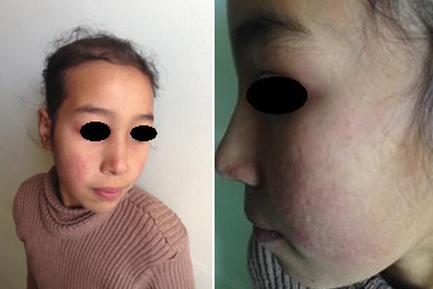
This clinical photograph displays a pediatric patient with dermatological manifestations consistent with Systemic Lupus Erythematosus (SLE). The image consists of two panels: a frontal view and a lateral close-up of the face. A distinctive malar rash, often referred to as a 'butterfly rash,' is visible across the nasal bridge and bilateral cheeks, notably sparing the nasolabial folds. The affected skin exhibits erythematous discoloration and an irregular texture. Higher magnification reveals atrophic scarring within the malar region, characterized by thin, slightly depressed skin with altered pigmentation. The forehead and peri-orbital regions appear relatively unaffected. These visual features are classic diagnostic markers for lupus-related skin involvement, illustrating the chronic inflammatory and sequela phases of the disease. The content is relevant for medical students and clinicians studying rheumatology, dermatology, and autoimmune connective tissue disorders.
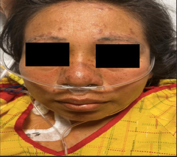
A clinical photograph of a female patient's face demonstrating classic dermatological manifestations of systemic lupus erythematosus (SLE). A confluent, erythematous malar rash is visible in a characteristic 'butterfly distribution,' spanning the bridge of the nose and both cheeks. Notably, the nasolabial folds are spared from the erythema. In addition to the malar rash, there are multiple discrete, raised, hyperpigmented, and round discoid lesions located along the hairline and eyebrows, suggesting discoid lupus involvement. Mild peri-orbital edema is present. The patient is shown with a nasal cannula in place, indicating respiratory support, consistent with potential systemic complications such as pleural effusion. This image serves as a teaching tool for identifying pathognomonic skin signs of autoimmune connective tissue diseases, specifically highlighting the distinction between the malar rash and lesions that involve the forehead and scalp.
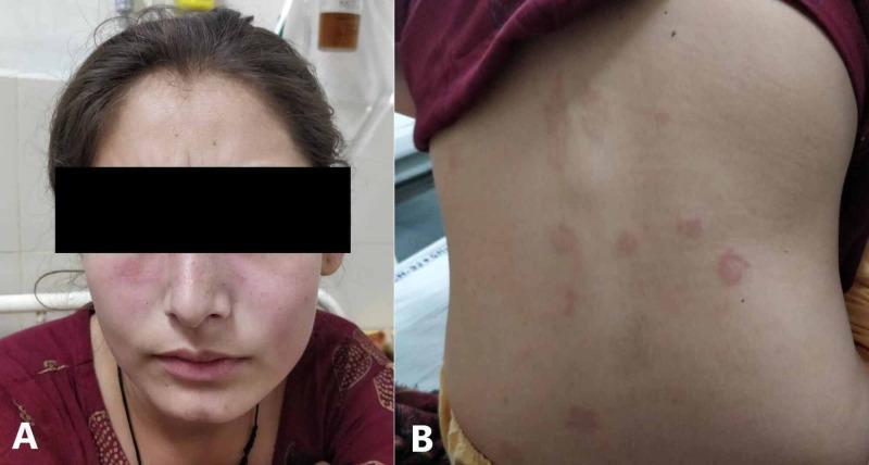
Two-panel clinical photograph illustrating dermatological manifestations of systemic lupus erythematosus (SLE) in a 20-year-old female. Image A presents a frontal view of the patient's face showing a classic malar or 'butterfly' rash. This is characterized by symmetric, erythematous, non-blanching patches across the malar eminences (cheeks) and bridge of the nose. Notably, there is distinct nasolabial sparing, a hallmark feature that differentiates this from other facial dermatoses like seborrheic dermatitis. Image B displays the patient's posterior trunk (back), exhibiting multiple discrete, erythematous, circular to oval maculopapular lesions. These lesions are scattered across the upper back and flank areas, appearing as distinct spots against otherwise clear skin, in contrast to the diffuse confluent redness on the face. These clinical findings are visually representative of acute cutaneous lupus erythematosus flares. The presentation is contextually associated with multisystem involvement, including pericarditis and hematologic abnormalities.
2019 ACR/EULAR Classification Criteria for SLE & Management
2019 EULAR/ACR Classification Criteria for SLE
Entry Criterion (Mandatory)
ANA titer ≥ 1:80 (on HEp-2 cells or equivalent positive test) - if absent, do not classify as SLE
- Note: Up to 15% of the healthy general population may have a positive ANA at 1:80; SLE patients typically have titers >1:320
Scoring System
CLINICAL DOMAINS
| Domain | Criterion | Points |
|---|---|---|
| Constitutional | Fever | 2 |
| Neuropsychiatric | Delirium | 2 |
| Psychosis | 3 | |
| Seizure | 5 | |
| Mucocutaneous | Non-scarring alopecia | 2 |
| Oral ulcers | 2 | |
| Subacute cutaneous OR discoid lupus | 4 | |
| Acute cutaneous lupus (malar rash) | 6 | |
| Musculoskeletal | Joint involvement (synovitis ≥2 joints OR morning stiffness ≥30 min) | 6 |
| Serosal | Pleural or pericardial effusion | 5 |
| Acute pericarditis | 6 | |
| Hematologic | Leukopenia (<4000/µL) | 3 |
| Thrombocytopenia (<100,000/µL) | 4 | |
| Autoimmune hemolysis | 4 | |
| Renal | Proteinuria >0.5 g/24 h | 4 |
| Renal biopsy: Class II or V lupus nephritis | 8 | |
| Renal biopsy: Class III or IV lupus nephritis | 10 |
IMMUNOLOGIC DOMAINS
| Domain | Criterion | Points |
|---|---|---|
| Antiphospholipid antibodies | Anticardiolipin Ab OR anti-β2GPI Ab OR lupus anticoagulant | 2 |
| Complement proteins | Low C3 OR low C4 | 3 |
| Low C3 AND low C4 | 4 | |
| SLE-specific antibodies | Anti-dsDNA OR anti-Smith | 6 |
A renal biopsy showing Class III or IV lupus nephritis alone (10 points) + ANA positivity = sufficient to classify as SLE.
Key Points about the Criteria
- ~5% of SLE patients are ANA-negative and are therefore excluded from clinical trial enrollment under this system
- Incomplete lupus (features of SLE without meeting the full criteria) is a recognized entity - some patients may warrant treatment even without reaching 10 points
- The criteria were derived and validated against expert clinical diagnosis as the reference standard
Management of SLE
General Principles
- Hydroxychloroquine (HCQ) for ALL patients unless contraindicated - reduces flares, organ damage, and mortality; also reduces risk of congenital heart block in fetuses of pregnant SLE mothers
- Minimize glucocorticoid exposure - chronic use is a major source of organ damage
- Treat to target: aim for remission or low disease activity
- Screen and manage cardiovascular risk factors aggressively (SLE carries 4-8x increased CV risk)
- Sun protection and photoprotection are important adjuncts
Non-Organ-Threatening Disease
| Manifestation | First-line | Add-on/Alternatives |
|---|---|---|
| Constitutional (fatigue, fever) | HCQ ± low-dose GC | NSAIDs (with caution) |
| Arthritis/Arthralgia | HCQ, NSAIDs (with caution) | Methotrexate (MTX), low-dose GC |
| Cutaneous (mild-moderate) | Topical GC/calcineurin inhibitors, HCQ | MTX, quinacrine as add-on to HCQ |
| Cutaneous (refractory/severe) | HCQ + MTX or MMF | Anifrolumab (add-on for severe skin disease) |
| Serositis | NSAIDs, colchicine, low-dose GC | - |
NSAIDs must be used cautiously in SLE due to risks of aseptic meningitis, hypertension, renal dysfunction, and elevated transaminases.
Organ-Threatening / Severe Disease
Lupus Nephritis
| Phase | Class III/IV LN | Class V (Membranous) |
|---|---|---|
| Induction | MMF (2-3 g/day) OR low-dose IV cyclophosphamide (Euro-Lupus) + GC | MMF ± calcineurin inhibitor (voclosporin or tacrolimus) |
| Add-on | Belimumab added to MMF (improved outcomes) | - |
| Maintenance | MMF OR azathioprine (AZA) + HCQ | MMF |
| Refractory | Rituximab (off-label); obinutuzumab (under study) | - |
- SGLT2 inhibitors are increasingly used for renal protection in LN
- ACE inhibitors or ARBs for renal and cardiovascular protection
- Class I/II: generally HCQ ± low-dose GC; no aggressive immunosuppression needed
Neuropsychiatric SLE
- Seizures: antiepileptics + immunosuppression (high-dose GC + CYC for severe cases)
- Anifrolumab and belimumab are not recommended for severe neuropsychiatric SLE
- Psychosis: requires high-dose GC; distinguish drug-induced (steroid psychosis) from SLE psychosis
Hematologic Manifestations
- Autoimmune hemolytic anemia / thrombocytopenia: high-dose GC; IVIG (2 g/kg over 3-5 days) for refractory cases
- Rituximab for refractory cytopenias
Biologic and Targeted Therapies
| Drug | Mechanism | Indication |
|---|---|---|
| Belimumab (Benlysta) | Anti-BLyS/BAFF; blocks B-cell survival | Moderately active SLE; active lupus nephritis (IV form) |
| Anifrolumab (Saphnelo) | Anti-type I IFN receptor (IFNAR1) | Moderate-to-severe SLE (add-on to standard of care); severe skin disease |
| Rituximab (anti-CD20) | B-cell depletion | Refractory LN, refractory cytopenias (off-label) |
| Voclosporin (Lupkynis) | Calcineurin inhibitor | Class III/IV/V LN (in combination with MMF) |
Glucocorticoid Strategy
- Acute severe flares: IV methylprednisolone (500-1000 mg/day x 3 days) then taper
- Maintenance: aim for prednisone ≤5 mg/day or complete taper when possible
- Chronic high-dose GC is associated with cataracts, osteoporosis, avascular necrosis, infections, and cardiovascular disease
- Osteoporosis prophylaxis (calcium, vitamin D, bisphosphonates) for patients on long-term GC
Antiphospholipid Syndrome (APS) Complicating SLE
- Positive aPL without thrombosis + primary prevention: low-dose aspirin ± HCQ
- First venous thrombosis: warfarin (target INR 2-3)
- Arterial thrombosis or recurrent thrombosis: warfarin (INR 3-4); avoid DOACs in high-risk aPL profiles
Pregnancy in SLE
- Plan pregnancy during remission/low disease activity
- HCQ should be continued throughout pregnancy (reduces flares and risk of fetal congenital heart block)
- Avoid teratogenic agents (MMF, MTX, CYC) - switch to AZA or calcineurin inhibitors if immunosuppression needed
- Low-dose aspirin + low-molecular-weight heparin for patients with aPL antibodies (to prevent pregnancy loss)
- Estrogen-containing contraceptives are avoided in active or moderate-severe disease and in those with aPL antibodies
Monitoring
- CBC, CMP, urinalysis, urine protein-creatinine ratio at each visit
- Anti-dsDNA and complement levels (C3, C4) for disease activity tracking - rising anti-dsDNA + falling complements predict flares
- Ophthalmologic screening for HCQ retinopathy (annual after 5 years of use)
- Cardiovascular risk assessment (lipids, BP, glucose)
- Bone densitometry for patients on chronic GC
Prognosis
- SLE shows a bimodal mortality pattern: early deaths from active disease and infections; late deaths from atherosclerotic cardiovascular disease
- Complete sustained remission is achieved in <10% of patients
- Poor prognostic factors: renal involvement, neuropsychiatric disease, Black/Hispanic ethnicity, lack of health insurance, positive anti-dsDNA, low complement
- Predictors of good prognosis: early diagnosis, HCQ use, low disease activity at 2 years
- Harrison's Principles of Internal Medicine, 22e (2025, McGraw Hill), Chapter 385-386 - primary source for criteria, pathogenesis, and management
- Goldman-Cecil Medicine, International Edition, Table 245 - criteria table
- Washington Manual of Medical Therapeutics - clinical pearls on workup
- Rheumatology, 2-Volume Set (2022, Elsevier) - classification comparison
- Based on the 2019 EULAR/ACR criteria (Aringer M et al., Arthritis Rheumatol 2019;71:1400-12) and 2023 EULAR SLE management recommendations (Fanouriakis A et al., Ann Rheum Dis 2024;83:15)