
How to use you
What I Can Help With
🩺 Medical & Clinical Questions
- "What are the causes of chest pain?"
- "How does metformin work?"
- "Explain the brachial plexus anatomy"
📚 Research & Literature
- "What does the latest evidence say about statins in primary prevention?"
- "Find recent meta-analyses on cognitive behavioral therapy for depression"
🌐 General Knowledge & Web Search
- "What's the latest on AI regulation in Europe?"
- "Explain how interest rates affect inflation"
💻 Code & Data
- "Write a Python script to clean this CSV"
- "Debug this JavaScript function"
📄 Documents & Files
- "Create a PowerPoint on diabetes management"
- "Make an Excel budget tracker"
🖼️ Images
- Find medical images, diagrams, ECGs, histology slides
- Analyze images you upload
- Generate original visuals, posters, or diagrams
💰 Tax & Finance (India)
Tips for Best Results
| Tip | Example |
|---|---|
| Be specific | "Dosing of amoxicillin for pediatric pneumonia" vs. "antibiotics" |
| Mention your audience | "Explain for a medical student" or "keep it simple" |
| Ask follow-ups | I remember our conversation context |
| Upload files | I can read PDFs, images, spreadsheets you share |
You have reference of mbbs book
📚 My Medical Textbook Library (MBBS & Beyond)
| Speciality | Books Available |
|---|---|
| Anatomy | Gray's Anatomy for Students, Netter's Atlas, Color Atlas of Human Anatomy (Vol 1-3), Ultrasound Anatomy, Imaging Anatomy |
| Biochemistry | Lippincott's (8e), Harper's (32e), Basic Medical Biochemistry (6e) |
| Physiology | Ganong's (26e), Guyton & Hall (14e), Boron & Boulpaep |
| Histology | Junqueira's Basic Histology (17e), Histology Text & Atlas |
| Embryology | Langman's Medical Embryology, The Developing Human (Moore) |
| Pathology | Robbins & Cotran (10e), Robbins Basic Pathology, Rubin's Pathology |
| Pharmacology | Katzung (16e), Goodman & Gilman (14e), Lippincott's Pharmacology |
| Microbiology | Murray's Medical Microbiology, Jawetz |
| Community Medicine | Park's Textbook (latest) |
| Forensic Medicine | Parikh's, PC Dikshit, Essentials of Forensic Medicine (2026 ed) |
| General Surgery | Bailey & Love (28e), Schwartz's (11e), Sabiston, S Das Manual |
| Medicine (Internal) | Harrison's (22e), Davidson's, Kumar & Clark |
| Pediatrics | Nelson's Textbook of Pediatrics |
| Obstetrics & Gynecology | Williams Obstetrics, Dutta's Obstetrics & Gynecology |
| Cardiology | Braunwald's Heart Disease |
| Dermatology | Fitzpatrick's, Andrews' Diseases of the Skin |
| ENT | Cummings Otolaryngology, Scott-Brown's |
| Radiology | Grainger & Allison's, Merrill's Atlas |
| Psychiatry | Kaplan & Sadock's |
| Ophthalmology | Kanski's Clinical Ophthalmology |
| Orthopedics | Campbell's Operative Orthopaedics |
| Anesthesiology | Miller's Anesthesia (10e), Morgan & Mikhail (7e) |
| Neurology | Adams & Victor's Principles of Neurology |
| + many more | Gastroenterology, Nephrology, Pulmonology, Genetics, Emergency Medicine, Rheumatology... |
How to Use It
- Search the textbooks for the most relevant content
- Pull exact passages with citations (book + page reference)
- Find diagrams or images from the books when helpful
- Cross-reference with PubMed for the latest research
Example Questions You Can Ask:
- "Explain the mechanism of action of beta blockers"
- "What are the stages of mitosis?" (Histology/Biochemistry)
- "Describe the brachial plexus" (Gray's Anatomy)
- "What is the pathophysiology of type 2 diabetes?" (Robbins + Guyton)
- "Treatment of community-acquired pneumonia" (Harrison's + guidelines)
Explain hernia topic with diagram and explanation from baily and love comoletely
hernia types inguinal femoral anatomy diagram
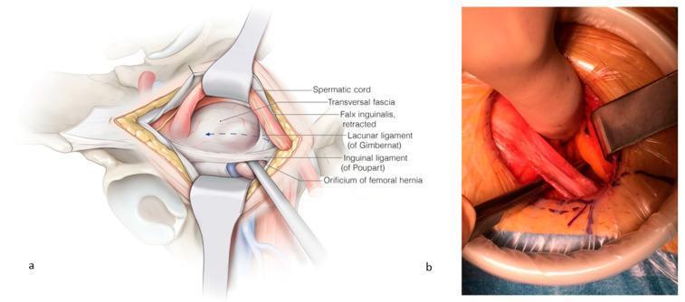
This composite visual contains an anatomical illustration (a) and a corresponding clinical intraoperative photograph (b) detailing the surgical repair of a femoral hernia. Panel (a) provides a labeled anatomical diagram of the right inguinal-femoral region. It depicts the spermatic cord and falx inguinalis retracted cranially, exposing the orificium of the femoral hernia. Key landmarks are identified: the transversalis fascia (opened), the inguinal ligament of Poupart (ventral border), and the lacunar ligament of Gimbernat (medial border). An instrument is shown passing through the femoral orifice to demonstrate the pathology. Panel (b) is a clinical photograph of the surgical field during an open repair. It demonstrates the clinical application of the anatomy shown in panel (a), with retractors maintaining exposure. A surgical instrument is seen incising the lacunar ligament to enlarge the femoral space for mesh placement. The image illustrates the relationship between the inguinal ligament, femoral vessels, and the hernia orifice. The content is intended for intermediate to advanced surgical education, specifically regarding open femoral hernia repair and the anatomy of the femoral canal.
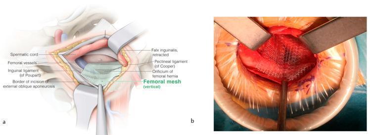
This composite image illustrates the surgical repair of a femoral hernia using a mesh-based approach. Figure (a) is an anatomical diagram of the inguinal and femoral regions during surgery. Key structures identified include the spermatic cord (superior-medial), femoral vessels (lateral), inguinal ligament of Poupart (ventral), and the pectineal ligament of Cooper (dorsal). The diagram shows the placement of a vertical femoral mesh covering the orificium of the femoral hernia, secured over Cooper’s ligament while maintaining an overlay medially and laterally over the femoral vessels. The falx inguinalis is shown in a retracted state. Figure (b) is an intraoperative clinical photograph corresponding to the diagram, showing the surgical field with metal retractors and a circular wound protector. A white, woven surgical mesh is visible, positioned over the femoral orifice and held in place with instruments. The image demonstrates the 'Lichtenstein-like' open technique for femoral hernia reinforcement, highlighting the spatial relationship between the prosthetic mesh and pelvic floor ligaments to prevent recurrence.
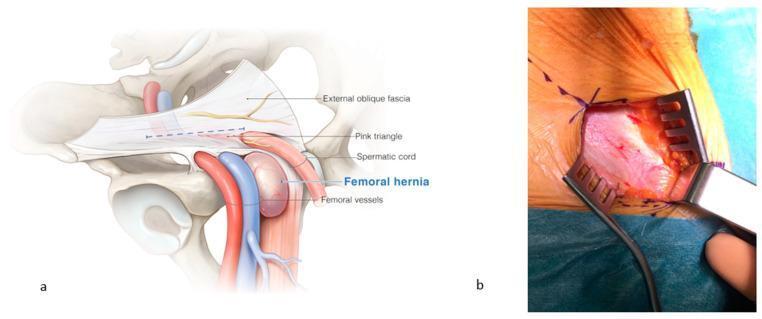
This dual-panel image provides an anatomical and clinical overview of a femoral hernia and its surgical exposure. Panel (a) is a detailed anatomical diagram of the right inguinal and femoral regions. It illustrates a femoral hernia as a distinct bulge emerging medially to the femoral vessels (artery and vein) and caudal to the inguinal canal. Labeled structures include the external oblique fascia, the 'pink triangle' (an area formed by the diversion of fascial vessels toward the external inguinal orifice), the spermatic cord, and femoral vessels. Panel (b) is a clinical photograph showing the intraoperative view after the incision of the external oblique fascia. The surgical field is exposed using three metal claw-style retractors, revealing the underlying fascia and fatty tissue in the femoral region. Sutures are visible along the superior aspect of the skin incision. This content is designed for medical education regarding hernia pathology and surgical repair, demonstrating the critical landmarks needed to differentiate femoral hernias from inguinal hernias and to safely navigate the femoral canal during surgery.
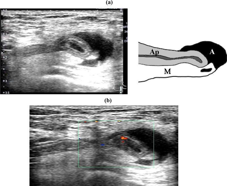
This composite educational resource features diagnostic ultrasound imaging and a corresponding schematic diagram of a de Garengeot hernia. Image (a) is a grayscale B-mode ultrasound showing a longitudinal view of the right inguinal region. It demonstrates a blind-ended tubular structure (the appendix, labeled 'Ap') protruding through the femoral canal. The appendix exhibits a preserved five-layered wall structure and a hyperechoic lumen, measuring approximately 4 mm at the body and 6 mm at the tip. Surrounding the appendix is a reticular hyperechoic area representing the mesoappendix ('M') and an adjacent anechoic region indicative of local ascites ('A'). Image (b) utilizes Color Doppler imaging to show pulsatile blood flow signals within the appendiceal wall, represented by red and blue color pixels. This finding confirms tissue viability and the absence of vascular compromise or strangulation within the hernia sac. A depth scale on the left provides a spatial reference up to 3.5 cm. These images serve as a key diagnostic reference for identifying an incarcerated but non-inflamed appendix within a femoral hernia.
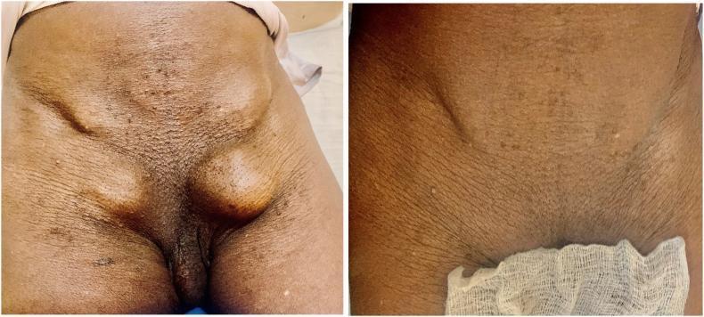
A clinical photograph comparison demonstrating bilateral inguinal and femoral hernias in an adult male, categorized under general surgery and anatomy. The preoperative image (left) shows four distinct ovoid swellings in the groin region. Superiorly, bilateral inguinal swellings are visible above the inguinal ligament. Inferior to these, bilateral femoral swellings are prominent in the femoral triangles, lateral to the pubic tubercle. The swellings are well-defined and raised, with skin stretched over the masses. The postoperative image (right) displays the resolution of these protrusions following surgical repair. The skin across the lower abdomen and inguinal region exhibits signs of aging, including pronounced skin folds, wrinkles, and scattered small seborrheic keratoses or skin tags. This visual serves as a clear educational example of complex pelvic floor hernias and the anatomical distinction between inguinal and femoral hernia locations.
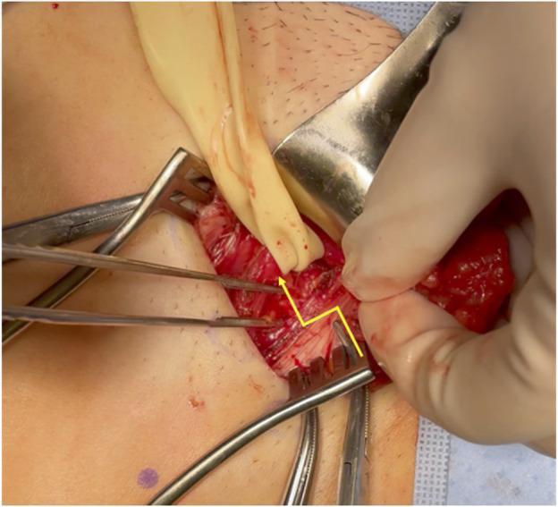
This clinical intraoperative photograph illustrates an open surgical repair of a femoral hernia, focusing on the releasing incision of the inguinal ligament. The surgical field is exposed using multiple instruments, including a metal retractor to pull back the superior tissue layer and two rake-style retractors maintaining lateral exposure. A gloved hand and surgical forceps are seen manipulating the dissected tissue. A yellow line is superimposed on the anatomy to indicate the precise path of the releasing incision through the inguinal ligament, which serves as the roof of the femoral canal. This maneuver is performed to reduce the tension on incarcerated or strangulated visceral contents, allowing for their safe reduction back into the peritoneal cavity. A Penrose drain or similar elastic loop is visible, likely used for traction on the spermatic cord or surrounding structures to improve visualization of the myopectineal orifice. The image demonstrates key landmarks for groin anatomy and the surgical management of strangulated hernias in a clinical setting.
HERNIA - Complete Notes from Bailey & Love's Short Practice of Surgery (28th Edition)
1. DEFINITION
"A hernia is an abnormal protrusion of an organ or tissue through an opening in the layer that normally confines it."
- Bailey & Love, p. 1080
2. PARTS OF A HERNIA
| Part | Description |
|---|---|
| Sac | A peritoneal diverticulum with a mouth (opening), neck (narrowest part), body, and fundus |
| Contents | Omentum (most common), bowel, bladder, ovary |
| Coverings | Attenuated layers of the abdominal wall dragged over the sac |
3. ANATOMICAL CAUSES OF ABDOMINAL WALL HERNIATION
- Natural areas of weakness - absence of muscle (lumbar triangles, posterior inguinal canal wall)
- Structural openings - inguinal canal (testis descent), femoral canal, oesophageal hiatus, umbilical ring
- Developmental abnormalities - patent processus vaginalis
- Acquired disruptions - surgical incisions (incisional hernia), trauma
4. DIAGRAM: SITES OF ABDOMINAL WALL HERNIAS
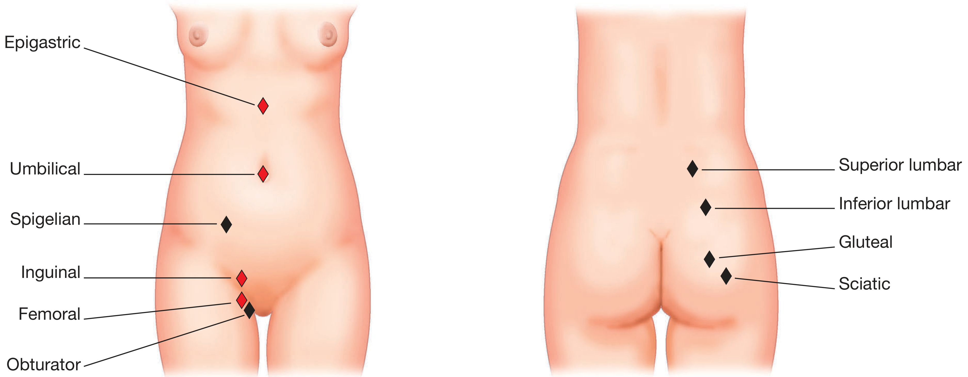
5. TYPES OF HERNIA
CLASSIFICATION
HERNIA
├── GROIN HERNIA
│ ├── Inguinal (most common)
│ │ ├── Indirect (lateral/oblique)
│ │ └── Direct (medial)
│ └── Femoral
│
├── VENTRAL HERNIA (anterior wall)
│ ├── Primary: Umbilical, Epigastric, Spigelian, Lumbar, Traumatic
│ └── Secondary: Incisional, Parastomal
│
└── OTHER: Obturator, Hiatus, Perineal
6. CLINICAL FEATURES (General)
History
- Reducible hernia - contents can return to peritoneal cavity; aching after exertion
- Irreducible hernia - cannot be reduced; obstruction/strangulation risk
- Obstructed hernia - bowel contents cannot pass; vomiting, distension, no strangulation yet
- Strangulated hernia - blood supply to contents is cut off; EMERGENCY - severe pain, vomiting, peritonism, overlying skin red/inflamed
Examination
- Examine lying AND standing (hernias may only appear standing)
- Ask patient to cough or Valsalva - expansile cough impulse is diagnostic
- Assess: reducibility, cough impulse, tenderness, skin colour, contralateral side
- Cellulitis over hernia = strangulation = surgical emergency
- Always check the opposite groin - occult contralateral hernia present in up to 20% of patients
7. INGUINAL HERNIA (Most Common)
"Inguinal hernia is the most common hernia in men and is around 10 times more common in men than in women."
- Bailey & Love, p. 1087
Key Diagram: Direct vs Indirect vs Femoral
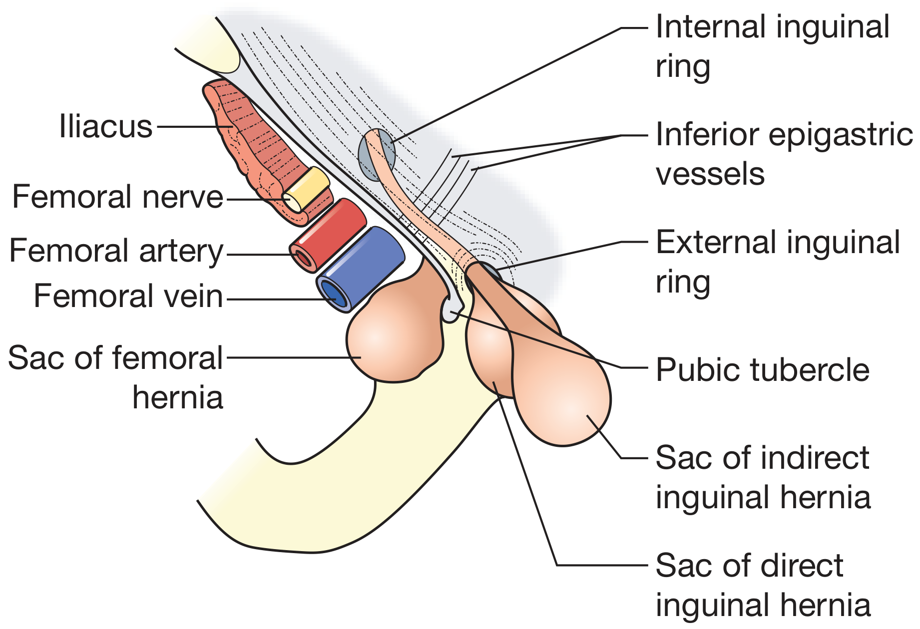
Anatomy of the Inguinal Canal
| Wall | Structure |
|---|---|
| Anterior | External oblique aponeurosis |
| Posterior | Transversalis fascia |
| Roof | Conjoint tendon (internal oblique + transversus abdominis arched fibres) |
| Floor | Inguinal (Poupart's) ligament - rolled inward free edge of external oblique |
- Deep (internal) inguinal ring - defect in transversalis fascia; lies midway between ASIS and pubic tubercle, 2-3 cm above femoral artery pulse; inferior epigastric vessels lie just medial to this ring
- Superficial (external) inguinal ring - inverted V-shaped defect in external oblique aponeurosis
- Testicular artery, veins, lymphatics
- Vas deferens
- Cremasteric muscle
- Ilioinguinal nerve, iliohypogastric nerve, genital branch of genitofemoral nerve
Indirect (Lateral/Oblique) Inguinal Hernia
- Hernia passes through the deep inguinal ring, lateral to the inferior epigastric vessels
- Travels along the inguinal canal and may reach the scrotum
- Congenital type: due to patent processus vaginalis (peritoneal tube that should obliterate after testicular descent but fails to)
- All neonatal and childhood inguinal hernias are of this type
- Acquired type also possible in adults if the deep ring weakens
- More common, more likely to strangulate (narrow neck)
Direct (Medial) Inguinal Hernia
- Hernia pushes through the posterior wall of the inguinal canal (Hesselbach's triangle), medial to the inferior epigastric vessels
- Always acquired; caused by weakness/stretching of transversalis fascia
- Hesselbach's triangle boundaries: inferior epigastric vessels (lateral), inguinal ligament (inferior), rectus abdominis (medial)
- Less likely to strangulate (broad neck)
- More common in elderly men with weak muscles
Laparoscopic View - Hernia Triangles
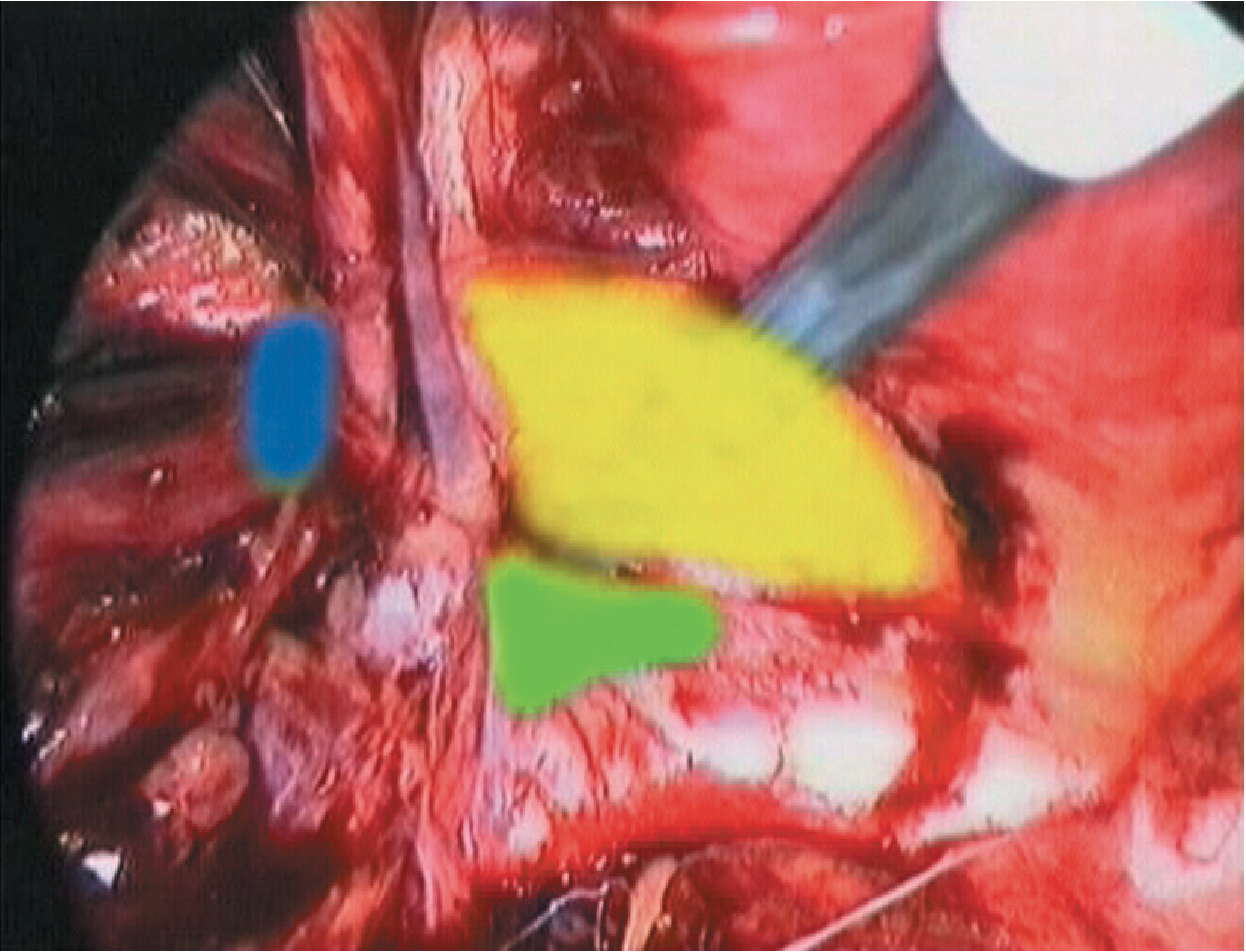
Comparison: Direct vs Indirect Inguinal Hernia
| Feature | Indirect (Lateral) | Direct (Medial) |
|---|---|---|
| Relation to inf. epigastric | Lateral | Medial |
| Origin | Congenital or acquired | Always acquired |
| Neck | Narrow | Wide |
| Strangulation risk | Higher | Lower |
| Age | Any, especially young | Elderly |
| Sex | Males >> females | Males |
| Goes to scrotum? | Yes | Rarely |
| Reducibility | May be irreducible | Usually reducible |
8. MANAGEMENT OF INGUINAL HERNIA
Conservative
- Watchful waiting safe in asymptomatic direct hernia, especially in elderly
- Surgical trusses NOT recommended
Surgery
Herniotomy
- In children only - excision and closure of the sac alone (no repair needed as muscles are healthy)
Open Suture Repair (Bassini's Repair)
- Open the anterior wall (external oblique aponeurosis)
- Dissect spermatic cord free, identify hernia type
- For indirect: separate and excise sac, close at neck
- For direct: invert sac, plicate transversalis fascia
- Sutures placed between conjoint tendon (above) and inguinal ligament (below) to strengthen posterior wall
- Shouldice modification: double-breasting of transversalis fascia; expert centres report <2% lifetime failure rate
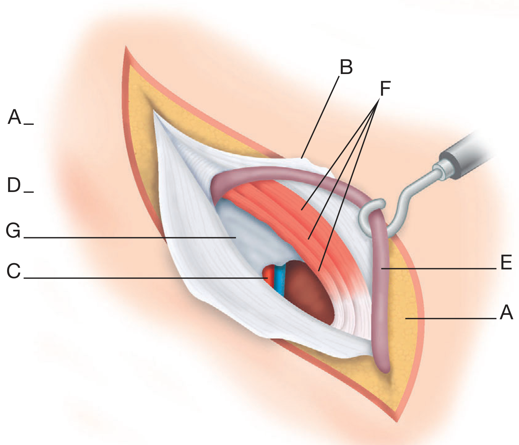
Mesh Repair (Gold Standard)
- Closure of defect alone has high recurrence; mesh reduces but does not eliminate it
- Lichtenstein tension-free repair - mesh placed flat over the posterior wall, most widely used open technique
- Mesh can bridge, plug, or augment the repair
- Current best practice: primary closure + mesh in tension-free manner with generous overlap
- Suturing mesh edge-to-edge (inlay with no overlap) = NOT recommended
- Synthetic meshes: polypropylene, polyester, PTFE - non-absorbable, allow tissue ingrowth
Laparoscopic Repair
- TAPP (Trans-Abdominal Pre-Peritoneal) or TEP (Totally ExtraPeritoneal)
- Mesh placed extraperitoneally over the entire myopectineal orifice
- Advantages: less post-op pain, faster recovery, better for bilateral or recurrent hernias
9. FEMORAL HERNIA
"50% present as an emergency with very high risk of strangulation."
- Bailey & Love, p. 1088
Anatomy of the Femoral Canal
- Lateral: Femoral vein
- Anterior: Inguinal ligament
- Posterior: Iliopectineal (Astley Cooper's) ligament
- Medial: Lacunar (Gimbernat's) ligament - sharp, unyielding edge that impedes reduction
Key Features
| Feature | Detail |
|---|---|
| Incidence | Less common than inguinal; commoner in women (female pelvis wider, larger femoral canal) |
| Typical patient | Thin, elderly woman |
| Position | Below and lateral to the pubic tubercle |
| Risk | Very high strangulation risk (50% emergency); tight unyielding neck (Gimbernat's ligament) |
| Cough impulse | Often absent (irreducible neck is tight) |
| Misdiagnosed as | Lymph node (1-2 cm, no cough impulse) |
Femoral vs Inguinal Hernia - Key Distinction
| Inguinal | Femoral | |
|---|---|---|
| Position | Above and medial to pubic tubercle | Below and lateral to pubic tubercle |
| Sex | Males | Females (more common) |
| Strangulation | Less frequent | Very frequent (50%) |
| Cough impulse | Present | Often absent |
10. VENTRAL HERNIAS
Umbilical Hernia
- Occurs in up to 10% of infants (higher in premature babies, and 8x more common in Black infants)
- Appears within weeks of birth; increases in size when crying
- Treatment: Conservative under age 2 (95% resolve spontaneously); surgical if persists beyond age 2
- Surgery: curved incision below umbilicus, reduce contents, close sac, close defect with interrupted absorbable sutures
- Umbilical ring stretches or reopens in adult life
- Associated with: obesity, ascites (cirrhosis), pregnancy, multiparity
- High risk of strangulation in cirrhotic patients (surgical repair required even if small)
- Surgery: Mayo's repair (overlapping fascial closure) or mesh for larger defects
Epigastric Hernia
- Through the linea alba between xiphisternum and umbilicus
- Usually small, often contains only extraperitoneal fat (pre-peritoneal fat)
- May be multiple
- Pain out of proportion to size; no peritoneal sac initially
- Surgery: repair with mesh for larger defects; recurrence risk even with mesh
Spigelian Hernia
- Through the spigelian fascia - along the lateral border of rectus abdominis, below the arcuate line
- Rare; often interparietal (between muscle layers) so no visible lump
- Diagnosed by ultrasound or CT
- High strangulation risk (narrow neck)
Incisional Hernia
- Through a previous surgical scar due to wound failure
- Causes: wound infection (most important), obesity, poor technique, abdominal distension
- Often large defects with multiple loculi
- Repair: Always use mesh; recurrence rate high even with mesh
- Surgical options: open repair (sublay mesh), laparoscopic IPOM (Intraperitoneal Onlay Mesh)
11. COMPLICATIONS OF HERNIA
| Complication | Description | Management |
|---|---|---|
| Reducible | Contents return spontaneously | Elective repair |
| Irreducible | Cannot be reduced manually | Urgent review |
| Obstructed | Bowel obstruction without ischaemia | Urgent surgery |
| Strangulated | Vascular compromise of contents | EMERGENCY surgery |
Strangulation - Important Points
- Signs: constant severe pain, vomiting, tachycardia, tender irreducible lump, overlying cellulitis/redness
- Femoral hernias strangulate most frequently
- Richter's hernia: only part of the bowel circumference is in the sac - can strangulate without obstruction
- Maydl's hernia (W-hernia): two loops in sac, middle loop in abdomen strangulates
- Treatment: Emergency laparotomy; resect non-viable bowel
12. CLINICAL EXAMINATION SUMMARY (Bailey & Love Summary Box 64.3)
- Reducibility
- Cough impulse
- Tenderness
- Overlying skin colour
- Multiple defects / contralateral side
- Signs of previous repair
- Scrotal content (for groin hernia)
13. CLINICAL PHOTO: Bilateral Inguinal + Femoral Hernias
Summary Table: All Hernia Types at a Glance
| Hernia | Location | Common in | Strangulation Risk | Notes |
|---|---|---|---|---|
| Indirect inguinal | Through deep inguinal ring, lateral to inf. epigastric | Young males | High | Congenital or acquired; can reach scrotum |
| Direct inguinal | Through Hesselbach's triangle, medial to inf. epigastric | Elderly males | Low | Always acquired; weak posterior wall |
| Femoral | Below inguinal ligament, through femoral canal | Elderly women | Very high (50%) | Below + lateral to pubic tubercle |
| Umbilical (child) | Through umbilical ring | Infants | Very low | 95% resolve by age 2 |
| Umbilical (adult) | Through umbilical ring | Obese/ascitic adults | Moderate | Cirrhosis = high risk |
| Epigastric | Through linea alba, above umbilicus | Middle-aged males | Low | Pain > size; often just fat |
| Spigelian | Lateral border of rectus, below arcuate line | Adults | High | Interparietal, diagnosed by US/CT |
| Incisional | Through surgical scar | Post-surgical, obese | Moderate | Always requires mesh repair |
| Parastomal | Around stoma | Stoma patients | Moderate | Common after colostomy |
| Obturator | Through obturator foramen | Elderly thin women | Very high | Howship-Romberg sign (thigh pain on extension) |