
Pathogenesis of mpgn
I'll start by reading the medical library skill, then search for MPGN pathogenesis content.
Searching sources...
Reading File
Now I'll search for MPGN pathogenesis in parallel across headings, paragraphs, and recent literature.
Finding Sources
Searching PubMed
membranoproliferative glomerulonephritis pathogenesis complement
Excellent results. Now I'll read the most detailed sections in parallel - Comprehensive Clinical Nephrology (dedicated chapter), Robbins Pathology, and Goldman-Cecil.
Reading File
Reading File
Reading File
Good content. Now let me read the detailed pathogenesis from the National Kidney Foundation Primer and search for complement-specific content, plus grab MPGN images.
Reading File
Finding Sources
Searching Images
membranoproliferative glomerulonephritis MPGN tram track GBM histology
Now let me get the detailed complement pathogenesis content from Comprehensive Clinical Nephrology and Robbins Pathology (C3 glomerulopathy section).
Reading File
Reading File
I now have comprehensive information from multiple authoritative sources. Let me also fetch the key MPGN histology image from Robbins.
Loading Image
Loading Image
I now have comprehensive, authoritative content from multiple nephrology and pathology textbooks, plus histological images. Here is the full synthesis:
Pathogenesis of Membranoproliferative Glomerulonephritis (MPGN)
Conceptual Framework
MPGN is not a single disease but a histopathologic pattern of glomerular injury. The unifying feature is dual injury to the mesangium and the subendothelial space, producing the classic "tram-track" (double contour) GBM appearance. The modern classification divides MPGN by the underlying immunopathologic mechanism into two major categories:
| Category | Key Mediator | IF Pattern | Complement Pathway |
|---|---|---|---|
| Immune complex-mediated (IC-MPGN) | IgG/IgM + complement | IgG + C3 + C1q/C4 (granular) | Classical + Alternative |
| Complement-mediated (C3 glomerulopathy) | Dysregulated alternative pathway | C3 dominant, no Ig | Alternative pathway only |
1. Immune Complex-Mediated MPGN (IC-MPGN)
Antigen-Antibody Deposition
The primary event is glomerular deposition of immune complexes, either by:
- Passive deposition of circulating preformed immune complexes from the bloodstream
- In situ immune complex formation - a circulating antibody binds to a "planted" antigen already lodged in the glomerular capillary wall
The antigens involved in so-called "idiopathic" IC-MPGN are often unknown. In secondary forms, they are typically proteins derived from infectious agents, especially:
- Hepatitis C virus (HCV) - the most common infectious trigger; often via cryoglobulinemia (type II/III mixed cryoglobulins with monoclonal IgM acting as a rheumatoid factor binding polyclonal IgG)
- Hepatitis B virus
- Other chronic bacterial/viral infections
Autoimmune diseases (SLE, with nucleosomes facilitating complex deposition on the anionic GBM) and monoclonal gammopathy (a monoclonal immunoglobulin acting as the depositing antibody) are other important causes.
Complement Activation
Once immune complexes lodge in the subendothelial space and mesangium, they activate both the classical pathway (via C1q binding to IgG/IgM) and the alternative pathway. This generates:
- C3a and C5a - potent anaphylatoxins and chemotactic factors
- Upregulation of endothelial adhesion molecules (ICAM-1, E-selectin, P-selectin)
- Local release of proinflammatory cytokines (IL-1, TNF-α)
Leukocyte Recruitment and GBM Injury
Neutrophils, monocytes/macrophages, and platelets are recruited to the glomerulus. They release:
- Oxidants - particularly hypohalous acids (e.g., hypochlorous acid) generated by neutrophil myeloperoxidase
- Proteases - elastase, cathepsin G, metalloproteinases
These cause direct GBM degradation and endothelial/mesangial cell injury.
GBM "Tram-Track" Formation
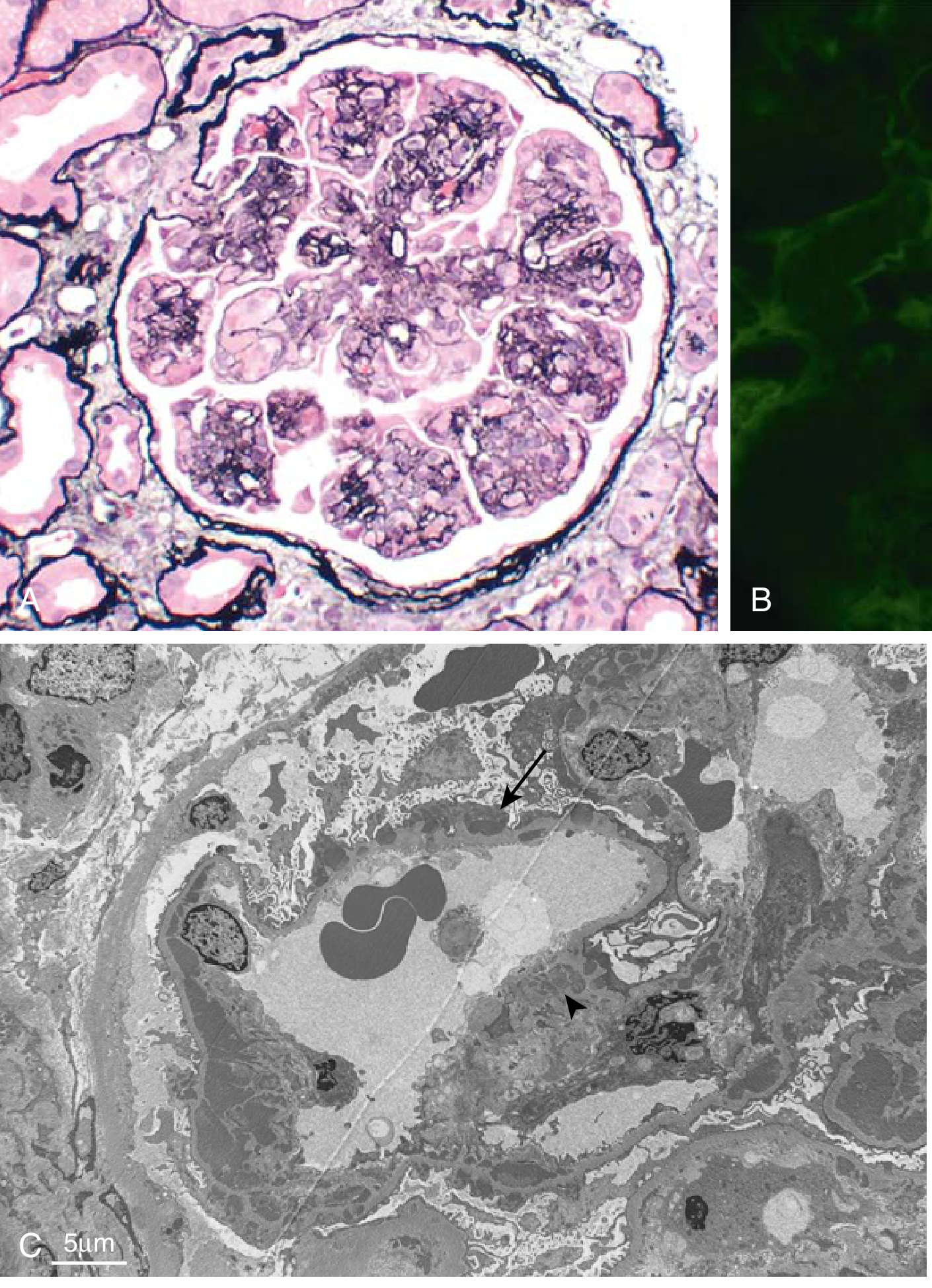
The subendothelial deposits stimulate new basement membrane synthesis by the glomerular cells. Mesangial cells interpose between the endothelium and the original GBM (mesangial interposition), laying down new matrix. The result is duplication ("splitting") of the GBM - the hallmark "tram-track" appearance on silver or PAS stains. The space between the two basement membrane layers is occupied by:
- Mesangial cell processes and matrix
- Endothelial cell processes
- Leucocytic elements
- Electron-dense immune deposits
Immunofluorescence shows granular IgG and C3 deposits, often with early complement components (C1q and C4), confirming classical pathway activation.
2. Complement-Mediated MPGN: C3 Glomerulopathy (C3G)
C3 glomerulopathy (C3G) includes Dense Deposit Disease (DDD) and C3 glomerulonephritis (C3GN). These are caused by dysregulated activation of the alternative complement pathway without immune complex deposition.
Normal Alternative Pathway Regulation
The alternative pathway undergoes constant low-level "tick-over" - spontaneous hydrolysis of C3 to C3(H₂O), which can associate with Factor B to form a fluid-phase C3 convertase. Normally this is tightly controlled by regulatory proteins:
- Factor H - binds C3b on host cells, accelerating decay of the C3 convertase (C3bBb) and acting as a cofactor for Factor I-mediated cleavage of C3b
- Factor I - serine protease that cleaves and inactivates C3b (requires Factor H or MCP as cofactor)
- Membrane Cofactor Protein (MCP/CD46) - surface-expressed; aids Factor I-mediated C3b cleavage on host cells
Mechanisms of Dysregulation in C3G
Acquired causes:
- C3 nephritic factor (C3NeF) - the most common cause; an IgG autoantibody that binds to and stabilizes the alternative pathway C3 convertase (C3bBb), preventing its normal decay. This leads to uncontrolled cleavage of C3 and persistent complement activation.
- Autoantibodies against Factor H, Factor B, or C3b itself
Genetic/hereditary causes:
- Loss-of-function mutations in Factor H, Factor I, or MCP - impair the normal braking mechanism on the alternative pathway
- Gain-of-function mutations in C3 or Factor B - promote C3 convertase formation
- Rearrangements in complement factor H-related (CFHR) genes - CFHR proteins compete with Factor H for C3b binding; abnormal CFHR complexes can disrupt normal Factor H-mediated regulation
Consequence of Dysregulation
Uncontrolled alternative pathway activation leads to:
- Massive C3 cleavage and C3b deposition along glomerular capillary walls and in the mesangium
- Low serum C3 levels (consumed)
- C5 convertase formation → terminal complement complex (C5b-9, MAC) assembly → glomerular cell injury
- No significant immunoglobulin deposition (IF shows C3 dominance without IgG/IgA/IgM or early complement components C1q/C4)
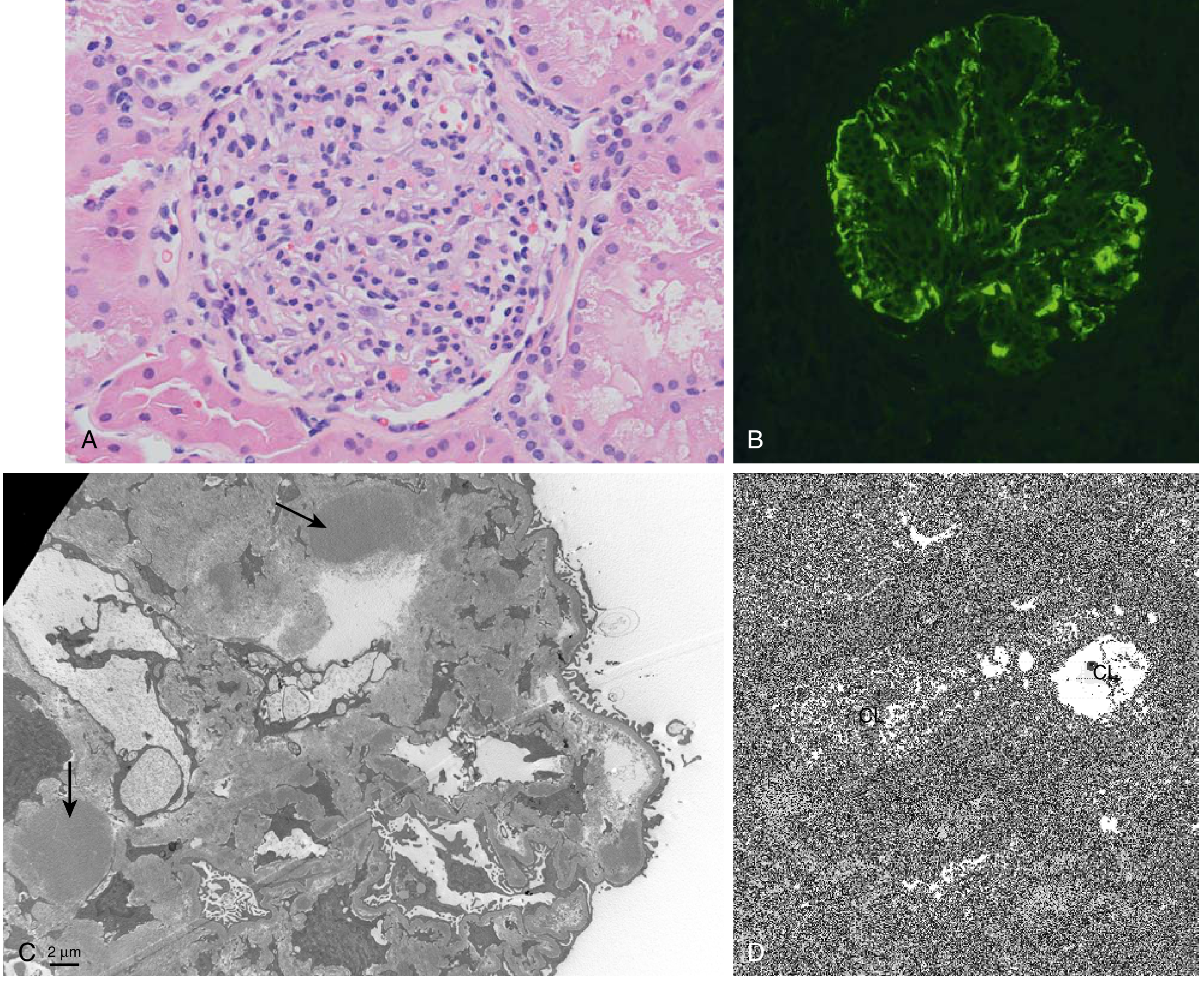
DDD vs. C3GN distinction on EM:
- DDD: Highly electron-dense, ribbon-like deposits transforming the GBM into an irregular, osmiophilic structure - deposits within the lamina densa itself
- C3GN: Subendothelial and mesangial "waxy" electron-dense deposits (similar pattern to IC-MPGN but with absent immunoglobulin on IF)
3. Downstream Mechanisms Common to Both Forms
Regardless of the initiating event, several shared mechanisms drive glomerular injury in the MPGN pattern:
| Step | Mechanism |
|---|---|
| Complement activation | Generation of C3a/C5a → mast cell degranulation, increased vascular permeability |
| Leukocyte recruitment | C5a + upregulated adhesion molecules → neutrophil, monocyte, platelet infiltration |
| Oxidative injury | MPO-derived HOCl, superoxide radicals damage GBM and endothelium |
| Protease release | Elastase, cathepsin G, MMPs → GBM degradation, mesangiolysis |
| Cytokine amplification | IL-1, TNF-α → perpetuate local inflammation |
| Mesangial interposition | New matrix/cell interposition between GBM layers → tram-track appearance |
| Podocyte injury | Disruption of slit diaphragm integrity → proteinuria (nephrotic component) |
| Endocapillary proliferation | Mesangial + endothelial cell proliferation + leukocytes → lobular accentuation, reduced GFR (nephritic component) |
4. Secondary Causes - Pathogenetic Summary
| Cause | Mechanism |
|---|---|
| HCV + cryoglobulinemia | Monoclonal IgM binds polyclonal IgG → immune complexes → IC-MPGN |
| HBV | HBsAg/HBeAg-containing immune complexes deposited in glomeruli |
| SLE | Nucleosome-containing immune complexes bind GBM via cationic nuclear proteins |
| Monoclonal gammopathy | Monoclonal Ig deposits directly or as part of immune complexes |
| DDD/C3GN | C3NeF, Factor H/I/MCP mutations → unregulated alternative pathway |
| Partial lipodystrophy | C3NeF often present → associated with DDD |
Key Diagnostic Differentiation
| Feature | IC-MPGN | C3G (DDD/C3GN) |
|---|---|---|
| IF | IgG + C3 + C1q + C4 (granular) | C3 dominant; no Ig, C1q, C4 |
| EM deposits | Subendothelial (discrete) | DDD: intramembranous ribbon; C3GN: subendothelial/mesangial "waxy" |
| Serum C3 | Often low | Typically very low (consumed) |
| C3NeF | Absent | Present in ~70% DDD, ~50% C3GN |
| Complement pathway | Classical + Alternative | Alternative only |
| Workup needed | HCV/HBV serology, ANA, SPEP | Genetic complement panel, Factor H/I/C3 levels, C3NeF assay |
Sources:
- Robbins & Kumar Basic Pathology, p. 507-509
- Robbins, Cotran & Kumar Pathologic Basis of Disease, p. 850
- Comprehensive Clinical Nephrology, 7th Edition, pp. 258, 1063-1070
- National Kidney Foundation Primer on Kidney Diseases, 8e, p. 3099
- A 2026 systematic review (PMID 40971335) on C3G and primary IC-MPGN confirms proteinuria as a key outcome predictor and highlights the shared disease burden across these mechanistically distinct entities.
How infection ie hcv leprosy malarie cause mpgn
Searching sources...
Finding Sources
Finding Sources
Finding Sources
Good leads. Now let me read those sections in full detail in parallel.
Reading File
Reading File
Reading File
Good. Let me now read the leprosy and malaria sections in detail, and get the HCV cryoglobulin mechanism from Henry's.
Reading File
Finding Sources
Reading File
Now let me get the malaria section and the HCV/HBV detailed mechanisms from Comprehensive Clinical Nephrology.
Reading File
Finding Sources
I need the malaria section more completely - let me get the full Brenner malaria block from the beginning.
Reading File
Finding Sources
Now let me get the HCV and HBV detailed pathogenesis from Comprehensive Clinical Nephrology chapter 22.
Finding Sources
Reading File
Let me now get the HBV-MPGN section and the HCV section in Comprehensive Clinical Nephrology.
Reading File
I now have rich content across all three infections. Let me compile the comprehensive answer.
How Infections Cause MPGN: HCV, Leprosy, and Malaria
All infection-associated MPGN falls under Immune Complex-Mediated MPGN (IC-MPGN). The shared principle is: persistent antigenic stimulation by a chronic infection → sustained antibody production → circulating immune complex formation → glomerular deposition → complement activation → the MPGN pattern of injury. Each pathogen has its own specific twist on this mechanism.
1. Hepatitis C Virus (HCV) - The Most Common Infectious Cause
HCV is the most frequent infectious trigger of IC-MPGN, and it operates primarily through cryoglobulinemia.
Step-by-Step Mechanism
Step 1 - Chronic viral antigenic drive
HCV infects hepatocytes and B lymphocytes. Persistent HCV viremia (core antigen, NS3, NS5 proteins) provides a continuous antigenic stimulus to the immune system over months to years.
Step 2 - B-cell dysregulation and cryoglobulin production
HCV infects B cells bearing CD81 (the HCV entry receptor), causing polyclonal then oligoclonal/monoclonal B-cell expansion. These B cells produce abnormal immunoglobulins that precipitate in the cold - cryoglobulins. In HCV, the type is almost always:
- Type II mixed cryoglobulinemia: a monoclonal IgM (with rheumatoid factor activity) + polyclonal IgG
- Type III: polyclonal IgM + polyclonal IgG
The monoclonal IgM has rheumatoid factor activity - it binds to the Fc region of the polyclonal IgG, creating large lattice-like immune complexes (IgM-IgG complexes containing HCV antigens).
Step 3 - Immune complex deposition in the glomerulus
These large, cold-precipitable complexes:
- Deposit in subendothelial and mesangial spaces (they are too large for subepithelial deposition)
- Can occlude capillary lumens, forming the characteristic intracapillary "cryoglobulin thrombi" seen on light microscopy - virtually pathognomonic of cryoglobulinemic GN
Step 4 - Complement activation
- Classical pathway: IgG in the complexes fixes C1q → classical cascade → C3a, C5a generation
- This explains the low C4 (a hallmark lab finding in cryoglobulinemic GN, unlike most other forms of MPGN where C3 is more suppressed)
- Rheumatoid factor in the IgM further amplifies complement consumption
Step 5 - Inflammatory injury producing MPGN pattern
C5a recruits neutrophils and monocytes → proteases, oxidants → GBM degradation + mesangial cell activation → mesangial interposition → tram-track formation.
Key Distinguishing Features of HCV-MPGN
| Feature | Finding |
|---|---|
| Serology | Anti-HCV Ab +, HCV RNA + in serum |
| Cryoglobulins | Positive (types II/III) |
| Rheumatoid factor | Elevated |
| C4 | Very low (classical pathway consumption) |
| C3 | Moderately low |
| IF | IgG + IgM + C3 (granular) |
| EM | Subendothelial deposits with curved microtubular or annular substructure (specific to cryoglobulins) |
| LM | MPGN pattern + intracapillary "cryoglobulin thrombi" |
HCV can also cause MPGN without cryoglobulinemia - through direct deposition of HCV antigen-antibody complexes in the glomerulus.
Treatment: Direct-acting antivirals (DAAs) achieve >95% viral remission and are the treatment of choice - eliminating the antigen drives remission of the GN.
2. Hepatitis B Virus (HBV)
HBV is a DNA virus containing three major antigens: HBsAg (surface), HBcAg (core), and HBeAg (e antigen). While MN is its most common glomerular lesion, MPGN is more common in adults with HBV (vs. MN which predominates in children).
Mechanism
Size and charge dictate deposit location:
- HBeAg is small and cationic → crosses GBM easily → subepithelial deposits → causes MN
- HBcAg and HBsAg are larger → lodge in subendothelial and mesangial regions → trigger MPGN pattern
Pathways of immune complex formation:
- Circulating immune complexes: HBV antigens + anti-HBV antibodies form complexes in the circulation that are passively deposited in the glomerulus
- In situ complex formation: HBcAg is locally expressed in glomerular cells; anti-HBcAg antibodies then bind to it within the glomerulus
- Direct cytopathic injury: HBV induces mesangial cell proliferation and increased type IV collagen production directly (in vitro evidence)
Once deposited, both classical and alternative complement pathways are activated, generating the full MPGN inflammatory cascade. Host genetic factors (MHC class II alleles) likely determine which patients develop GN.
3. Malaria (Plasmodium species)
Which Species Causes MPGN?
| Species | Renal Pattern |
|---|---|
| Plasmodium malariae (quartan malaria) | MPGN/membranoproliferative - classic "quartan nephropathy" |
| Plasmodium falciparum | Mesangial proliferation or MPGN; AKI in severe malaria (from hemoglobinuria) |
| P. vivax, P. ovale | Rarely cause significant glomerular disease |
Mechanism of P. malariae Quartan Nephropathy
Step 1 - Parasitic antigen release
Plasmodium malariae undergoes schizogony (rupture of infected RBCs) on a 72-hour cycle, releasing merozoite antigens, soluble malarial antigens, and altered erythrocyte membrane proteins into the bloodstream.
Step 2 - Immune complex formation and deposition
- Host antibodies (IgG, IgM) form complexes with P. malariae antigens
- These complexes deposit in the glomerulus: subendothelial, mesangial, and within the GBM
- Biopsy confirmation: IgG, IgM, C3, and P. malariae antigen co-localize in glomeruli
- EM shows electron-dense material within the irregularly thickened GBM
Step 3 - Impaired immune complex clearance (key amplifying mechanism)
Malaria has a unique trick: the parasite downregulates complement receptor 1 (CR1) expression on monocytes and macrophages. CR1 normally binds complement-opsonized immune complexes, which is the primary mechanism for clearing them from the circulation. With CR1 deficiency, immune complexes accumulate in the blood and deposit in the glomerulus at higher concentrations.
Step 4 - MPGN pattern injury
Subendothelial deposits → complement activation → neutrophil/monocyte recruitment → mesangial interposition → tram-track GBM duplication.
Clinical Features of Quartan Nephropathy
- Affects children in endemic areas (sub-Saharan Africa, Nigeria, Uganda)
- Nephrotic syndrome + progressive renal failure within 3-5 years
- Does NOT respond to antimalarials or steroids - the MPGN process becomes autonomous once established
- The most common LM lesion in Nigerian children: diffuse capillary wall thickening with focal double-contouring and segmental sclerosis
4. Leprosy (Mycobacterium leprae)
Leprosy causes glomerular disease in up to 45% of patients, particularly those with the lepromatous (LL) and borderline lepromatous (BL) forms. The GN is immune complex-mediated with frequent hypocomplementemia.
Glomerular patterns in leprosy:
- Mesangioproliferative GN (most common)
- MPGN (less common but well described)
- FSGS
- Renal amyloidosis (AA type, from chronic inflammation)
- Diffuse proliferative GN + crescents (especially in erythema nodosum leprosum reactions)
Mechanism
Step 1 - Mycobacterium leprae antigen release
M. leprae antigens (glycolipid-I, lipoarabinomannan, PGL-1) are released during bacterial multiplication. In lepromatous leprosy, the organism multiplies freely due to defective cell-mediated immunity, providing a large and sustained antigenic load.
Step 2 - Immune complex formation
Host antibodies (IgG, IgM) form complexes with M. leprae antigens. Patients with lepromatous leprosy have high levels of circulating immune complexes and hypergammaglobulinemia.
Step 3 - Glomerular deposition and complement activation
- Immune complexes deposit in mesangial and subendothelial zones
- IF shows IgG, IgM, C3, and fibrin deposition
- Complement consumption leads to low serum C3 (hypocomplementemia)
Step 4 - MPGN pattern injury
Same downstream pathway: complement activation → chemotaxis → leukocyte infiltration → mesangial interposition → GBM duplication.
Special trigger - Erythema Nodosum Leprosum (ENL) reactions:
During ENL (type II lepra reaction), there is massive immune complex deposition from sudden release of M. leprae antigens during treatment. This can precipitate rapidly progressive (crescentic) GN in some patients.
Treatment: Multidrug therapy (MDT) - the incidence of reduced kidney function decreases to 9% after 8 months of MDT as the antigen load is eliminated.
Unified Summary: Why Chronic Infections Cause MPGN
Chronic infection
       ↓
Persistent antigenic stimulation
       ↓
Sustained antibody production → Circulating immune complexes
       ↓
Glomerular IC deposition (subendothelial + mesangial)
       ↓
Classical ± alternative complement pathway activation
       ↓
C3a / C5a generation → chemotaxis
       ↓
Neutrophil / monocyte infiltration
       ↓
Proteases + oxidants → GBM degradation
       ↓
Mesangial cell activation → interposition + new BM synthesis
       ↓
GBM duplication ("tram-track")  ← THE MPGN PATTERN
| Infection | Antigen | IC Type | Special Feature |
|---|---|---|---|
| HCV | HCV core/NS proteins | IgM (RF+) + IgG cryoglobulins | Cryoglobulin thrombi; low C4 |
| HBV | HBsAg, HBcAg, HBeAg | HBV-Ag/Ab complexes | Antigen size/charge determines deposit site |
| P. malariae | Malarial antigens | IgG/IgM + parasitic Ag | Impaired CR1-mediated clearance |
| M. leprae | Leprosal glycolipids | IgG/IgM + M. leprae Ag | ENL reactions → acute flares |
Sources:
- Comprehensive Clinical Nephrology, 7th Edition, pp. 809-810 (leprosy, viral GN)
- Brenner and Rector's The Kidney, pp. 1512-1513 (malaria, parasitic GN)
- Henry's Clinical Diagnosis and Management by Laboratory Methods, pp. 2961-2966 (MPGN classification, cryoglobulins)
- Harrison's Principles of Internal Medicine, 22E, p. 2472 (HCV, HBV mechanisms)
How sle sjogren and scleroderma and monoclonal ab cause mpgn
Searching sources...
Finding Sources
Finding Sources
Finding Sources
Finding Sources
Reading File
Reading File
Excellent content. Now let me read deeper - SLE lupus nephritis MPGN mechanism, scleroderma, and the PGNMID/monoclonal section in full detail.
Reading File
Finding Sources
Finding Sources
Let me now get the lupus nephritis pathogenesis detail and check for scleroderma kidney involvement.
Finding Sources
Finding Sources
Finding Sources
Finding Sources
I've now hit the query limit. Let me read those two flagged sections for scleroderma and SLE.
Reading File
Reading File
Let me now fetch the PGNMID biopsy image and get the scleroderma kidney section from Brenner.
Loading Image
Reading File
I now have comprehensive content from multiple authoritative sources. Let me compile the full, well-structured answer.
How SLE, Sjögren Syndrome, Scleroderma, and Monoclonal Antibodies Cause MPGN
All of these conditions cause Immune Complex-Mediated MPGN (IC-MPGN) - the pathway where immunoglobulins (autoantibodies or abnormal monoclonal proteins) form deposits in the mesangium and subendothelial space, activate complement, and drive the structural MPGN injury pattern.
1. Systemic Lupus Erythematosus (SLE)
SLE is one of the richest and most mechanistically detailed causes of IC-MPGN.
The Core Trigger: Failure to Clear Apoptotic Debris
In SLE, defective clearance of apoptotic cells leads to accumulation of nuclear material (dsDNA, histones, nucleosomes, Sm, Ro, La antigens) in the circulation. The immune system loses tolerance to these self-antigens and generates a broad array of pathogenic autoantibodies - particularly anti-dsDNA and anti-nucleosome antibodies.
Mechanism of Glomerular Deposition
Immune complexes form and deposit in the glomerulus by two routes:
- Circulating immune complex deposition - Preformed anti-dsDNA/nucleosome complexes are passively deposited in the subendothelial and mesangial zones
- In situ formation facilitated by nucleosomes - Nucleosomes are cationic nuclear proteins (histones) that can bind directly to the negatively charged glomerular basement membrane. Once planted, they act as "sticky traps" for anti-dsDNA antibodies, forming immune complexes in situ within the capillary wall
The size and charge of the immune complexes determines the deposit location:
- Large complexes → subendothelial + mesangial → MPGN pattern (class III/IV lupus nephritis)
- Small cationic complexes → cross GBM → subepithelial → membranous pattern (class V lupus nephritis)
Complement Activation
SLE activates the classical complement pathway maximally - the hallmark is the "full house" immunofluorescence pattern (IgG + IgA + IgM + C3 + C1q all positive), reflecting robust classical pathway activation. Low both C3 and C4 (unlike C3G where only C3 is low).
The classical cascade generates:
- C3a and C5a → chemotaxis of neutrophils, monocytes
- C1q also directly activates leukocytes
- Terminal membrane attack complex (C5b-9) injures endothelial and mesangial cells
Tubuloreticular Inclusions
A characteristic feature of lupus GN (and helpful diagnostically): tubuloreticular inclusions (TRIs) in glomerular endothelial cells on EM - viral-like structures induced by high levels of interferon-α in SLE.
Histologic Pattern in SLE
MPGN corresponds most closely to Class III (focal proliferative) and Class IV (diffuse proliferative) lupus nephritis - particularly the "active" lesions with endocapillary proliferation, subendothelial deposits, and wire-loop lesions. An MPGN pattern (lobular accentuation, tram-tracks) may also appear in Class V (membranous) overlap.
| IF Feature | SLE MPGN |
|---|---|
| IgG | +++ |
| IgA, IgM | + (full house) |
| C1q | + (distinctive, usually absent in other MPGN) |
| C3 | ++ |
| C4 | + |
| TRIs on EM | Highly characteristic |
2. Sjögren Syndrome
Sjögren syndrome is one of the most common non-HCV causes of cryoglobulinemic GN, and operates through a mechanism that closely parallels HCV.
Mechanism: B-cell Dysregulation and Cryoglobulin Production
Step 1 - Chronic B-cell stimulation
In Sjögren syndrome, the glandular immune infiltrate consists predominantly of B cells and T cells. Chronic autoantigen stimulation (Ro/SSA, La/SSB antigens) drives B-cell proliferation and activation, leading to polyclonal then oligoclonal/monoclonal B-cell expansion.
Step 2 - Cryoglobulin formation
Up to 80% of Sjögren patients with glomerulonephritis develop Type II mixed cryoglobulinemia - a monoclonal IgM with rheumatoid factor activity (binds polyclonal IgG Fc region), forming large cold-precipitable immune complexes. Sjögren syndrome is the most common non-HCV cause of type II cryoglobulinemia.
Step 3 - Glomerular deposition and MPGN
These IgM-IgG cryoglobulin complexes deposit in the subendothelial space and mesangium, forming the MPGN pattern. The monoclonal IgM is the dominant immunoglobulin on IF (unlike SLE's full house).
Step 4 - Classical complement activation
The IgG in the complexes activates C1q → classical pathway → very low C4 (characteristic lab finding, as in HCV cryoglobulinemia). C3 is variably reduced.
Key Laboratory Clue
Low C4 with positive cryoglobulins + anti-Ro/anti-La antibodies + sicca symptoms → Sjögren-associated cryoglobulinemic MPGN.
| IF Feature | Sjögren MPGN |
|---|---|
| Dominant Ig | IgM (monoclonal) |
| IgG | + (polyclonal, part of cryo complex) |
| C3 | + |
| C4 | Very low |
| EM | Microtubular/annular substructure in deposits |
3. Scleroderma (Systemic Sclerosis)
Scleroderma is fundamentally different from SLE and Sjögren - its primary renal injury mechanism is vascular/microangiopathic, not immune complex-mediated. However, understanding this distinction is important because scleroderma can still produce an MPGN-like pattern by a different route.
Primary Mechanism: Scleroderma Renal Crisis (TMA, Not Immune Complex)
The dominant renal lesion in scleroderma is thrombotic microangiopathy (TMA) - intimal proliferation ("onion-skinning") of interlobular arteries and afferent arterioles, fibrinoid necrosis, and microthrombi. This can produce a chronic vascular MPGN-like pattern (endothelial injury → mesangial interposition → tram-tracks) without immune complex deposition - IF shows absent or sparse immunoglobulins and complement.
How It Still Produces the MPGN Histologic Pattern
Persistent endothelial injury in scleroderma (from:
- Vascular autoantibodies - anti-endothelial cell antibodies
- Vasospasm (Raynaud phenomenon affecting renal microvasculature)
- Complement activation via anti-endothelial antibodies
→ Chronic endothelial damage → mesangial interposition and new GBM synthesis → double contour tram-track appearance on silver stain → MPGN pattern on LM without immune complex deposits
This distinguishes scleroderma-TMA from IC-MPGN: IF will be negative (no IgG/C3 deposits), and EM shows no discrete electron-dense deposits - just endothelial swelling and subendothelial flocculent material.
Overlap with Other Diseases
- When scleroderma overlaps with SLE (undifferentiated connective tissue disease, MCTD), full IC-MPGN with immune deposits can occur
- Vascular lesions resembling "scleroderma kidney" can also appear in MCTD (interlobular artery intimal mucoid edema and fibrous sclerosis)
Key point: Scleroderma causes an MPGN-like structural pattern but by endothelial injury/TMA rather than immune complex deposition - a vascular MPGN without immune deposits.
4. Monoclonal Antibodies / Monoclonal Gammopathy of Renal Significance (MGRS)
This is arguably the most mechanistically distinct and diagnostically challenging cause of MPGN.
The Concept of MGRS
Not all clonal plasma cell or B-cell disorders reach the threshold for myeloma or lymphoma. A small clone producing a monoclonal immunoglobulin (M-protein) can damage the kidney through the direct pathologic properties of that abnormal protein - without causing systemic tumor burden. These are grouped under MGRS (Monoclonal Gammopathy of Renal Significance).
MPGN is the most common glomerular pattern seen with monoclonal immunoglobulin deposits.
The Key Entity: PGNMID (Proliferative GN With Monoclonal Immunoglobulin Deposits)
PGNMID is the prototype monoclonal MPGN. Its distinguishing features:
Mechanism:
- A small B-cell or plasma cell clone produces a single monoclonal immunoglobulin (usually IgG)
- This M-protein is deposited in the glomerular mesangium and subendothelial space - not as part of a conventional antigen-antibody immune complex, but as a direct deposit of the monoclonal protein itself due to its physicochemical properties (abnormal charge, tendency to self-aggregate, or binding to glomerular matrix components)
- Once deposited, the monoclonal IgG activates complement (particularly via IgG3, which is the most complement-activating IgG subclass and the predominant subclass in PGNMID) → classical pathway → MPGN injury cascade
Unusual Features:
- In most patients, no circulating M-protein is detectable by routine serum/urine electrophoresis - the clone is too small
- Bone marrow biopsy is negative in many cases
- Only 30% have a detectable monoclonal or biclonal band on serum immunofixation
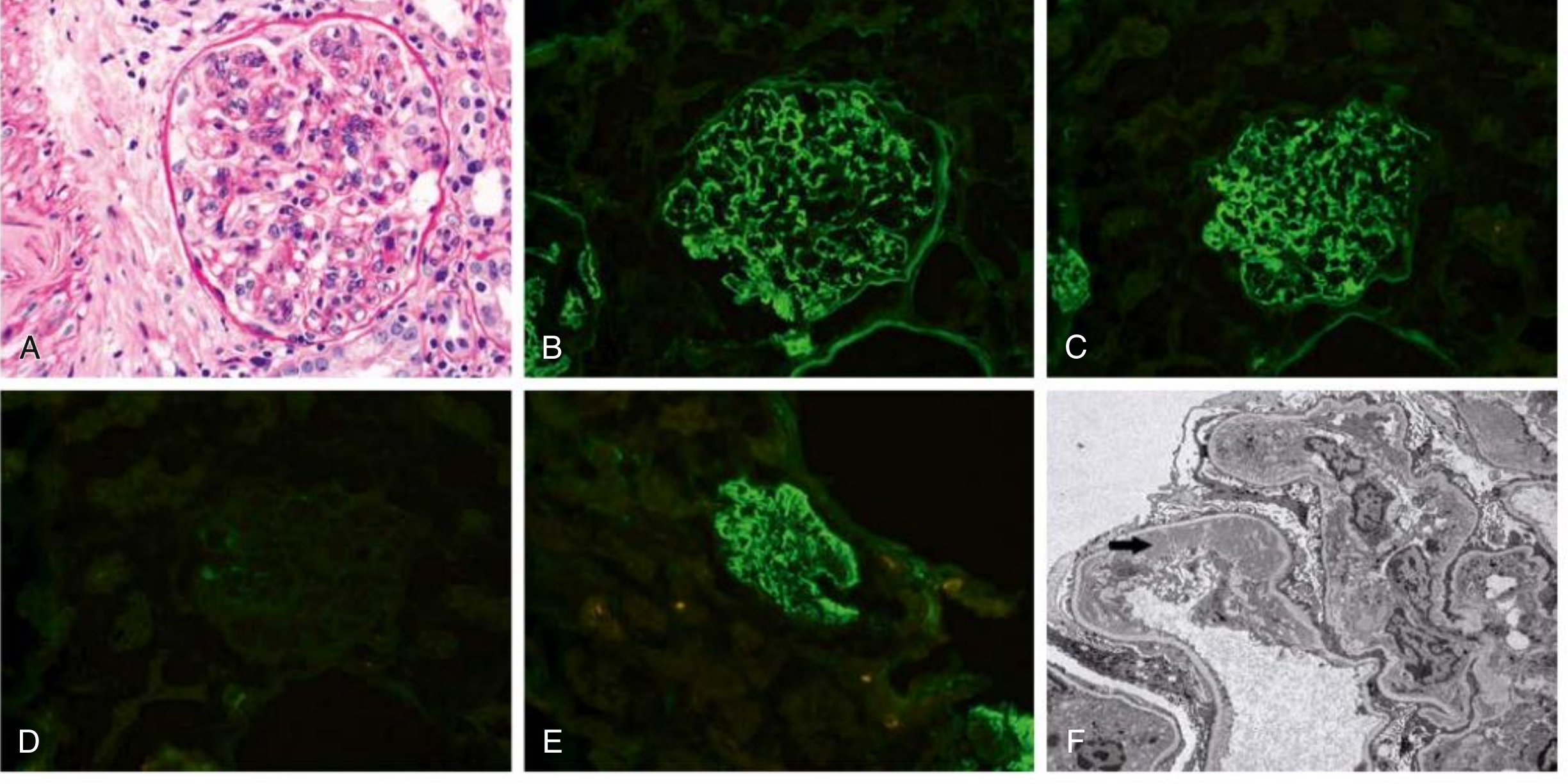
Biopsy is the key diagnostic tool:
The Diagnostic Clue: Light Chain Restriction on IF
In all other causes of IC-MPGN (SLE, infections, Sjögren), IF shows both kappa AND lambda light chains (polyclonal deposits). In PGNMID/MGRS-MPGN, IF shows only kappa OR only lambda - light chain restriction - proving the monoclonal nature.
| Feature | Polyclonal IC-MPGN | Monoclonal MPGN (MGRS/PGNMID) |
|---|---|---|
| IF light chains | Both κ + λ (equal) | Only κ OR only λ (restricted) |
| IF heavy chain | IgG + IgM + IgA (mixed) | Single heavy chain (IgG >> IgM > IgA) |
| IgG subclass | Mixed | IgG3 dominant |
| Circulating M-protein | Absent | Present in only 30% |
| Bone marrow | Normal | May show small clone |
Other Monoclonal MPGN Entities Within MGRS
| Entity | Mechanism | Hallmark |
|---|---|---|
| PGNMID | Direct deposit of monoclonal IgG | IgG3 κ restriction, MPGN pattern |
| Cryoglobulinemia Type I | Monoclonal IgM alone (from myeloma/WM) precipitates in cold | Pure IgM deposits, associated lymphoma |
| Immunotactoid GN | Microtubular organized deposits of monoclonal IgG | Large-bore microtubules (>30 nm) on EM |
| Fibrillary GN (DNAJB9+) | Randomly arranged fibrils of polyclonal Ig | 10-20 nm fibrils on EM; may show MPGN pattern |
| MIDD (Monoclonal Ig Deposition Disease) | Linear (not granular) deposits of κ or λ light chains | Linear IF; no tram-tracks (more like DN nodules) |
Comparison: How Each Autoimmune/Monoclonal Cause Differs
| Cause | Antigen Driving IC Formation | IF Pattern | Key Lab Clue | EM Hallmark |
|---|---|---|---|---|
| SLE | Nuclear Ag (dsDNA, nucleosomes) | Full house (IgG+IgA+IgM+C1q+C3) | ↓C3+C4; anti-dsDNA+ | TRIs in endothelial cells |
| Sjögren | Ro/La → B-cell driven IgM-RF production | IgM dominant + IgG + C3 | ↓C4; cryoglobulins+; anti-Ro+ | Microtubular deposits |
| Scleroderma | Anti-endothelial Ab → vascular TMA | Absent/sparse (non-IC mechanism) | Normal C3/C4; ANA+, anti-Scl-70 | No discrete deposits; endothelial swelling |
| Monoclonal (MGRS/PGNMID) | M-protein direct glomerular deposition | Single Ig class, light chain restricted (κ only or λ only) | SPEP/IFE ± ; κ/λ free light chains | Subendothelial deposits, IgG3 dominant |
Sources:
- Comprehensive Clinical Nephrology, 7th Edition, pp. 253-254, 313-315 (autoimmune-MPGN, MGRS, PGNMID)
- Henry's Clinical Diagnosis and Management by Laboratory Methods, pp. 2965-2967 (SLE, Sjögren, monoclonal gammopathy MPGN)
- Brenner and Rector's The Kidney, pp. 1467-1468 (scleroderma kidney, MCTD)
This is a shared conversation. Sign in to Orris to start your own chat.