
Cerebellum
Reading File
"Cerebellum" is broad - it could mean anatomy, physiology, circuits, clinical syndromes, or pathology. Let me search the library across multiple angles simultaneously.
Finding Sources
Finding Sources
Excellent sources found. Now let me read the most comprehensive dedicated cerebellum sections in parallel - from Neuroscience: Exploring the Brain, Costanzo Physiology, and Neuroanatomy through Clinical Cases.
Reading File
Reading File
Reading File
Good start. Now let me read more detailed content - the full cerebellum section from Neuroscience: Exploring the Brain (block 4), the detailed Costanzo section, and the Neuroanatomy through Clinical Cases chapter.
Reading File
Reading File
Reading File
I now have rich content. Let me fetch the key anatomical images and read a bit more from Neuroanatomy through Clinical Cases for the clinical correlates section.
Loading Image
Loading Image
Reading File
I have comprehensive content. Now let me compile the full, detailed answer on the cerebellum.
The Cerebellum
Overview
The cerebellum ("little brain") is a foliated, highly convoluted structure located in the posterior fossa, dorsal to the pons and rostral medulla. It forms the roof of the fourth ventricle and is situated just below the tentorium cerebelli and the occipital lobes. Despite comprising only about 10% of the brain's total volume, it contains more than half of all the brain's neurons - largely due to its densely packed granule cells.
Its core roles are:
- Coordination and smoothing of movement (synergy)
- Motor planning and initiation
- Maintenance of posture and balance
- Coordination of head and eye movements
- Motor learning and certain higher cognitive functions
The cerebellum does not generate movement directly - it has no direct connections to lower motor neurons. Instead, it fine-tunes movement by acting through connections to the motor cortex and brainstem motor systems. - Neuroanatomy through Clinical Cases 3rd Ed., p. 726
Gross Anatomy
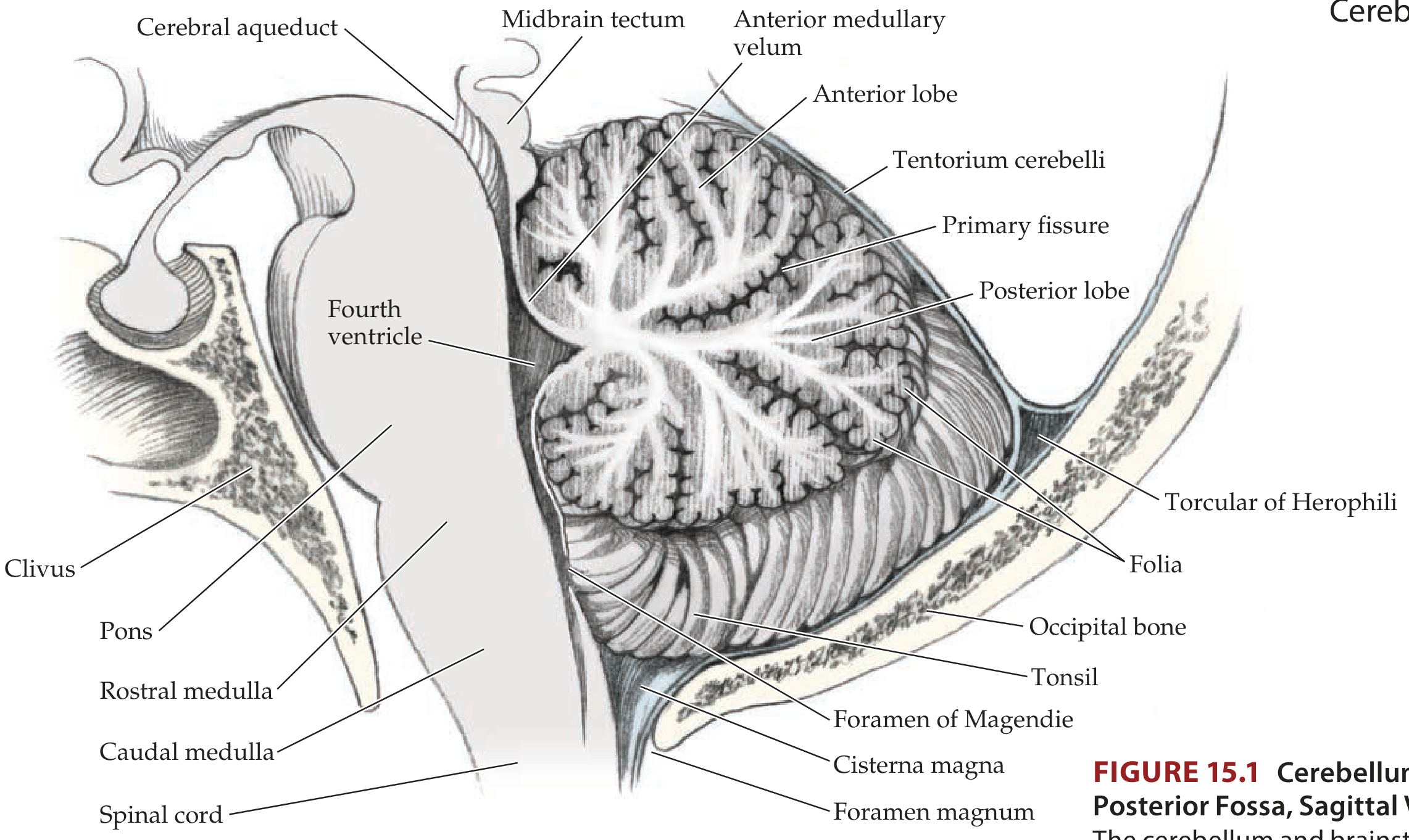
Lobes
The cerebellum is divided by deep fissures into three lobes:
| Lobe | Separated By | Key Structures |
|---|---|---|
| Anterior lobe | Primary fissure (superiorly) | - |
| Posterior lobe | Primary fissure / Posterolateral fissure | Largest lobe |
| Flocculonodular lobe | Posterolateral fissure | Flocculi + nodulus; vestibular connections |
The vermis is the midline worm-like structure connecting the two cerebellar hemispheres. The cerebellar tonsils hang inferiorly near the foramen magnum - clinically important because elevated intracranial pressure can force them downward (tonsillar herniation), compressing the medulla and causing death.
Three Cerebellar Peduncles
The cerebellum connects to the brainstem via three paired white matter stalks:
| Peduncle | Direction | Key Pathways |
|---|---|---|
| Superior (brachium conjunctivum) | Primarily efferent | Output to thalamus (dentatorubrothalamic tract) |
| Middle (brachium pontis) | Primarily afferent | Pontocerebellar fibers (from contralateral cortex via pons) |
| Inferior (restiform body) | Mixed | Spinocerebellar tracts (afferent); vestibulocerebellar fibers (both) |
Deep Cerebellar Nuclei (medial to lateral)
All output from the cerebellar cortex is relayed through these nuclei, which project excitatory signals outward:
- Fastigial nucleus - vestibulocerebellum output (balance, posture)
- Globose and emboliform nuclei (together = interposed nuclei) - spinocerebellum output (limb movements)
- Dentate nucleus - largest; pontocerebellum output (motor planning, cognitive functions)
Functional Divisions
The cerebellum is organized into three functional zones based on their dominant inputs and outputs:
| Division | Anatomical Region | Primary Input | Function |
|---|---|---|---|
| Vestibulocerebellum (archicerebellum) | Flocculonodular lobe + inferior vermis | Vestibular nuclei | Balance, eye movements, postural stability |
| Spinocerebellum (paleocerebellum) | Vermis + intermediate hemispheres | Spinal cord (proprioception, touch) | Synergy of limb and trunk movements; correction of ongoing movement |
| Pontocerebellum / Cerebrocerebellum (neocerebellum) | Lateral cerebellar hemispheres | Cerebral cortex via pontine nuclei | Motor planning, initiation; timing; cognition |
- Costanzo Physiology 7th Ed., p. 113-114
Cortical Layers and Cellular Architecture
The cerebellar cortex has a uniform, three-layer structure everywhere:
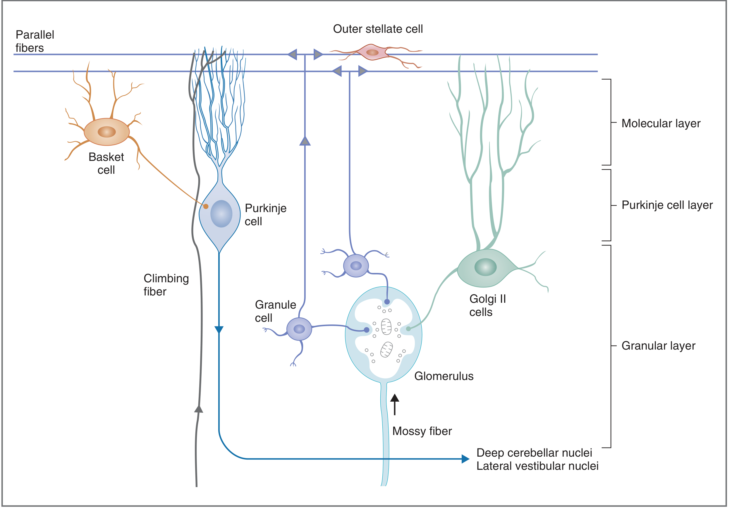
The Three Layers (inside to outside)
1. Granular Layer (innermost)
- Contains granule cells - the most numerous neurons in the entire CNS
- Also contains Golgi type II cells
- Mossy fiber axon terminals synapse on granule cell dendrites within specialized structures called glomeruli (also receive inhibitory input from Golgi cells)
- Granule cell axons ascend into the molecular layer and bifurcate into parallel fibers, which run along the length of the folia
2. Purkinje Cell Layer (middle)
- A single layer of large, flask-shaped Purkinje cells with elaborate fan-like dendritic trees extending into the molecular layer
- The only output neurons of the cerebellar cortex
- Output is always inhibitory (GABAergic), targeting the deep cerebellar nuclei and vestibular nuclei
3. Molecular Layer (outermost)
- Contains: parallel fibers (granule cell axons), Purkinje cell dendrites, stellate cells, and basket cells
- Parallel fibers run perpendicular to Purkinje dendrites, forming excitatory synapses on hundreds of Purkinje cells as they pass
Input Systems: Mossy Fibers vs. Climbing Fibers
| Feature | Mossy Fibers | Climbing Fibers |
|---|---|---|
| Origin | Spinal cord (spinocerebellar), pontine nuclei, vestibular nuclei, reticular formation | Contralateral inferior olivary nucleus (medulla) ONLY |
| Target | Granule cell dendrites (via glomeruli) | Wrap directly around Purkinje cell body and proximal dendrites |
| Effect | Excitatory; indirect to Purkinje cells (via granule cells → parallel fibers) | Excitatory; each Purkinje cell receives input from only ONE climbing fiber |
| Collaterals | Direct excitatory collaterals to deep cerebellar nuclei | Direct excitatory collaterals to deep cerebellar nuclei |
| Role | Ongoing sensorimotor information | Error signals; motor learning (long-term depression, LTD) |
- Neuroanatomy through Clinical Cases 3rd Ed., p. 730-731
- Costanzo Physiology 7th Ed., p. 114
Interneurons (all inhibitory)
- Basket cells: inhibit Purkinje cells (surround axon initial segment)
- Stellate cells: inhibit Purkinje cell dendrites
- Golgi cells: inhibit granule cells (feedback inhibition at glomeruli)
Output Circuit (The Basic Loop)
Cerebral cortex
      ↓ (corticospinal / corticopontine)
Pontine nuclei → [mossy fibers] → Granule cells → Parallel fibers
                                                         ↓
                                                  Purkinje cells (inhibitory)
                                                         ↓
                                              Deep cerebellar nuclei
                                    (fastigial / interposed / dentate)
                                                         ↓
                                              Thalamus (VL nucleus)
                                                         ↓
                                              Motor cortex (feedback)
The net effect: cerebellar output (via deep nuclei → thalamus → motor cortex) modulates and refines descending motor commands, achieving smooth, coordinated movement.
Motor Control Hierarchy - Where Cerebellum Fits
The central motor system operates as a three-level hierarchy (Neuroscience: Exploring the Brain, p. 1318):
| Level | Structures | Role |
|---|---|---|
| Strategy | Association cortex, basal ganglia | What movement to make |
| Tactics | Motor cortex, cerebellum | How to sequence muscle contractions |
| Execution | Brainstem, spinal cord | Activate lower motor neurons |
The cerebellum operates at the tactics level - ensuring smooth, accurate timing of multi-joint movements.
Clinical Correlates
A key principle: cerebellar lesions are ipsilateral (information does not cross until it leaves the cerebellum, unlike the corticospinal system which crosses twice and returns to the same side).
Classic Signs of Cerebellar Dysfunction
| Sign | Description |
|---|---|
| Ataxia | Irregular, uncoordinated movement - the cardinal sign |
| Dyssynergia | Decomposition of movement - joints move sequentially instead of simultaneously |
| Dysmetria | Misjudging distances (overshoot = hypermetria, undershoot = hypometria); seen in finger-nose test |
| Intention tremor | Tremor that appears/worsens during voluntary movement (unlike Parkinson's resting tremor) |
| Dysdiadochokinesia | Inability to perform rapid alternating movements |
| Nystagmus | Involuntary eye oscillation (especially with vestibulocerebellum/flocculonodular lesions) |
| Dysarthria | Slurred, scanning speech |
| Truncal ataxia | Wide-based, unsteady gait; vermis/midline lesions |
Lesion Localization Rules
- Flocculonodular lobe / inferior vermis lesion → truncal ataxia, gait instability, nystagmus, vertigo, nausea/vomiting (vestibular disruption)
- Lateral hemisphere lesion → ipsilateral limb (appendicular) ataxia - dysmetria, intention tremor, dyssynergia
- Midline vermis lesion → truncal/gait ataxia predominantly
Tonsillar Herniation
Mass lesions or severe ICP elevation can push the cerebellar tonsils through the foramen magnum, compressing the medulla's respiratory centers - a life-threatening emergency.
Clinically Important: Alcohol and the Cerebellum
Ethanol preferentially depresses cerebellar circuits, producing the classic signs mimicking cerebellar disease: staggering gait, dysmetria (failed sobriety tests), and slurred speech. This illustrates how sensitive cerebellar circuits are to pharmacological disruption. - Neuroscience: Exploring the Brain, p. 1379
Summary Table
| Feature | Detail |
|---|---|
| Location | Posterior fossa, dorsal to pons/medulla; roof of 4th ventricle |
| Cortical output cell | Purkinje cell (always INHIBITORY, GABAergic) |
| Input fibers | Mossy fibers (spinal cord, pons) + Climbing fibers (inferior olive only) |
| Deep nuclei (medial→lateral) | Fastigial → Globose/Emboliform → Dentate |
| Output pathway | Deep nuclei → Superior peduncle → Thalamus (VL) → Motor cortex |
| Key function | Coordination, timing, motor learning, balance |
| Lesion sign | Ipsilateral ataxia |
Sources: Neuroanatomy through Clinical Cases 3rd Ed. | Costanzo Physiology 7th Ed. | Neuroscience: Exploring the Brain 5th Ed.
This is a shared conversation. Sign in to Orris to start your own chat.