
CA cervix
carcinoma cervix colposcopy histology
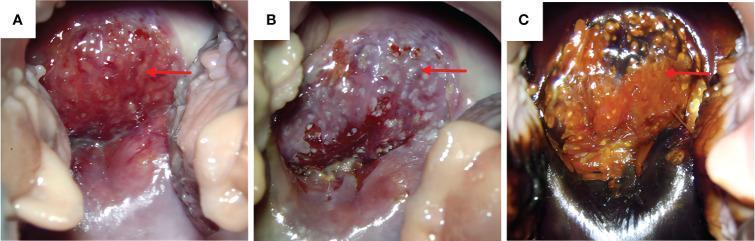
A series of colposcopy clinical photographs (labeled A, B, and C) depicting a uterine cervix with high-grade pathology. (A) Baseline colposcopy shows significant erosion-like changes in the anterior cervical lip with pronounced vascular proliferation and a friable, reddened surface (red arrow). (B) Following the application of acetic acid (vinegar test), the lesion demonstrates positive acetowhite changes, appearing as opaque white spots. In this same region, atypical vascular patterns including mosaic blood vessels are clearly visible. (C) Following the application of Lugol's iodine (Schiller test), the anterior cervical lip remains iodine-negative (unstained), retaining its original color while the surrounding normal tissue or vaginal vault displays dark staining. This lack of iodine uptake indicates a lack of glycogen, which is characteristic of dysplastic or malignant squamous epithelium. These visual findings are consistent with High-grade Squamous Intraepithelial Lesion (HSIL) or invasive Squamous Cell Carcinoma (SCC).
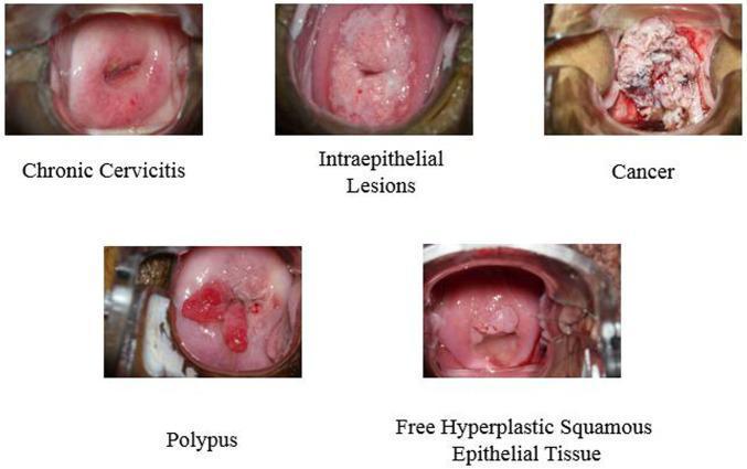
This composite of clinical colposcopy photographs illustrates five distinct pathological states of the human cervix. 1. Chronic Cervicitis: Shows a relatively smooth, erythematous ectocervix with a linear lesion and punctate hemorrhages around the external os. 2. Intraepithelial Lesions: Features a thickened, opaque, whitish acetowhite-like area covering the transformation zone, suggesting dysplastic changes. 3. Cervical Cancer: Displays an advanced neoplastic state characterized by a large, exophytic, friable mass with an irregular, cauliflower-like texture and variegated color (whitish and necrotic red areas) obscuring normal anatomy. 4. Polypus (Cervical Polyp): Exhibits a distinct, pedunculated, smooth, fleshy red growth protruding from the cervical canal. 5. Free Hyperplastic Squamous Epithelial Tissue: Shows a more subtle, uniform, pinkish elevation of the squamous epithelium without the severe disruption seen in malignancy. These images serve as educational references for gynecological oncology and colposcopic diagnosis, highlighting key morphological differences in texture, color, and tissue architecture used to distinguish benign inflammation from precancerous lesions and invasive carcinoma.
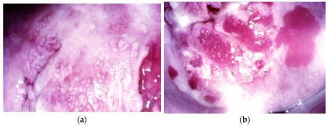
This set of clinical photographs obtained via colposcopy (14x magnification) illustrates early-stage cervical cancer presentations. Image (a) depicts a microinvasive lesion of the cervix, showing a complex, textured surface with prominent acetowhite patterns, reticular mosaicism, and areas of punctate vascularity. The tissue appears uneven with alternating pink and pale white zones, indicating dysplastic epithelial changes. Image (b) shows a stage IA2 squamous cell carcinoma characterized by significant vascular atypia. Visible features include irregular, deep pink atypical vessels with varying diameters and corkscrew or comma-like morphologies. The surface is nodular and heterogeneous, featuring raised exophytic areas and intense focal hyperemia. These images serve as educational examples for identifying high-grade cervical intraepithelial lesions and early invasive squamous cell carcinoma during diagnostic colposcopic evaluation, highlighting the importance of vessel morphology and surface texture in oncologic staging.
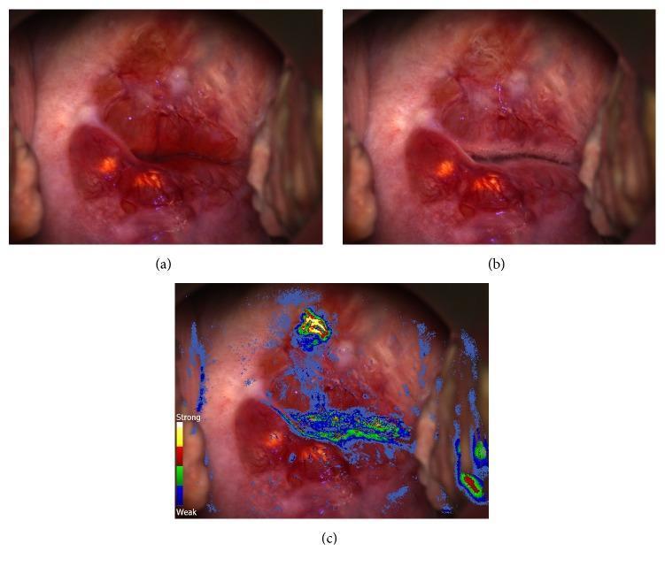
This set of clinical photographs illustrates a gynecological colposcopy examination for cervical cancer screening. Image (a) shows the native cervix prior to acetic acid application, revealing a reddish, smooth surface with Nabothian cysts visible at the 6 and 8 o’clock positions. Image (b) shows the same anatomical region 30 seconds after the application of 3% acetic acid; it demonstrates a minimal, subtle acetowhitening reaction near the external os, which might be missed on conventional visual inspection. Image (c) presents a Dynamic Spectral Imaging (DSI) color-coded map overlaid on the colposcopic view. The DSI scale (ranging from blue for weak to red/yellow for strong) identifies a focal area of persistent, intense acetowhitening at the 11-12 o'clock position. This digital mapping provides an objective assessment of tissue kinetics, highlighting a high-risk lesion that was histologically confirmed as invasive squamous cell carcinoma. This comparison demonstrates the clinical utility of adjunctive digital imaging in identifying neoplastic lesions that lack distinctive classic colposcopic features.
"cervical cancer"[MeSH Terms] AND "treatment"[MeSH Terms]
"cervical cancer"[MeSH Terms] AND "chemoradiotherapy"[MeSH Terms]
Carcinoma of the Cervix
Epidemiology
- Third most common gynecologic neoplasm in the United States (behind corpus and ovarian cancers), primarily due to effective screening programs.
- In developing countries, it remains the second most frequent cause of cancer death in women.
- Mean age at diagnosis: 47 years; bimodal peaks at 35-39 years and 60-64 years.
- Lifetime probability in the US: 1:128.
- ~30% of US cases occur in women who have never had a Pap smear; in developing countries this approaches 60%.
Etiology and Risk Factors
- High-risk HPV types: 16, 18, 31, 33, 35, 45, 51, 52, 56 (15 high-risk types total)
- HPV 16 and 18 account for ~70% of cases
- HPV 16 predominates in squamous cell carcinoma; HPV 18 more commonly implicated in adenocarcinoma
- Herpes simplex virus and Chlamydia trachomatis act as cofactors
- HIV infection (immune suppression) - cervical cancer is an AIDS-defining illness
- Young age at first intercourse (<16 years)
- Multiple sexual partners
- Cigarette smoking
- High parity
- Low socioeconomic status
- Chronic immune suppression
Pathology
Histologic Types
| Type | Frequency | Notes |
|---|---|---|
| Squamous cell carcinoma | ~80% | Most common; develops at transformation zone |
| Adenocarcinoma + mixed adenosquamous | ~15% | Increasing proportion; less amenable to Pap screening |
| Small cell neuroendocrine carcinoma | <5% | Very aggressive; resembles oat cell carcinoma of the lung |
Squamous Cell Carcinoma Variants
- Large cell keratinizing - forms keratin pearls; best prognosis
- Large cell nonkeratinizing - individual cell keratinization, no keratin pearls
- Small cell anaplastic - scanty cytoplasm, coarsely granular chromatin, no nucleoli; most aggressive
Gross Morphology
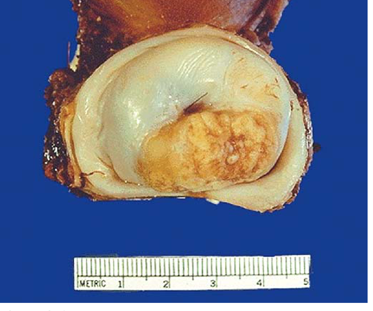
Lymph Node Metastasis Risk
- <1% for tumors <3 mm depth
-
10% when invasion exceeds 3 mm
Clinical Features
- Asymptomatic early (detected on screening)
- Abnormal vaginal bleeding (most common symptom) - postcoital, intermenstrual, or postmenopausal bleeding
- Leukorrhea (watery, blood-stained, malodorous discharge)
- Dyspareunia
- Dysuria, frequency (bladder involvement)
- Rectal symptoms (advanced disease)
- Advanced disease: hydronephrosis, renal failure, fistulae
Diagnosis
- Pap smear (screening)
- Colposcopy with directed biopsy
- Cone biopsy / LEEP for microinvasive disease
- Examination under anesthesia (EUA) for clinical staging
Colposcopic Findings
FIGO Staging (2018 - Current)
| Stage | Description |
|---|---|
| I | Strictly confined to the cervix |
| IA | Microscopic only, depth <5 mm |
| IA1 | Stromal invasion <3 mm |
| IA2 | Stromal invasion ≥3 mm and <5 mm |
| IB | Deepest invasion ≥5 mm, limited to cervix |
| IB1 | <2 cm greatest dimension |
| IB2 | ≥2 cm and <4 cm |
| IB3 | ≥4 cm |
| II | Beyond uterus, not to pelvic wall or lower 1/3 vagina |
| IIA | Upper 2/3 vagina involved, no parametria |
| IIA1 | <4 cm |
| IIA2 | ≥4 cm |
| IIB | Parametrial involvement (not to pelvic wall) |
| III | Lower 1/3 vagina, pelvic wall, hydronephrosis, or LN+ |
| IIIA | Lower 1/3 vagina, no pelvic wall extension |
| IIIB | Pelvic wall and/or hydronephrosis/nonfunctioning kidney |
| IIIC1 | Pelvic LN metastasis only |
| IIIC2 | Para-aortic LN metastasis |
| IV | Beyond true pelvis or bladder/rectal mucosal involvement |
| IVA | Bladder/rectal mucosa |
| IVB | Distant metastases |
- Stage IA: horizontal spread removed; depth only used
- Stage IB split into 3 substages (IB1, IB2, IB3)
- New Stage IIIC added for LN metastasis (regardless of tumor size)
Treatment
Stage IA1
- Cone biopsy (if fertility desired, negative margins, no LVSI)
- Simple (extrafascial) hysterectomy if no fertility desired
Stage IA2
- Modified radical hysterectomy (Type II / Wertheim's) + pelvic lymphadenectomy
- OR radical trachelectomy (fertility-sparing in selected patients)
Stage IB1 - IIA1
- Radical hysterectomy (Type III / Meigs) + pelvic lymphadenectomy - preferred for younger patients
- OR definitive chemoradiation (equally effective; surgery preferred to preserve ovarian function and avoid radiation late effects)
Stage IB2 - IIA2 ("Bulky" tumors)
- Concurrent chemoradiation (cisplatin-based) - primary treatment
- Neoadjuvant chemotherapy followed by surgery in selected centers
Stage IIB - IVA
- Concurrent cisplatin-based chemoradiation (external beam + brachytherapy)
- Cisplatin 40 mg/m² weekly during radiation is standard
Stage IVB / Recurrent
- Palliative chemotherapy (cisplatin + paclitaxel ± bevacizumab)
- Bevacizumab (anti-VEGF) improves survival in metastatic disease
Surgery: Radical Hysterectomy (Piver-Rutledge Classification)
| Type | Procedure | Indication |
|---|---|---|
| Type I | Extrafascial hysterectomy | Stage IA1 |
| Type II (Wertheim's) | Medial 1/2 cardinal + uterosacral ligaments; uterine artery divided at ureter | IA2, small IB1 |
| Type III (Meigs) | Most of cardinal + uterosacral + upper 1/3 vagina; full pelvic LND | IB1, IIA |
| Type IV | Periureteral tissue + 3/4 vagina removed | Rare |
| Type V | Distal ureter and bladder resection | Very rarely done |
Prognosis (5-Year Survival by Stage)
| Stage | 5-Year Survival |
|---|---|
| Stage I | ~85-90% |
| Stage II | ~60-75% |
| Stage III | ~30-35% |
| Stage IV | ~5-15% |
- Stage (most important)
- Lymph node status
- Tumor size
- LVSI (lymphovascular space invasion)
- Histologic subtype (small cell = worst prognosis)
- Parametrial involvement
Prevention
- HPV vaccination (Gardasil 9 - covers HPV 6, 11, 16, 18, 31, 33, 45, 52, 58): recommended for girls and boys ages 9-26 years; most effective before sexual debut
- Cervical screening: Pap smear (cytology) +/- HPV co-testing every 3-5 years depending on guidelines
- Colposcopy and treatment of CIN lesions (LEEP, cryotherapy, cone biopsy)
Key Points to Remember
- HPV 16 and 18 cause ~70% of cases - HPV testing is both diagnostic and screening
- Squamous cell carcinoma is most common (80%); arises at the squamocolumnar junction/transformation zone
- FIGO staging is clinical (updated 2018 to allow imaging and pathological LN findings)
- Chemoradiation (cisplatin + RT) is the backbone of treatment for stages IIB and above
- Radical hysterectomy = Wertheim's (Type II) for early; Meigs (Type III) for IB/IIA
- Cervical cancer is a preventable disease - vaccination + screening can dramatically reduce incidence