
Highlight the contrast in parameters of pulmonary function test between obstructive and restrictive conditions answer this according to 10 marker question in cvts for physiotherapy students
Reading File
Now let me search the medical library for PFT content on obstructive and restrictive conditions.
Finding Sources
Finding Sources
Excellent finds. Now let me read the most relevant sections in parallel:
Reading File
Reading File
Reading File
Now let me get more detailed content on the obstructive and restrictive patterns from Murray & Nadel and Goldman-Cecil:
Reading File
Reading File
Excellent! I have very rich content. Let me get the restrictive defect section from Murray & Nadel and more details from Goldman-Cecil:
Reading File
Reading File
I now have comprehensive material. Let me also fetch the flow-volume curve images for inclusion in the answer:
Reading File
I now have all the content needed for a comprehensive 10-marker answer. Let me compile the full response.
Pulmonary Function Tests (PFT): Obstructive vs. Restrictive Conditions
Introduction (1 mark)
Pulmonary function tests (PFTs) are a core diagnostic tool used to classify respiratory disorders and monitor disease progression. They measure respiratory mechanics (lung volumes, flow rates), and gas exchange. Most pulmonary disorders are broadly categorized as obstructive or restrictive based on their characteristic PFT pattern. This classification directly guides physiotherapy management in cardiorespiratory conditions.
Definitions (1 mark)
| Type | Definition |
|---|---|
| Obstructive | Small airway obstruction to expiratory flow - characterized by increased resistance to airflow, primarily affecting exhalation |
| Restrictive | Proportional decreases in all lung volumes - characterized by reduced lung compliance or mechanical limitation of chest expansion |
Key PFT Parameters - Contrast Table (3 marks)
| Parameter | Obstructive | Restrictive |
|---|---|---|
| FVC (Forced Vital Capacity) | Normal or slightly decreased | Markedly decreased (↓↓↓) |
| FEV₁ (Forced Expiratory Volume in 1 sec) | Normal or slightly decreased | Markedly decreased (↓↓↓) |
| FEV₁/FVC Ratio | Decreased (↓↓↓) - KEY finding | Normal or increased - KEY finding |
| FEF₂₅₋₇₅% (Mid-expiratory flow) | Markedly decreased (↓↓↓) | Normal |
| FRC (Functional Residual Capacity) | Normal or increased (gas trapping) | Markedly decreased (↓↓↓) |
| TLC (Total Lung Capacity) | Normal or increased (hyperinflation) | Markedly decreased (↓↓↓) |
| RV (Residual Volume) | Increased (air trapping) | Normal or decreased |
| RV/TLC Ratio | Increased | Normal |
| DLCO (Diffusing capacity for CO) | Decreased (in emphysema due to alveolar surface area loss) | Decreased (due to thickened alveolar-capillary membrane in fibrosis) |
| Airway Resistance (RAW) | Increased | Normal |
| Bronchodilator Response | Significant reversibility possible | Minimal or no reversal |
- Barash's Clinical Anesthesia, 9e, p. 1139 (Table 15-2)
- Goldman-Cecil Medicine, Table 73-2
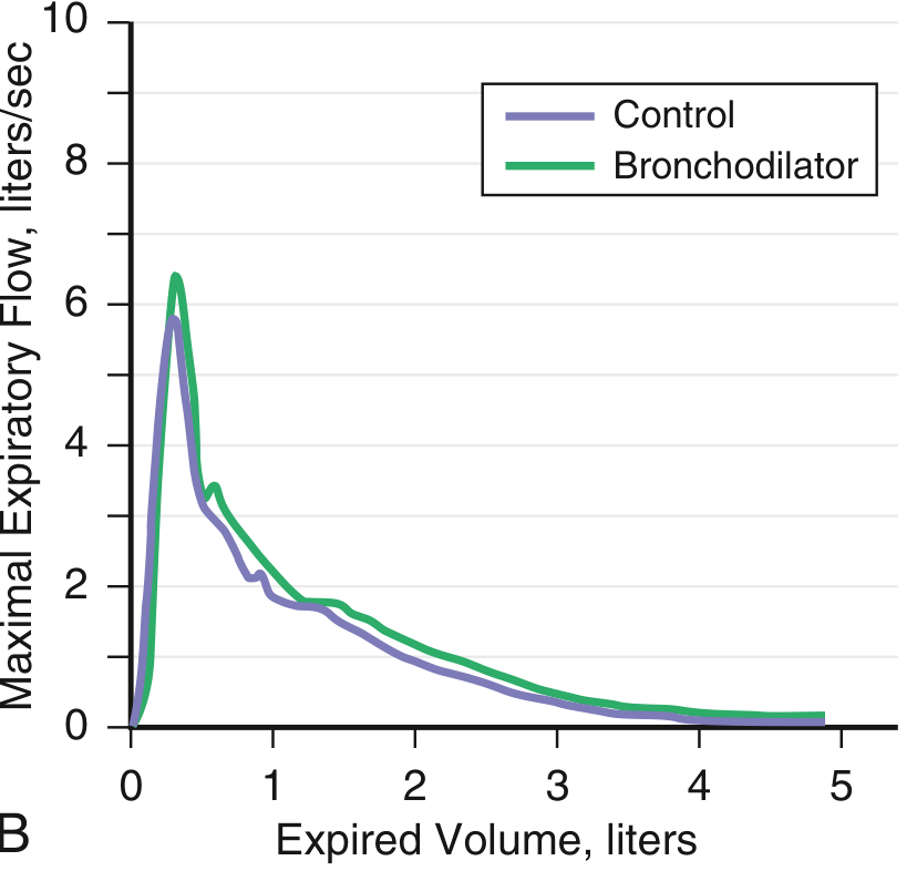
Flow-Volume Curve Patterns (1.5 marks)
The flow-volume curve is a highly informative graphical representation:
Obstructive:
- The expiratory limb shows a characteristic "scooped out" or concave (curvilinear) appearance - reflecting reduced flow rates at low lung volumes
- Peak expiratory flow is reached early, but flow decays rapidly
- The curve is wider (increased TLC and RV shift the curve rightward)
Restrictive:
- The expiratory limb shows a characteristic "witch's hat" or tall peaked shape - a narrower curve due to reduced volumes
- Peak expiratory flow may be relatively preserved, but the total exhaled volume is small
- The curve is narrow and shifted leftward
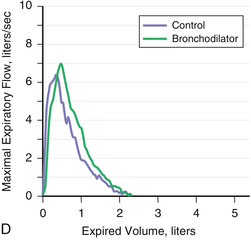
- Goldman-Cecil Medicine, p. 867
Pathophysiological Basis (1.5 marks)
Obstructive
- Airflow limitation is due to airway narrowing, increased collapsibility, or loss of elastic recoil (as in emphysema)
- Air trapping occurs because small airways collapse prematurely during exhalation - RV and FRC increase
- Hyperinflation is a compensatory mechanism - TLC increases in severe cases
- Diseases: COPD, bronchial asthma, bronchiolitis, bronchiectasis, cystic fibrosis
Restrictive
-
Lung volumes are reduced because of either:
- Parenchymal causes - reduced lung compliance (interstitial lung disease, pulmonary fibrosis, sarcoidosis, asbestosis)
- Extra-parenchymal causes - mechanical limitation of chest expansion (obesity, kyphoscoliosis, ankylosing spondylitis, neuromuscular disorders: Guillain-Barre syndrome, muscular dystrophy, myasthenia gravis, diaphragmatic paralysis)
-
FEV₁/FVC is preserved or elevated because both FEV₁ and FVC fall proportionally - the lungs are small but the airways are not obstructed
-
Diseases: Idiopathic pulmonary fibrosis, ILD, pleural disease, neuromuscular disease
-
Murray & Nadel's Textbook of Respiratory Medicine, p. 759-760
Severity Grading Based on FEV₁ (1 mark)
Severity in both patterns is graded by FEV₁ (% predicted):
| Severity | FEV₁ % Predicted |
|---|---|
| Mild | ≥70% |
| Moderate | 50-69% |
| Moderately severe | 35-49% |
| Severe | <35% |
In restrictive disease, severity is graded by TLC (% predicted):
- Mild: TLC ≥70%
- Moderate: TLC 51-69%
- Severe: TLC ≤50%
Physiotherapy Implications (1 mark)
| Pattern | Physiotherapy Focus |
|---|---|
| Obstructive | Pursed-lip breathing (to prevent dynamic airway collapse), diaphragmatic breathing, airway clearance techniques (ACBT, PEP therapy, autogenic drainage), bronchodilator inhalation prior to exercise, energy conservation |
| Restrictive | Chest expansion exercises, incentive spirometry, positioning (sitting upright to maximize FRC), strengthening of respiratory muscles, mobilization, postural correction (especially in kyphoscoliosis) |
Summary Mnemonic
OBSTRUCTIVE = "Can't get air OUT" → FEV₁/FVC DOWN, TLC/RV UP, scooped curve RESTRICTIVE = "Can't get air IN" → FEV₁/FVC NORMAL/UP, TLC DOWN, peaked narrow curve
Quick Reference Comparison (for revision)
| Feature | Obstructive | Restrictive |
|---|---|---|
| Pathology | Airway obstruction | Reduced lung/thoracic compliance |
| FEV₁/FVC | Low (<0.70) | Normal or High (≥0.70) |
| TLC | Normal / High | Low |
| RV | High | Normal / Low |
| Flow-volume curve | Concave/scooped | Peaked/narrow |
| Examples | COPD, Asthma | IPF, Kyphoscoliosis, Neuromuscular |
| Bronchodilator response | Yes (asthma especially) | No |
Sources: Murray & Nadel's Textbook of Respiratory Medicine (2-Volume); Goldman-Cecil Medicine International Edition; Barash's Clinical Anesthesia, 9e
This is a shared conversation. Sign in to Orris to start your own chat.