
Femoral triangle
The femoral triangle is a subfascial space in the upper anterior thigh with important neurovascular contents. Here's a quick overview while I pull from the medical library:
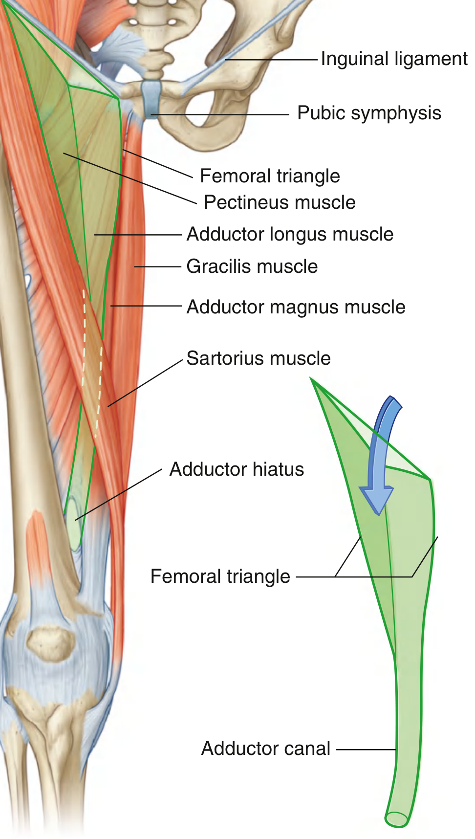
- Boundaries: inguinal ligament (base/superior), medial border of sartorius (lateral), medial border of adductor longus (medial), floor formed by iliopsoas and pectineus
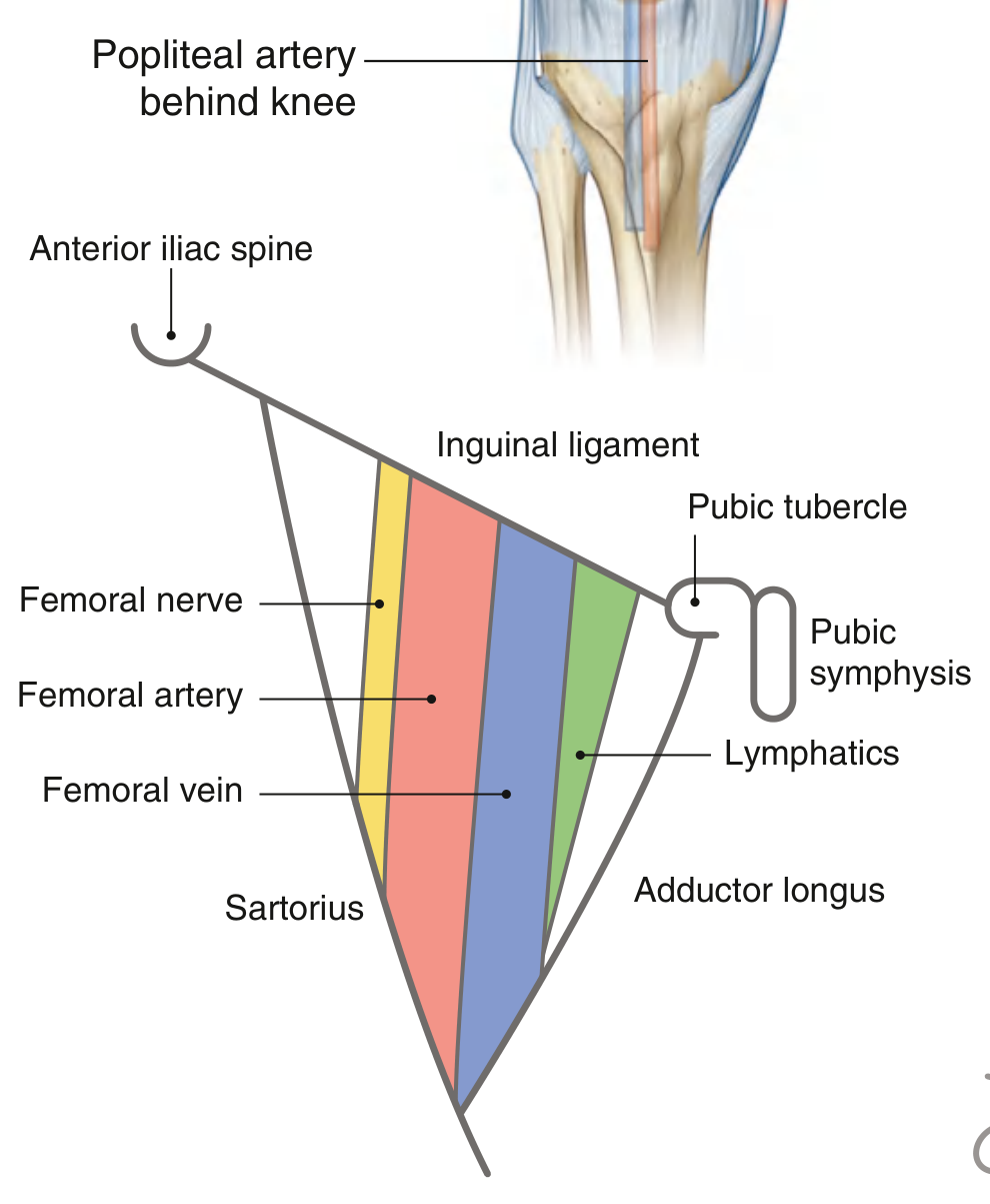
- Contents (lateral to medial): femoral nerve, femoral artery, femoral vein, femoral canal (containing lymphatics/Cloquet's node) - remembered as NAVY (Nerve, Artery, Vein, Y-fronts/lymphatics)
- Roof: fascia lata (with the saphenous opening/fossa ovalis)
- Apex: leads into the adductor canal (Hunter's canal)
Searching sources...
Reading File
Finding Sources
Finding Sources
Reading File
Reading File
Reading File
Reading File
Now let me fetch the key diagrams:
Loading Image
Loading Image
Loading Image
Here is a complete, textbook-sourced overview of the femoral triangle.
Femoral Triangle
The femoral triangle is a wedge-shaped (pyramid-shaped) depression in the upper anterior thigh, at the junction between the anterior abdominal wall and the lower limb. It provides a gateway for major neurovascular structures entering the limb.
Boundaries
| Boundary | Structure |
|---|---|
| Base (superior) | Inguinal ligament (from ASIS to pubic tubercle) |
| Lateral border | Medial margin of sartorius muscle |
| Medial border | Medial margin of adductor longus muscle |
| Roof | Fascia lata (with the saphenous opening - fossa ovalis) |
| Floor | Laterally - iliopsoas; medially - pectineus and adductor longus |
| Apex | Junction of sartorius and adductor longus (points inferiorly, leads into the adductor canal) |
Contents (Lateral to Medial - "NAVY")
N - A - V - Y: Nerve, Artery, Vein, Y-fronts (lymphatics/canal)
From lateral to medial:
- Femoral nerve (L2, L3, L4) - lies in the muscular (lateral) compartment, outside the femoral sheath
- Femoral artery - continuation of the external iliac artery below the inguinal ligament; palpable midway between the ASIS and pubic symphysis
- Femoral vein - medial to the artery
- Femoral canal - most medial compartment; contains lymphatic vessels and the deep inguinal (Cloquet's) lymph node at its upper end
The Femoral Sheath
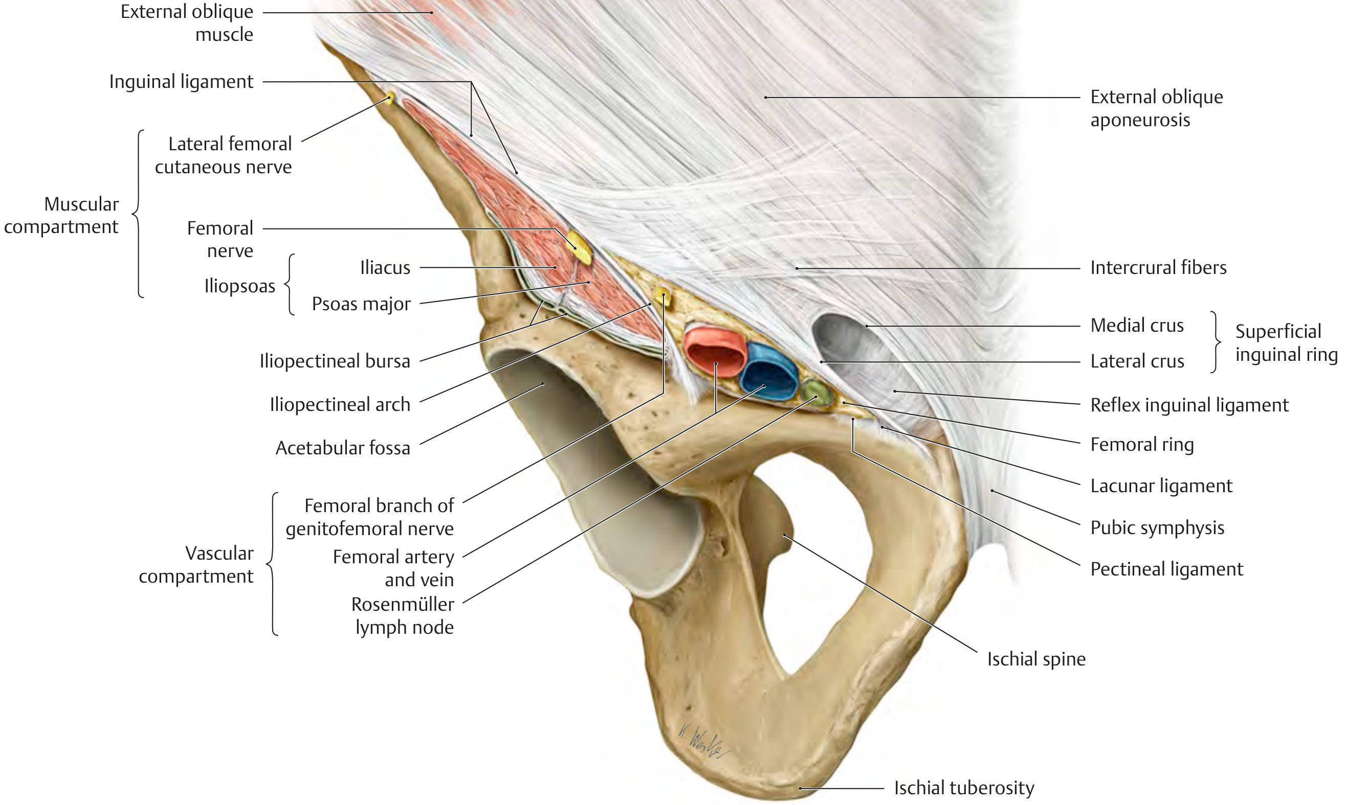
The femoral artery, vein, and lymphatics are enclosed in a funnel-shaped fascial sleeve called the femoral sheath:
- Continuous superiorly with transversalis fascia (anterior) and iliac fascia (posterior)
- Merges inferiorly with connective tissue of the vessels
- Divided into three compartments:
- Lateral: femoral artery
- Middle: femoral vein
- Medial: femoral canal (lymphatics)
- The femoral nerve is NOT inside the sheath - it lies lateral to it in the muscular compartment
The Femoral Canal
- Most medial compartment of the femoral sheath
- Conical shape, approximately 1.25 cm long
- Femoral ring = the superior opening of the canal (covered by femoral septum)
- Contains: lymphatic vessels, areolar tissue, Cloquet's (Rosenmüller) node
- The femoral ring is bounded by: inguinal ligament (anteriorly), femoral vein (laterally), lacunar ligament (medially), pectineal ligament (posteriorly)
- Clinical significance: the femoral ring is a potential weak point - site of femoral hernias (more common in women due to wider pelvis)
Apex and Continuation
- The apex leads into the adductor canal (Hunter's canal), which descends medially down the thigh
- The femoral artery and vein travel through the adductor canal and exit via the adductor hiatus in adductor magnus to become the popliteal artery and vein in the popliteal fossa behind the knee
Femoral Artery Pulse Point
The femoral artery is palpable (and easily located with ultrasound) just inferior to the inguinal ligament, midway between the ASIS and pubic symphysis. This is the standard landmark for:
- Femoral artery cannulation (cardiac catheterization, coronary angiography)
- Femoral vein access (right heart catheterization, pulmonary artery catheters, renal vein sampling)
- Emergency vascular access
Clinical Relevance
| Condition | Relevance |
|---|---|
| Femoral hernia | Viscera protrudes through the femoral ring into the femoral canal; appears below and lateral to the pubic tubercle (vs. inguinal hernia which is above/medial) |
| Femoral artery access | Key site for cardiac catheterization and angiography |
| Femoral nerve block | Anaesthesia for anterior thigh and knee surgery |
| Femoral vein thrombosis | Can propagate to iliofemoral DVT |
| Lymphadenopathy | Inguinal lymph nodes drain the lower limb, perineum, and lower abdominal wall; enlarged nodes palpable in this region |
| Psoas abscess | Can track along iliopsoas into the femoral triangle |
Sources: Gray's Anatomy for Students, pp. 675-676; THIEME Atlas of Anatomy - General Anatomy and Musculoskeletal System, p. 573
This is a shared conversation. Sign in to Orris to start your own chat.