
Molecular basis of apoptosis mechanism and morphological changes md pathology exam question Robbins based answer
Reading File
Finding Sources
Finding Sources
Searching PubMed
apoptosis mechanisms
Searching the Web
Robbins Basic Pathology apoptosis intrinsic extrinsic pathway morphology caspases
Finding Sources
Finding Sources
Finding Sources
Reading File
Reading File
Searching Images
apoptosis morphology apoptotic bodies chromatin condensation histology Robbins
Finding Sources
Finding Sources
query_images_db
Apoptosis: molecular basis, mechanism and morphological changes
Definition:
Apoptosis is a regulated, energy-dependent form of cell death in which a cell activates enzymes that degrade its own nuclear DNA and cytoplasmic proteins. The cell fragments into membrane-bound apoptotic bodies, which are rapidly phagocytosed without leakage of cellular contents or inflammation.
Robbins & Kumar Basic Pathology, p. 24.
Apoptosis is a regulated, energy-dependent form of cell death in which a cell activates enzymes that degrade its own nuclear DNA and cytoplasmic proteins. The cell fragments into membrane-bound apoptotic bodies, which are rapidly phagocytosed without leakage of cellular contents or inflammation.
Robbins & Kumar Basic Pathology, p. 24.
Molecular basis
The central mediators are caspases: cysteine proteases that cleave proteins after aspartate residues. They are synthesized as inactive procaspases and become activated in a cascade.
- Initiator caspases: caspase-8, caspase-9 (and caspase-10)
- Executioner caspases: caspase-3, caspase-6, caspase-7
Apoptosis proceeds through two main pathways that converge on executioner caspase activation.
1. Intrinsic or mitochondrial pathway
This is the major pathway in most physiological and pathological apoptosis.
Stimuli
- Withdrawal of growth factors or hormones
- DNA damage by radiation, cytotoxic drugs, hypoxia, or oxidative stress
- Accumulation of misfolded proteins causing ER stress
- Severe cellular injury
Mechanism
- Cellular stress activates BH3-only proteins such as Bim, Bid, Bad, Puma, and Noxa.
- BH3-only proteins inhibit anti-apoptotic BCL-2 family members:
- BCL-2
- BCL-XL
- MCL-1
- This permits activation and oligomerization of pro-apoptotic proteins BAX and BAK in the outer mitochondrial membrane.
- BAX and BAK cause mitochondrial outer membrane permeabilization.
- Cytochrome c is released into cytosol.
- Cytochrome c combines with Apaf-1 and procaspase-9 to form the apoptosome.
- Apoptosome activates caspase-9, which activates executioner caspases, especially caspase-3 and caspase-7.
- Executioner caspases produce the structural and nuclear changes of apoptosis.
Flow chart:
DNA damage / growth factor withdrawal / ER stress→
BH3-only proteins→ inhibition of
BCL-2, BCL-XL→ activation of
BAX and BAK→ mitochondrial permeability
→
cytochrome c release→
Apaf-1 apoptosome→
caspase-9→
caspase-3, -6, -7→ apoptosis
Robbins & Kumar Basic Pathology, p. 24.
2. Extrinsic or death receptor pathway
This pathway is initiated by extracellular death signals acting through cell-surface death receptors of the TNF receptor family.
Important receptors and ligands
- Fas (CD95) receptor and Fas ligand (FasL)
- TNF receptor 1 and TNF
- TRAIL receptors
Mechanism
- FasL on activated cytotoxic T cells binds Fas/CD95 on the target cell.
- Fas receptors aggregate and recruit adaptor proteins, especially FADD (Fas-associated death domain).
- This produces the death-inducing signaling complex (DISC).
- DISC activates initiator caspase-8.
- Caspase-8 activates executioner caspases, mainly caspase-3 and caspase-6.
- The target cell undergoes apoptosis.
Flow chart:
FasL or TNF→
Fas/CD95 or TNF receptor→
FADD + DISC formation→
caspase-8→
executioner caspases→ apoptosis
Caspase-8 can also cleave Bid, a BH3-only protein, thereby amplifying apoptosis through the mitochondrial pathway.
Robbins & Kumar Basic Pathology, p. 24.
3. Cytotoxic T-cell-mediated apoptosis
Cytotoxic T lymphocytes and NK cells kill virus-infected, tumor, and graft cells by:
- FasL-Fas interaction, and
- Release of perforin and granzyme B.
Perforin forms pores in the target-cell membrane, allowing granzyme B to enter and activate caspases directly. This produces apoptosis.
Execution phase: how caspases cause cell death
Activated executioner caspases cause:
-
Cleavage of cytoskeletal and nuclear proteins
Causes cell shrinkage, membrane blebbing, and fragmentation. -
Activation of endonucleases
Caspase-3 cleaves ICAD, the inhibitor of caspase-activated DNase (CAD).
Free CAD fragments DNA into nucleosomal units, producing the characteristic DNA ladder pattern. -
Inactivation of DNA repair proteins
For example, cleavage of PARP prevents DNA repair. -
Alteration of plasma membrane
Phosphatidylserine becomes exposed on the outer membrane surface, acting as an “eat-me” signal for macrophages and adjacent cells.
Morphological changes in apoptosis
1. Cell shrinkage
- The cell becomes smaller.
- Cytoplasm becomes dense and intensely eosinophilic.
- Organelles remain relatively intact but are closely packed.
2. Chromatin condensation
- This is the most characteristic morphologic feature.
- Chromatin aggregates beneath the nuclear membrane as dense peripheral crescents.
- The nucleus may later fragment into multiple pieces, called karyorrhexis.
3. Membrane blebbing
- The plasma membrane remains intact but forms protrusions or blebs.
- There is no early loss of membrane integrity.
4. Formation of apoptotic bodies
- The cell breaks into small, membrane-bound fragments.
- These contain cytoplasm, organelles, and sometimes nuclear fragments.
5. Rapid phagocytosis
- Macrophages or neighboring parenchymal cells engulf apoptotic bodies.
- Lysosomal enzymes degrade them.
6. Absence of inflammation
- Cellular constituents do not leak into extracellular tissue.
- Therefore, apoptosis typically does not cause an inflammatory reaction.
Appearance on H&E stain
An apoptotic cell appears as a round or oval shrunken cell with:
- Intensely eosinophilic cytoplasm
- Condensed, dark basophilic nuclear chromatin
- Nuclear fragments or apoptotic bodies
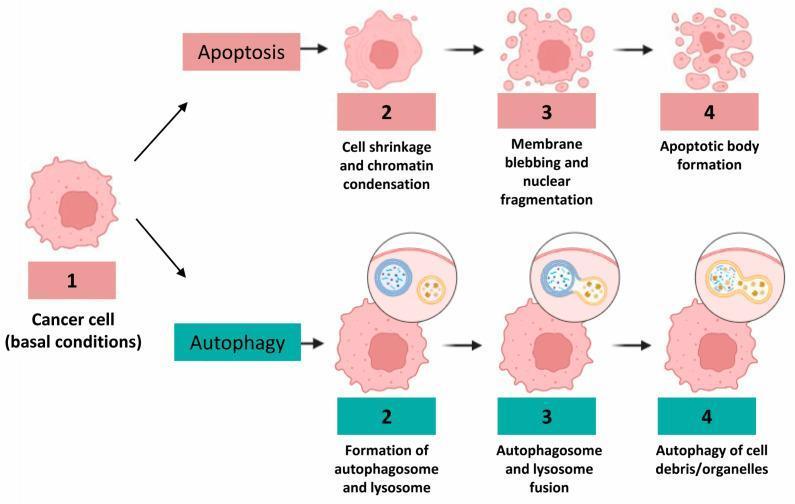
Robbins, Cotran & Kumar Pathologic Basis of Disease, Fig. 2.15.
Apoptosis versus necrosis
| Feature | Apoptosis | Necrosis |
|---|---|---|
| Cell size | Shrinkage | Swelling |
| Cell membrane | Intact, with blebs | Disrupted and ruptured |
| Nuclear change | Condensation and fragmentation | Pyknosis, karyorrhexis, then karyolysis |
| Cell contents | Retained within apoptotic bodies | Leak into extracellular space |
| Inflammation | Absent or minimal | Marked inflammation |
| Mechanism | Caspase-mediated programmed death | Severe injury with membrane damage and ATP depletion |
One-line conclusion
Apoptosis is caspase-mediated programmed cell death occurring through intrinsic mitochondrial or extrinsic death-receptor pathways, morphologically characterized by cell shrinkage, chromatin condensation, membrane blebbing, apoptotic-body formation, rapid phagocytosis, and no inflammation.
This is a shared conversation. Sign in to Orris to start your own chat.