
Chest expansion measurements according to tape
chest expansion measurement tape respiratory examination
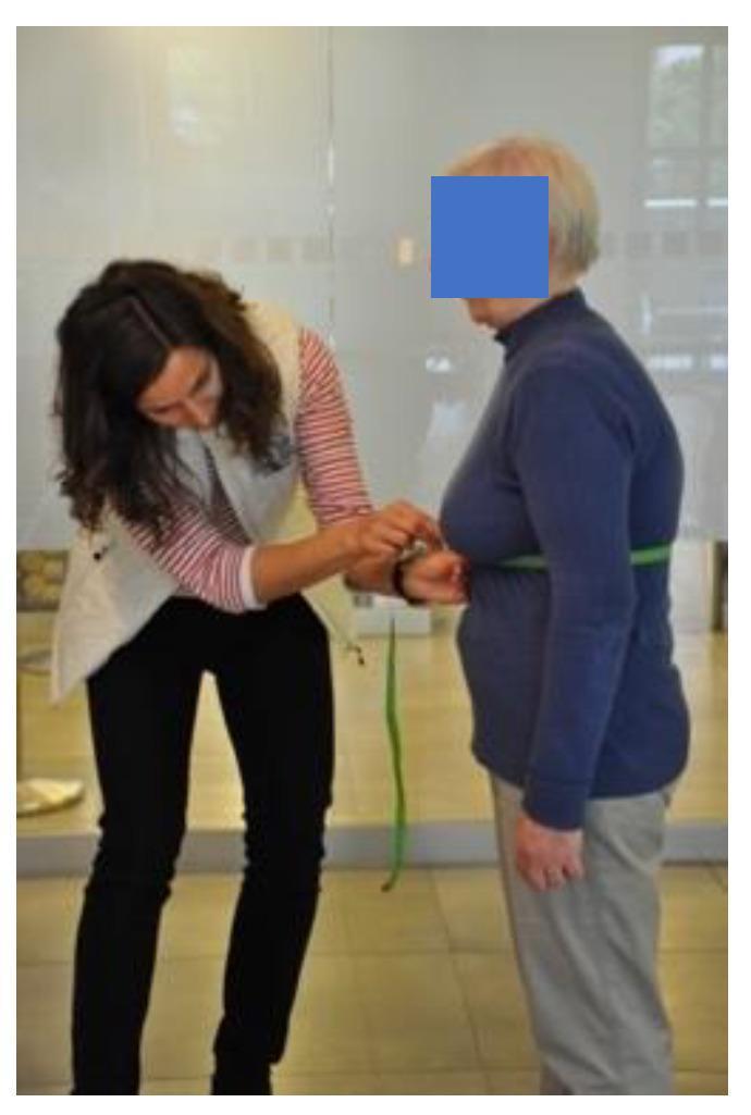
A clinical photograph demonstrating a circumferential chest expansion measurement, a physical assessment used to evaluate chest wall mobility and respiratory function. A clinician on the left is using a flexible green measuring tape to determine the thoracic circumference of an elderly patient. The tape is positioned horizontally around the patient's torso at the level of the xiphoid process of the sternum. This clinical procedure typically involves recording the difference in circumference between maximal inspiration and maximal expiration to assess lung excursion and rib cage flexibility. The measurement is relevant in the context of pulmonary rehabilitation for chronic respiratory diseases and the monitoring of conditions such as ankylosing spondylitis. The patient is shown standing in thin, close-fitting clothing with arms relaxed at the sides to ensure measurement accuracy.
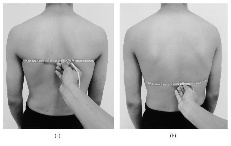
This clinical photograph illustrates the standardized procedure for measuring chest expansion (CE) using a tape measure on a human subject in a standing position. The image is divided into two panels: (a) Upper chest expansion measurement, where the measuring tape is positioned at the level of the fifth thoracic vertebra (T5) posteriorly, crossing the mid-clavicular line and the third intercostal space anteriorly. (b) Lower chest expansion measurement, showing the tape aligned with the 10th thoracic vertebra (T10) posteriorly and the xiphoid process anteriorly. In both panels, an examiner's hand is visible securing the '0' point of the tape measure against the midline of the spine using the index finger to ensure stability and accuracy during respiratory cycles. This technique is used in clinical physical therapy and pulmonary assessments to quantify thoracic mobility and assess lung function parameters such as FEV1 and FVC. The visual emphasizes correct anatomical landmark alignment for reproducible measurement of inspiratory and expiratory thoracic diameter changes.
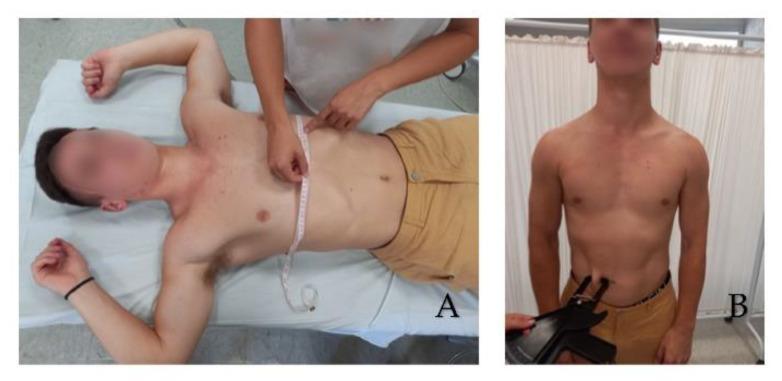
This clinical procedural image consists of two panels demonstrating anthropometric measurement techniques for assessing body composition and respiratory mechanics. Panel A (left) shows a subject in a supine position on an examination table with arms abducted to 90 degrees. A clinician is using a flexible, non-elastic measuring tape to determine the thorax circumference. The tape is positioned horizontally at the level of the xiphoid process, captured during maximum inspiration to evaluate chest expansion. Panel B (right) depicts a subject standing in an erect, neutral posture while a clinician performs a skinfold thickness test on the abdomen. A specialized skinfold caliper is used to pinch and measure the subcutaneous fat layer near the umbilical region. These procedures are common in physical therapy, sports medicine, and nutritional assessments to monitor physiological changes, calculate body density, or assess respiratory function. The images emphasize proper anatomical landmarking—the xiphoid process for thoracic girth and the abdominal wall for skinfold measurement—essential for clinical reliability.
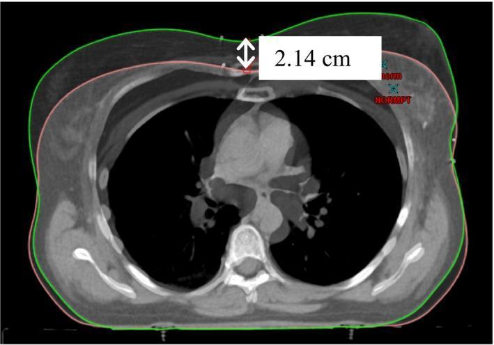
A diagnostic CT scan image in a transverse axial view of the human chest, demonstrating a comparison between two different respiratory states. The image features overlapping body contours: a pink outline representing the patient's body during free breathing (FB) and a green outline representing deep inspiration breath hold (DIBH). A specific measurement of 2.14 cm is labeled with a white arrow on the anterior chest wall, quantifying the outward expansion of the thoracic cavity during DIBH. Within the chest cavity, clear anatomical structures are visible, including the spinal column, ribs, bilateral lungs (low density/black), and the heart and mediastinal vessels (intermediate density). This visualization is used in radiation oncology planning, specifically for heart-sparing techniques in left-sided breast cancer treatment, where DIBH increases the distance between the chest wall and the cardiac surface to reduce incidental radiation dose to the heart.
Chest Expansion Measurement by Tape
Technique
| Level | Posterior landmark | Anterior landmark |
|---|---|---|
| Upper chest | T5 vertebra | 3rd intercostal space / 4th ICS (males) |
| Lower chest | T10 vertebra | Xiphoid process / xiphisternal junction |
- In males: tape is placed at the level of the 4th intercostal space
- In females: tape is placed just below the breasts at the xiphisternal (xiphoid) level
- Apply the tape snugly but not tightly around the chest at the chosen level
- Ask the patient to perform a forced maximal expiration (breathe out fully)
- Record the circumference at full expiration
- Ask the patient to take a maximal deep inspiration (breathe in fully)
- Record the circumference at full inspiration
- Chest expansion = Inspiration measurement - Expiration measurement
Normal Values
| Category | Normal chest expansion |
|---|---|
| Healthy adults | 5-7 cm (some sources: >5 cm) |
| Minimum acceptable | >2.5 cm (Washington Manual) |
| Young adults (<45 yrs) | Generally ≥5 cm |
| Older adults | Decreases with age |
- Values are age- and sex-dependent with overlap between normal and pathological ranges
- Values <5 cm in young persons with chronic inflammatory back pain strongly suggest Ankylosing Spondylitis (AS)
- Values <2.5 cm have high specificity (but low sensitivity) for AS
Clinical Images
Causes of Reduced Chest Expansion
- Ankylosing Spondylitis - costovertebral joint fusion is the classic cause; reduced expansion is a formal diagnostic criterion
- Pleural effusion / empyema (asymmetric reduction)
- Pneumonia (reduced + basal crackles)
- Pneumothorax / hemothorax
- Rib fractures (voluntary splinting)
- COPD / emphysema (barrel chest)
- Shrinking lung syndrome (SLE)
- Neuromuscular weakness (reduced respiratory muscle strength)
Key Points
- Normal values must be corrected for age and sex - there is significant overlap with disease
- Measurement should always be taken at the same anatomical level for reproducibility (especially for serial follow-up in AS)
- The BASMI (Bath Ankylosing Spondylitis Metrology Index) includes chest expansion as one of its 5 components alongside lumbar flexion, lateral flexion, cervical rotation, and occiput-to-wall distance
- Chest expansion <5 cm is included in the modified New York criteria and Rome criteria for AS diagnosis
At axillary Nipple and xipisternal level
chest expansion tape measurement axillary nipple xiphisternal level normal values clinical examination
https://www.ijhsr.org/IJHSR_Vol.11_Issue.7_July2021/IJHSR03.…
chest expansion measurement three levels axillary nipple xiphisternal tape
This clinical photograph illustrates the standardized procedure for measuring chest expansion (CE) using a tape measure on a human subject in a standing position. The image is divided into two panels: (a) Upper chest expansion measurement, where the measuring tape is positioned at the level of the fifth thoracic vertebra (T5) posteriorly, crossing the mid-clavicular line and the third intercostal space anteriorly. (b) Lower chest expansion measurement, showing the tape aligned with the 10th thoracic vertebra (T10) posteriorly and the xiphoid process anteriorly. In both panels, an examiner's hand is visible securing the '0' point of the tape measure against the midline of the spine using the index finger to ensure stability and accuracy during respiratory cycles. This technique is used in clinical physical therapy and pulmonary assessments to quantify thoracic mobility and assess lung function parameters such as FEV1 and FVC. The visual emphasizes correct anatomical landmark alignment for reproducible measurement of inspiratory and expiratory thoracic diameter changes.
A clinical photograph demonstrating a circumferential chest expansion measurement, a physical assessment used to evaluate chest wall mobility and respiratory function. A clinician on the left is using a flexible green measuring tape to determine the thoracic circumference of an elderly patient. The tape is positioned horizontally around the patient's torso at the level of the xiphoid process of the sternum. This clinical procedure typically involves recording the difference in circumference between maximal inspiration and maximal expiration to assess lung excursion and rib cage flexibility. The measurement is relevant in the context of pulmonary rehabilitation for chronic respiratory diseases and the monitoring of conditions such as ankylosing spondylitis. The patient is shown standing in thin, close-fitting clothing with arms relaxed at the sides to ensure measurement accuracy.
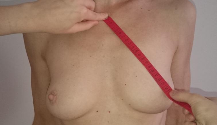
A clinical photograph demonstrating the measurement of the Jugulum-Nipple Distance (JND) on a female subject. The image shows a red measuring tape being held diagonally across the chest. The superior endpoint of the tape is positioned at the suprasternal notch (jugulum), while the inferior endpoint is held at the apex of the left nipple. This anthropometric measurement is commonly used in plastic and reconstructive breast surgery to assess breast ptosis, determine symmetry, and plan surgical interventions such as mastopexy or breast reduction. The subject's breasts are visible in a frontal view, exhibiting natural rounded morphology with visible mammary structures and unremarkable skin with minor nevi. Hands of an examiner are visible at both ends of the tape to ensure precise anatomical placement. This visual serves as an educational reference for standardized preoperative or postoperative clinical assessment in mammary surgery.
Chest Expansion by Tape - Three Levels
Anatomical Landmarks
| Level | Anterior landmark | Posterior landmark | Intercostal space equivalent |
|---|---|---|---|
| Axillary | Axilla / anterior axillary fold | T2-T3 level | ~2nd intercostal space (ICS) |
| Nipple | Nipple line | T4-T5 level | ~4th intercostal space (ICS) |
| Xiphisternal | Xiphoid process / xiphisternal junction | T10 level | Lower thorax |
Normal Values (Adults)
| Level | Males | Females | General range |
|---|---|---|---|
| Axillary | ~3.5 - 4.0 cm | ~3.2 - 3.6 cm | 3-5 cm |
| Nipple | ~4.0 - 4.6 cm | ~3.7 - 4.1 cm | 3.5-5 cm |
| Xiphisternal | ~4.5 - 5.3 cm | ~4.1 - 4.7 cm | 4-6 cm |
Key pattern: Expansion progressively increases from axilla → nipple → xiphisternal level, because the lower chest has greater excursion (diaphragmatic breathing contributes more at the lower levels).
Technique (same for all three levels)
- Patient stands erect, arms slightly abducted
- Place flexible cloth tape snugly around chest at the chosen level
- Record circumference at full forced expiration
- Record circumference at full deep inspiration
- Expansion = Inspiration - Expiration
- Take best of 3 readings at each level
Important Notes
- Males > Females at all three levels (anatomical rib cage differences; males use more costal breathing, females more diaphragmatic)
- Values decline with age - can fall by 50-60% between ages 15 and 75
- The nipple level (4th ICS) is the most commonly used single level in clinical practice and in studies such as the Moll & Wright (1972) reference standard
- In females, measurements at the nipple level can be unreliable due to breast tissue; the xiphisternal level is preferred (as also noted in Firestein & Kelley's Rheumatology)
- Ankylosing spondylitis criterion: <2.5 cm at the 4th ICS has high specificity; <5 cm in young patients with inflammatory back pain is strongly suggestive
Summary Table (Young Adults, Traditional Instruction)
| Level | Males (mean) | Females (mean) |
|---|---|---|
| Axilla | 3.5 cm | 3.2 cm |
| Nipple | 4.0 cm | 3.7 cm |
| Xiphisternal | 4.5 cm | 4.1 cm |