

9c71a992-b718-4c66-a2fc-dd76fc227286.jpeg
Solve this
Reading File
This is a clinical case about post-thyroidectomy hypoparathyroidism. Let me search the medical library for comprehensive content on calcium regulation and parathyroid hormone.
Searching sources...
Finding Sources
Finding Sources
Finding Sources
Excellent sources found. Now let me read the key sections on PTH/calcium regulation and the clinical details.
Reading File
Reading File
Reading File
Finding Sources
Finding Sources
Finding Sources
Finding Sources
Reading File
Finding Sources
Reading File
Finding Sources
I now have comprehensive information to write a full, detailed answer. Let me also fetch the calcium regulation diagram from the textbook.
Loading Image
I have everything needed. Here is the complete, structured answer to all five sub-questions of this clinical case.
Q1 - Post-Thyroidectomy Hypoparathyroidism & Hypocalcemia
Clinical Diagnosis: Hypoparathyroidism leading to Hypocalcemia (Tetany)
1. What is the cause of this condition? [2 marks]
The cause is surgical hypoparathyroidism following total thyroidectomy.
During total thyroidectomy, the four parathyroid glands (which lie embedded in or very close to the posterior thyroid capsule) are highly vulnerable to accidental removal, devascularization, or ischemic damage. Loss of these glands removes the source of parathyroid hormone (PTH), which is the principal hormone maintaining serum calcium.
Without PTH:
- Calcium reabsorption in the renal distal tubule falls
- Bone resorption by osteoclasts decreases
- Renal activation of Vitamin D (1,25-diOH-D3/calcitriol) is impaired, reducing intestinal calcium absorption
- Serum calcium drops acutely - hypocalcemia results
This is the most common cause of hypoparathyroidism. As stated in Sabiston Textbook of Surgery: "Hypoparathyroidism, the most common cause of hypocalcemia, often develops because of surgery in the central neck, such as radical resection of head and neck cancers or incidentally after thyroidectomy."
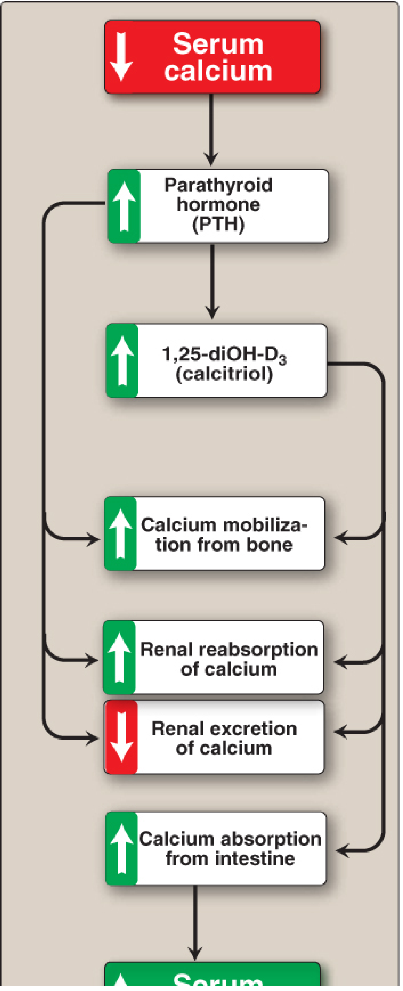
2. What are the hormones regulating serum calcium? Discuss their MOA. [3 marks]
Three hormones regulate serum calcium:
A. Parathyroid Hormone (PTH) - HYPERCALCEMIC hormone
PTH is the major calcium-elevating hormone, secreted by chief cells of the parathyroid glands in response to low ionized serum calcium (detected via the calcium-sensing receptor, a G-protein-coupled receptor).
Mechanism of Action:
| Target Organ | Action | Effect |
|---|---|---|
| Bone | Activates osteoclasts via RANK-L pathway → bone resorption | Releases Ca²⁺ and PO₄³⁻ into blood |
| Kidney (distal tubule) | Increases Ca²⁺ reabsorption; inhibits phosphate reabsorption (phosphaturia) in proximal tubule | Raises serum Ca²⁺, lowers serum PO₄³⁻ |
| Kidney (proximal tubule) | Stimulates 1-alpha-hydroxylase enzyme | Converts 25-OH-D3 → active calcitriol (1,25-diOH-D3) |
| Intestine (indirect) | Via calcitriol → increases calbindin expression | Increases intestinal Ca²⁺ absorption |
PTH secretion uses a negative feedback loop: as serum Ca²⁺ rises, PTH release is suppressed.
B. Calcitriol (1,25-dihydroxyvitamin D3) - HYPERCALCEMIC hormone
Calcitriol is the active form of Vitamin D, formed in the kidney under PTH stimulation.
Mechanism of Action:
- Acts like a steroid hormone - enters cells, binds cytosolic receptor, the complex moves to the nucleus and acts on DNA response elements
- Intestine: Increases expression of calbindin (calcium-binding protein) → enhances intestinal Ca²⁺ absorption (major effect)
- Bone: Stimulates bone resorption (mobilizes Ca²⁺ reserve), synergistic with PTH
- Kidney: Increases Ca²⁺ and phosphate reabsorption
C. Calcitonin - HYPOCALCEMIC hormone
Calcitonin is secreted by the parafollicular C-cells of the thyroid gland in response to high serum calcium.
Mechanism of Action:
- Bone: Inhibits osteoclast activity → decreases bone resorption → lowers serum Ca²⁺
- Kidney: Inhibits tubular reabsorption of Ca²⁺ → increases urinary calcium excretion
- Intestine: Reduces calcium absorption
- Net effect: Opposes PTH - lowers serum calcium
Note: In this patient, the thyroid was removed for malignancy, so calcitonin from C-cells is also lost - but since calcitonin is hypocalcemic, its loss would actually tend to raise calcium. The dominant problem is PTH loss.
3. What is the cause for symptoms seen in this case? [4 marks]
The patient developed:
- Cramps in hand muscles
- Continuous twitching (fasciculations)
- Laryngeal spasm
All of these are manifestations of tetany due to hypocalcemia.
Pathophysiology:
Normal ionized calcium stabilizes the neuronal membrane by blocking sodium (Na⁺) channels - it raises the threshold for action potential firing.
When serum calcium falls (plasma Ca²⁺ = 6 mg/dL; normal = 8.8-10.4 mg/dL):
- Less Ca²⁺ is available to plug Na⁺ channels
- Na⁺ channels become hyperpermeable
- The threshold for nerve depolarization drops
- Spontaneous repetitive firing of motor and sensory neurons occurs without any stimulus
- This drives sustained, uncontrolled muscle contraction = tetany
Specific symptom causes:
| Symptom | Mechanism |
|---|---|
| Hand muscle cramps | Hypocalcemia → hyperexcitability of peripheral motor nerves → carpopedal spasm |
| Continuous twitching | Spontaneous depolarization of neuromuscular junctions due to low Ca²⁺ threshold |
| Laryngeal spasm | Most dangerous - hyperexcitability of the recurrent laryngeal nerve → spasm of laryngeal muscles → stridor, risk of asphyxia |
Signs of latent tetany (before overt spasm):
- Chvostek's sign: Tapping the facial nerve (just anterior to the ear) causes twitching of facial muscles
- Trousseau's sign: Inflating a BP cuff above systolic pressure for 3 minutes causes carpal spasm (main d'accoucheur / "obstetrician's hand" posture)
As stated in Robbins & Cotran Pathologic Basis of Disease: "The hallmark of hypocalcemia is tetany, which is characterized by neuromuscular irritability resulting from decreased blood calcium levels. Symptoms range from circumoral numbness or paresthesia of the distal extremities and carpopedal spasm, to life-threatening laryngospasm and generalized seizures."
Treatment with calcium gluconate (IV/oral) and Vitamin D restores ionized calcium and immediately reverses neuromuscular hyperexcitability - which explains why the patient's symptoms resolved.
4. Comment on the values obtained during blood and urine tests. [4 marks]
| Parameter | Patient's Value | Normal Range | Interpretation |
|---|---|---|---|
| Plasma calcium | 6 mg/dL | 8.8-10.4 mg/dL | Low - Hypocalcemia |
| Plasma phosphorus | 5 mg/dL | 2.5-4.5 mg/dL | High - Hyperphosphatemia |
| Urine calcium | 25 ng/dL | 100-300 mg/day | Low - Hypocalciuria |
| Urine phosphorus | 0.1 g/dl | 0.4-1.3 g/day | Low - Hypophosphaturia |
Explanation of the pattern:
The classic biochemical signature of hypoparathyroidism is:
-
Low plasma Ca²⁺ + High plasma PO₄³⁻: PTH normally promotes urinary phosphate excretion (phosphaturia) and calcium reabsorption. Without PTH, phosphate is retained in blood (hyperphosphatemia) and calcium is not reabsorbed (hypocalcemia). As Comprehensive Clinical Nephrology states: "Loss of PTH (hypoparathyroidism) leads to hypocalcemia and hyperphosphatemia."
-
Low urine calcium: Even though serum calcium is low, the kidney still excretes some calcium. However, in hypoparathyroidism, absence of PTH means there is no signal to reabsorb calcium in the distal tubule - yet the filtered load of calcium is also very low (because serum Ca²⁺ is low). The net result is reduced urinary calcium. Additionally, there is no calcitriol-driven increased gut absorption to replenish the pool.
-
Low urine phosphorus: Without PTH, the proximal tubule reabsorbs phosphate avidly (phosphate reabsorption is the default state; PTH normally inhibits it). So despite elevated serum phosphorus, phosphaturia is low because PTH is absent and renal tubular phosphate reabsorption is high.
Summary of biochemical pattern in this case:
- Plasma: ↓ Ca²⁺ + ↑ PO₄³⁻ = Hypoparathyroidism
- Urine: ↓ Ca²⁺ + ↓ PO₄³⁻ = consistent with absent PTH
5. What are the tests done to detect this condition in its latent phase? [2 marks]
Latent (subclinical) hypocalcemia/tetany can be detected before overt symptoms appear by:
A. Bedside Clinical Tests
-
Chvostek's Sign
- Tap the facial nerve just anterior to the ear lobe (over the parotid)
- Positive: ipsilateral twitching of the corner of the mouth, nose, or eye
- Indicates subclinical neuromuscular hyperexcitability
-
Trousseau's Sign
- Inflate a sphygmomanometer above systolic BP on the arm for 3 minutes
- Positive: carpal spasm = flexion of wrist and MCP joints, extension of IP joints, adduction of thumb ("obstetrician's hand")
- More specific than Chvostek's for latent tetany
-
Peroneal Sign (Lust's Sign)
- Tap over the peroneal nerve → dorsiflexion and eversion of foot
B. Laboratory Tests
-
Serum calcium (total and ionized): Serum Ca²⁺ below 8.8 mg/dL confirms hypocalcemia; ionized Ca²⁺ < 4.6 mg/dL is diagnostic
-
Serum PTH (intact PTH assay): Low or undetectable PTH in the setting of low calcium confirms hypoparathyroidism
-
Sulkowitch Test (Urine calcium): A simple bedside test - adding Sulkowitch reagent (oxalic acid) to urine generates a precipitate proportional to calcium content; absent/fine precipitate = low urine calcium = hypocalcemia
-
ECG: Prolonged QT interval (due to prolonged phase 2 of cardiac action potential from hypocalcemia) is an objective indicator
-
Serum phosphorus: Elevated in hypoparathyroidism
References:
- Robbins & Cotran Pathologic Basis of Disease - Hypoparathyroidism, Clinical Features
- Barash's Clinical Anesthesiology, 9e - Calcium Physiology
- Lippincott's Biochemistry, 8th ed - Vitamin D and calcitriol regulation
- Comprehensive Clinical Nephrology, 7th ed - Hypoparathyroidism
- Sabiston Textbook of Surgery - Hypocalcemia
- Brenner & Rector's The Kidney - PTH-Vitamin D Endocrine System
This is a shared conversation. Sign in to Orris to start your own chat.