
Mangement of laceration on forehead and knee
forehead laceration repair wound closure suture
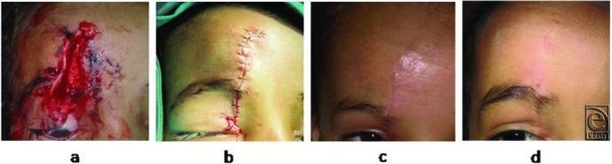
This clinical progression series comprises four photographs documenting the repair and healing of a traumatic facial laceration. Panel (a) shows the initial injury: a vertical, deep 6-cm laceration on the forehead involving the skin, subcutaneous fat, and muscle, characterized by tissue gaping and fresh blood. Panel (b) depicts the wound immediately postoperative after primary closure using autologous hair filament sutures; the edges are well-approximated with visible interrupted stitches and minimal peri-wound erythema. Panel (c) shows the follow-up at 5 weeks, demonstrating a maturing, slightly erythematous linear scar without suture track marks or infection. Panel (d) illustrates the 6-month long-term outcome, displaying a soft, flat, hypopigmented scar that blends with the surrounding skin texture. The series highlights surgical techniques aimed at minimizing permanent scarring and suture-induced ‘railroad’ marks in pediatric plastic surgery and emergency medicine contexts.
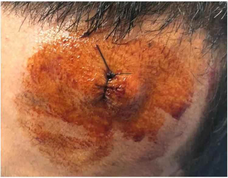
This clinical photograph displays a post-operative sutured wound on the frontal region (forehead) of a human patient near the hairline. The primary focus is a linear laceration that has been primarily closed using multiple simple interrupted, black, non-absorbable sutures. The wound edges appear well-approximated. The surrounding cutaneous tissue exhibits significant trauma-induced changes, most notably a large area of perilesional ecchymosis and bruising, characterized by a spectrum of discoloration ranging from reddish-brown and dark purple to faint yellow-blue. The skin surface is coated with a topical antiseptic solution (likely povidone-iodine), giving it a glossy, orange-tinted appearance. There is evidence of mild localized swelling and dried serosanguinous exudate around the suture entry points. This image serves as a clinical example of soft tissue repair following foreign body removal and illustrates the expected inflammatory and traumatic responses in the immediate post-surgical phase.
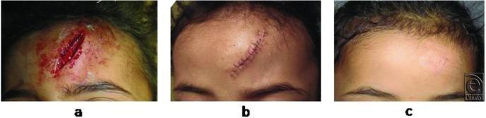
This clinical photograph sequence documents the progression of facial wound healing in a pediatric patient across three stages. Panel (a) shows an acute, deep vertical laceration on the forehead approximately 4 cm in length. The wound exhibits disrupted skin edges, visible subcutaneous tissue, and active bleeding with surrounding erythema. Panel (b) illustrates the same region 6 days post-repair using autologous hair sutures. The wound edges are well-approximated with visible simple interrupted sutures, and there is localized postoperative inflammation and crusting along the incision line. Panel (c) displays the outcomes at 3 months post-procedure, demonstrating a matured, fine-line scar. The scar is flat and hypopigmented, showing excellent integration with the surrounding skin texture and a complete absence of cross-hatch suture marks ('railroad tracking'). This series serves as an educational comparison of surgical repair outcomes and long-term cicatrization in aesthetic facial regions.
Management of Lacerations: Forehead and Knee
General Principles (Both Sites)
| Phase | Actions |
|---|---|
| Preparation | Antibiotic prophylaxis (if contaminated/high-risk), tetanus prophylaxis, adequate analgesia/anaesthesia, wound irrigation |
| Wound | Haemostasis, exploration, early debridement, repair of deeper structures |
| Closure | Skin closure without tension, appropriate suture choice, dressings |
| Follow-up | Suture/splint removal, physiotherapy (knee), scar management |
Step 1 - Haemostasis
- Apply direct pressure first.
- For scalp/forehead: the area is highly vascular. Control brisk bleeding by clamping vessels at wound edges (e.g. Raney clips) or placing a figure-of-eight suture at the bleeding edge.
- Local anaesthetics with epinephrine help control haemorrhage from small vessels in highly vascular wounds (face, scalp).
Step 2 - Assessment
- Assess for intracranial injury before wound care - a laceration may be part of a bigger injury.
- Palpate gently with a gloved finger to detect galeal involvement or depressed skull fracture. If a palpable depression is found, obtain CT.
- Classify as superficial (frontalis muscle not involved) or deep (frontalis muscle involved) - this determines repair technique.
- Note the orientation relative to skin tension lines. Horizontal forehead lacerations (parallel to tension lines, perpendicular to frontalis) give the best cosmetic results.
- Assess for joint capsule penetration - if the wound communicates with the joint, it requires urgent orthopaedic referral and washout.
- Assess neurovascular status (sensation, capillary refill, pulses) before giving local anaesthetic.
- Look for tendon injury (assess active extension against resistance).
- Exclude foreign bodies, fractures or dislocations with imaging as appropriate.
Step 3 - Anaesthesia
- Topical: lidocaine-epinephrine-tetracaine (LET) or lidocaine-prilocaine (EMLA) - especially useful in children to reduce pain of injection.
- Local infiltration: most commonly used for both sites.
- Nerve block: supraorbital/supratrochlear nerve block for forehead lacerations.
Step 4 - Irrigation and Debridement
- Irrigate all wounds as early as possible with warm normal saline to reduce bacterial contamination and allow better visualisation.
- Debride non-viable tissue until healthy bleeding occurs at wound edges.
- Healthy subcutaneous fat: yellow and soft.
- Muscle viability: judged by colour, capacity to bleed, and contractility.
- Do not shave hair on the scalp or eyebrows before closure - it increases infection risk and removes important anatomical landmarks.
- For the forehead, if hair obscures the field, brush it aside or matt it with petrolatum/bacitracin ointment.
Step 5 - Wound Closure
Forehead Laceration
- Close with 6-0 nonabsorbable monofilament simple interrupted sutures or tissue adhesive.
- Place key stitches first to align skin tension lines and the hairline.
- Remove sutures at 5 days to minimize scarring (early removal on the face is important).
- First close muscle fascia with 4-0 absorbable suture (not removed).
- Then close skin with 5-0 or 6-0 nonabsorbable monofilament or tissue adhesive.
- Suture removal at 5 days.
- Suitable for superficial, low-tension wounds on the forehead.
- May need a deep layer first if there is any tension.
- The forehead is one of the sites particularly suited to tape closure.
- Primary indication: superficial, straight laceration under little or no tension.
- Can also be applied after early suture removal to maintain wound edge approximation while reducing risk of permanent suture marks.
| Layer | Suture Type | Size | Removal |
|---|---|---|---|
| Muscle fascia | Absorbable | 4-0 | Not removed |
| Skin | Nonabsorbable monofilament | 5-0 or 6-0 | 5 days |
| Skin alt. | Tissue adhesive | - | Not removed |
Knee Laceration
- The knee is a high-tension, mobile site - closure must account for movement.
- Simple superficial lacerations: 4-0 or 5-0 nonabsorbable monofilament interrupted sutures.
- Deeper lacerations with subcutaneous tissue involvement: layered closure using absorbable suture for subcutaneous layer, nonabsorbable for skin.
- Avoid excessive tension on skin edges - undermining may be needed.
- Do not tape-close over the knee as primary closure (tape fails over joints due to movement).
- If joint capsule is involved: do NOT close in the ED - requires orthopaedic washout.
- Suture removal: typically 10-14 days (longer than face, due to tension and movement).
Step 6 - Tetanus Prophylaxis
- Puncture-type injuries in a contaminated environment
- Bites, compound fractures, wounds containing foreign bodies
- Wounds with systemic sepsis
- Heavy contamination (soil, manure)
-
6-hour delay before surgery
- Extensive devitalised tissue
Step 7 - Dressings and Aftercare
- Apply a non-adherent wound dressing.
- Forehead: pressure dressing over deep scalp lacerations for the first 24 hours post-repair helps prevent wound hematoma formation.
- Knee: padded dressing; advise patient to limit flexion initially to avoid wound dehiscence.
- Keep wound clean and dry for 24-48 hours.
- Wound check at 48 hours in contaminated or complex wounds.
- Antibiotic prophylaxis: for contaminated/dirty wounds, high-risk patients, or wounds with delayed presentation. Not routinely needed for clean lacerations.
Step 8 - Suture Removal Timing
| Site | Removal |
|---|---|
| Forehead/face | 5 days |
| Scalp | 10-14 days |
| Knee (lower limb) | 10-14 days |