Now I have all the material needed. Let me compile the comprehensive viva answer.
🧠 BRAINSTEM SYNDROMES — Comprehensive Neurosurgery Viva Guide
Sources: Localization in Clinical Neurology 8e (Brazis), Adams & Victor's Principles of Neurology 12e, Harrison's Principles of Internal Medicine 21e, Bradley & Daroff's Neurology in Clinical Practice
📌 CONCEPT: What are Brainstem (Alternating / Crossed) Syndromes?
Definition: Ipsilateral cranial nerve deficit + contralateral long tract (motor/sensory) deficit = lesion in the BRAINSTEM. The cranial nerve points to the level; the crossing deficit tells you which side.
The Golden Rule of Localisation:
- Ipsilateral CN palsy + Contralateral hemiplegia = Brainstem lesion
- All syndromes named after the cranial nerve involved at that level
🗺️ BRAINSTEM LEVELS & SYNDROMES AT A GLANCE
| Level | Syndrome | CN Involved | Key Feature |
|---|
| Midbrain (ventral) | Weber | III | CN III + contralateral hemiplegia |
| Midbrain (tegmentum) | Benedikt | III | CN III + contralateral hemiataxia + tremor |
| Midbrain (tegmentum dorsal) | Claude | III | CN III + contralateral cerebellar ataxia |
| Midbrain (tectum) | Parinaud | — | Vertical gaze palsy, no CN III palsy |
| Midbrain (tectum + III) | Nothnagel | III (bilateral) | CN III + ataxia, no hemiplegia |
| Pons (ventral) | Millard-Gubler | VI + VII | Lateral gaze + facial palsy + contralateral hemiplegia |
| Pons (tegmental) | Foville/Raymond-Foville | VI + VII | Ipsilateral gaze palsy + facial palsy + contralateral hemiplegia |
| Medulla (lateral) | Wallenberg | IX, X, V (spinal) | PICA territory; crossed pain/temp, Horner, dysphagia |
| Medulla (medial) | Dejerine (Medial Medullary) | XII | Tongue palsy + contralateral hemiplegia + dorsal column loss |
| Medulla (dorsolateral) | Avellis | X | Palate + vocal cord palsy + spinothalamic loss |
| Medulla | Jackson | X + XII | Avellis + ipsilateral tongue palsy |
| Medulla | Babinski-Nageotte | Multiple | Crossed cerebellar + sympathetic + spinothalamic |
🔴 SECTION A: MIDBRAIN SYNDROMES
1. WEBER SYNDROME (Ventral Midbrain)
📊 Demography
- Age: Any adult; peak 50–70 years (ischaemic); younger if aneurysm or tumour
- Sex: M > F (vascular causes)
- Aetiology: Posterior cerebral artery or basilar artery perforators occlusion; posterior communicating artery aneurysm (compresses CN III from outside); midbrain tumour; tuberculoma
🧬 Anatomy / Pathology
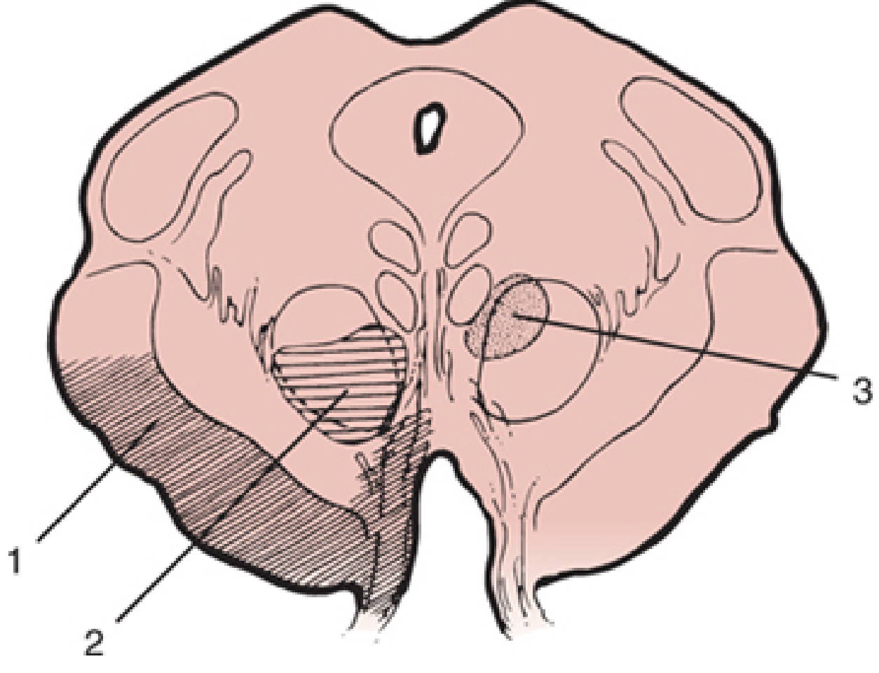
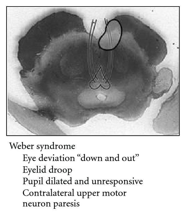
The lesion is in the cerebral peduncle (crus cerebri) — ventromedial midbrain, damaging:
- CN III fascicle (exits through crus cerebri)
- Corticospinal + corticobulbar fibres (cerebral peduncle)
Pathology: The parasympathetic fibres of CN III travel on the outside of the nerve → compressive lesions (aneurysm, herniation) hit parasympathetics FIRST → dilated pupil as the earliest sign.
Fig 1 — Mesencephalic cross-section. Zone 1 (Weber): cerebral peduncle + CN III fascicle. Zone 2 (Benedikt): tegmentum + red nucleus + CN III. Zone 3 (Claude): dorsal tegmentum. — Localization in Clinical Neurology, 8e
🩺 Clinical Features (Memorise as 2 + 2)
Ipsilateral (same side as lesion):
- CN III palsy — complete ptosis, eye "down and out," mydriasis (dilated fixed pupil), diplopia
- Supranuclear gaze palsy toward opposite side (if medial peduncle involved)
Contralateral (opposite side):
3. Hemiplegia (including lower face) — UMN type; corticospinal + corticobulbar
4. Hyperreflexia + Babinski sign
⚠️ Viva Trap: In Weber, eye goes DOWN and OUT (LR6, SO4 intact, CN III out). Ask: "Why is the face involved?" → Corticobulbar fibres cross; facial nucleus for lower face is contralateral UMN.
🔬 Investigations
MRI Brain (Gold Standard)
- Indication: Any suspected acute brainstem stroke or mass
- Sequence: DWI (hyperintense = acute infarct within hours), ADC map (hypointense = restricted diffusion), FLAIR (hyperintense after 24h), T1 + T2
- Positive finding: Hyperintense signal in ventromedial midbrain on DWI/FLAIR; can see PCA territory infarction
CT Brain
- Indication: Emergency if MRI not available; rule out haemorrhage
- Positive: Hyperdense (blood) or hypodense (infarct after 24h) lesion in midbrain
CT/MR Angiography
- Indication: Suspect posterior communicating artery aneurysm, PCA occlusion
- Positive: Aneurysm at PCoA-ICA junction; PCA occlusion
DSA (Digital Subtraction Angiography)
- Indication: Aneurysm confirmed on CTA, pre-surgical planning
- Gold standard for vascular anatomy
Lumbar Puncture
- Indication: If xanthochromia suspected (SAH with false-negative CT)
Scoring/Grading:
- NIHSS (NIH Stroke Scale) — baseline and serial monitoring
- Hunt & Hess (if SAH from aneurysm): Grade 1–5
- Fisher Scale (SAH, CT based): Grade 1–4
- GCS for consciousness level
💊 Management
A. General/Acute
- Call for help — activate stroke team; ABCDE
- Position — 30° head elevation if raised ICP; flat if BP low
- Oxygen, IV access, cardiac monitor
- BP management — in acute ischaemic stroke: allow up to 220/120 if not thrombolysing; target <180/105 if thrombolysing
- Blood glucose control (4–10 mmol/L)
- NPO if dysphagia
B. Specific Medical Management
If Ischaemic Stroke:
- tPA (Alteplase) within 4.5 hours
- MOA: Tissue plasminogen activator → activates plasminogen → plasmin → fibrinolysis
- Dose: 0.9 mg/kg IV (max 90 mg); 10% as bolus, rest over 60 min
- Contraindications: Haemorrhagic stroke, BP >185/110, INR >1.7, recent surgery <14 days, platelets <100,000
- Aspirin 325 mg (if no thrombolysis, within 24h)
- MOA: Irreversible inhibition of COX-1 → ↓ TXA2 → ↓ platelet aggregation
- Dose: 160–325 mg loading, then 75–100 mg/day
- Dual antiplatelet (Aspirin + Clopidogrel) for TIA/minor stroke (POINT trial) × 21 days, then single antiplatelet
- Clopidogrel MOA: Prodrug → active metabolite blocks P2Y12 ADP receptor on platelets irreversibly
- Statins: Atorvastatin 80 mg/day
- MOA: HMG-CoA reductase inhibitor → ↓ LDL → plaque stabilisation + anti-inflammatory
- Anticoagulation (if cardioembolic): Heparin or NOAC after 24h
- Heparin MOA: Binds antithrombin III → inhibits thrombin (IIa) and Xa
- Warfarin MOA: Inhibits Vitamin K epoxide reductase → ↓ Factors II, VII, IX, X, Protein C & S
If Posterior Communicating Artery Aneurysm:
→ See surgical management below
C. Surgical Options
1. Mechanical Thrombectomy (MT)
- Indication: Large vessel occlusion (LVO) of basilar artery or PCA; NIHSS ≥6; within 6–24h (DAWN/DEFUSE criteria); CT/CTA confirms occlusion
- Contraindication: No salvageable penumbra; large established infarct (ASPECTS <6)
- Position: Supine, femoral access (groin)
Steps:
- GA or conscious sedation
- Femoral artery access → guide catheter to vertebrobasilar system
- Microcatheter through clot → Stentriever deployment
- Aspiration (ADAPT technique) or combination
- Thrombus retrieval
- Post-procedure angiography to confirm recanalization
- ICU transfer
2. Surgical Clipping (for PCoA Aneurysm causing Weber)
- Indication: Unruptured PCoA aneurysm with CN III palsy (high risk of rupture); ruptured aneurysm (Hunt & Hess I–III)
- Contraindication: Poor-grade SAH (H&H IV–V) — may delay; elderly with poor medical status
- Position: Supine, head turned 15–30° to contralateral side, Mayfield 3-pin head holder
Steps (Pterional Craniotomy + Clipping):
- Position: Supine, head fixed in Mayfield clamp, turned 30° contralateral
- Scalp incision: Pterional (frontotemporal)
- Craniotomy: Frontotemporal bone flap
- Open sylvian fissure
- Identify ICA, CN II, follow to PCoA origin
- Temporary clip on parent artery (if needed)
- Permanent clip across aneurysm neck
- Confirm clip with ICG videoangiography or Doppler
- Closure in layers
3. Endovascular Coiling (alternative to clipping for PCoA aneurysm)
- Indication: Poor surgical candidate; aneurysm anatomy favourable for coiling (narrow neck, fundus:neck ratio >2); unruptured
- Steps: Femoral access → microcatheter into aneurysm sac → platinum coils deployed → sac thrombosis
2. BENEDIKT SYNDROME (Midbrain Tegmentum — Ventral)
Anatomy/Pathology
Lesion in midbrain tegmentum, involving:
- CN III fascicle → ipsilateral CN III palsy
- Red nucleus → contralateral involuntary movements (rubral tremor = Holmes tremor, choreoathetosis)
- Corticospinal tract (partial) → contralateral hemiparesis
- Medial lemniscus may be involved
Cause: Posterior cerebral artery (interpeduncular branches) infarction; tuberculoma; tumour; haemorrhage
Clinical Features
| Sign | Side | Structure Damaged |
|---|
| CN III palsy (ptosis, mydriasis, down-out eye) | Ipsilateral | CN III fascicle |
| Rubral tremor / Holmes tremor (coarse, intention/rest tremor, 2–5 Hz) | Contralateral | Red nucleus + dentatorubrothalamic fibres |
| Hemiparesis | Contralateral | Corticospinal tract |
| Choreoathetosis | Contralateral | Red nucleus + subthalamic connections |
⚠️ Key differentiator: Benedikt = CN III + tremor (red nucleus). Weber = CN III + hemiplegia only.
Investigations: Same as Weber (MRI DWI, MRA)
Management
- Stroke management as above
- Holmes/Rubral tremor — notoriously drug-resistant
- Levodopa/carbidopa (dopaminergic pathway restoration)
- Clonazepam, propranolol (partial benefit)
- Deep Brain Stimulation (DBS) of VIM thalamus — most effective for refractory tremor
- DBS Indication: Disabling tremor >12 months, failed ≥2 medications
- Target: Ventral Intermediate (VIM) nucleus of thalamus, or caudal zona incerta
3. CLAUDE SYNDROME (Midbrain Tegmentum — Dorsal)
Pathology
More dorsal than Benedikt — involves:
- Dorsal red nucleus + brachium conjunctivum (superior cerebellar peduncle after decussation)
- CN III fascicle
Clinical Features
- Ipsilateral CN III palsy
- Contralateral cerebellar ataxia (asynergia, dysmetria, dysdiadochokinesis) — more prominent than Benedikt, without hemiballismus
- Minimal or no hemiparesis
Mnemonic: Claude = Cerebellar (dorsal); Benedikt = Both (cerebellar + pyramidal + tremor)
4. NOTHNAGEL SYNDROME (Midbrain Tectum)
- Lesion: Superior cerebellar peduncle + CN III (unilateral or bilateral)
- Features: Bilateral CN III palsies + cerebellar ataxia + gaze palsies
- Cause: Tumour (pinealoma, collicular glioma) — commonest
- No hemiplegia (pyramidal tract spared)
5. PARINAUD SYNDROME (Dorsal Midbrain / Pretectal)
Demography
- Age: Young adults (pineal tumours 15–35 years); children (aqueductal stenosis); any age (MS, vascular)
- Aetiology: Pinealoma/germinoma (most common), hydrocephalus (aqueductal dilation), MS, vascular, trauma
Pathology
Lesion in pretectal area (posterior commissure + superior colliculi + rostral interstitial nucleus of MLF). NO CN III palsy (nuclear level spared). Supranuclear vertical gaze pathways damaged.
Clinical Features (DEWDROP mnemonic)
| Feature | Mechanism |
|---|
| Dissociated light-near reflex (light-near dissociation) — pupils react to accommodation but NOT light | Pretectal lesion spares EW nucleus but disrupts afferent light arc via posterior commissure |
| Elevation palsy (upgaze palsy) | Damage to rostral interstitial nucleus of MLF (riMLF) and posterior commissure |
| Widened palpebral fissures (Collier's sign / "setting sun") | Lid retraction from dorsal midbrain compression |
| Downbeat or convergence-retraction nystagmus on attempted upgaze | Pretectal area damage |
| Retraction nystagmus + convergence spasm | Damage to posterior commissure |
| Obstructive hydrocephalus (common with pineal tumours) | Aqueductal compression |
| Papilloedema (if hydrocephalus) | Raised ICP |
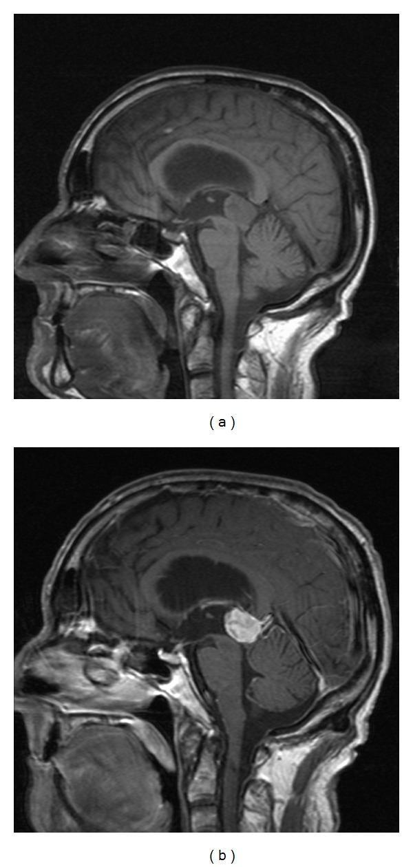
Fig 2 — Sagittal T1 MRI (a) pre-contrast; (b) post-gadolinium showing homogeneous enhancing pineal mass compressing the tectum with secondary aqueductal obstruction and hydrocephalus.
Investigations
- MRI Brain: Pineal mass, tectal plate compression, aqueductal stenosis, hydrocephalus
- Serum tumour markers: AFP (yolk sac), β-hCG (germinoma), LDH
- CSF tumour markers (after relieving hydrocephalus)
- Visual field testing + VEP
- Ophthalmology: Slit lamp for Kayser-Fleischer (Wilson's, if young)
Management
Step 1: Treat hydrocephalus
- Endoscopic Third Ventriculostomy (ETV)
- Indication: Obstructive hydrocephalus from pineal/tectal mass
- Position: Supine, Mayfield clamp
- Steps: Burr hole (Kocher's point, 2.5 cm from midline, 1 cm anterior to coronal suture) → rigid endoscope into lateral ventricle → through foramen of Monro → floor of 3rd ventricle → fenestrate floor with monopolar electrode → balloons dilate ostium → CSF flows into prepontine cistern
- Advantages: Avoids VP shunt; simultaneous endoscopic biopsy
- Contraindication: Previous infection, thickened floor, unfavourable anatomy
Step 2: Tumour-specific therapy
- Germinoma: Radio-sensitive → craniospinal radiation ± chemotherapy (BEP — Bleomycin, Etoposide, Cisplatin) — NO primary surgery needed
- Teratoma / Non-germinomatous GCT: Surgery first (posterior interhemispheric or supracerebellar infratentorial approach) then radiation + chemo
- Pineocytoma (WHO Grade I): Surgical resection → EFS >90%
- Pineoblastoma (WHO Grade IV): Surgery + craniospinal RT + chemo (poor prognosis)
- MS/vascular: Treat primary cause
Surgical Approach (Pineal Region):
- Supracerebellar Infratentorial (Krause approach): Most common
- Position: Sitting (risk: VAE) or prone/concorde (safer, preferred now)
- Steps: Midline posterior fossa craniotomy below tentorium → dissect between tentorium and cerebellum → expose quadrigeminal plate → identify tumour → microsurgical excision
- Occipital Transtentorial: For superior extension
- Posterior Interhemispheric: For tumours extending into corpus callosum
🔵 SECTION B: PONTINE SYNDROMES
6. MILLARD-GUBLER SYNDROME (Ventral Pons)
Demography
- Age: 50–70 years (vascular); any age (tumour, MS)
- Cause: Basilar artery perforators occlusion; pontine glioma; MS; haemorrhage
Anatomy/Pathology
Lesion in ventral pons (base of pons), involving:
- CN VI nucleus or fascicle → ipsilateral lateral gaze palsy / abducens palsy
- CN VII fascicle (as it loops around CN VI nucleus) → ipsilateral facial palsy
- Corticospinal tract (basis pontis) → contralateral hemiplegia
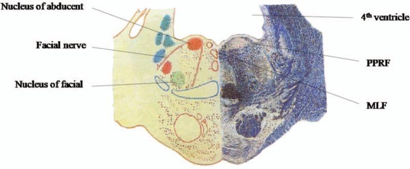
Fig 3 — Transverse section of caudal pons showing CN VII looping around CN VI nucleus (facial colliculus), PPRF, and MLF — anatomical substrate of pontine gaze palsy syndromes.
Clinical Features
| Sign | Side | Structure |
|---|
| Abducens palsy (inability to abduct eye) | Ipsilateral | CN VI fascicle |
| LMN facial palsy (all divisions — includes forehead, no Bell's phenomenon) | Ipsilateral | CN VII fascicle |
| Hemiplegia (UMN) | Contralateral | Corticospinal tract |
⚠️ Key point: Facial palsy here is LMN type (involves forehead) because the facial nucleus itself or its intrapontine fascicle is involved — distinct from UMN (central) facial palsy which spares the forehead.
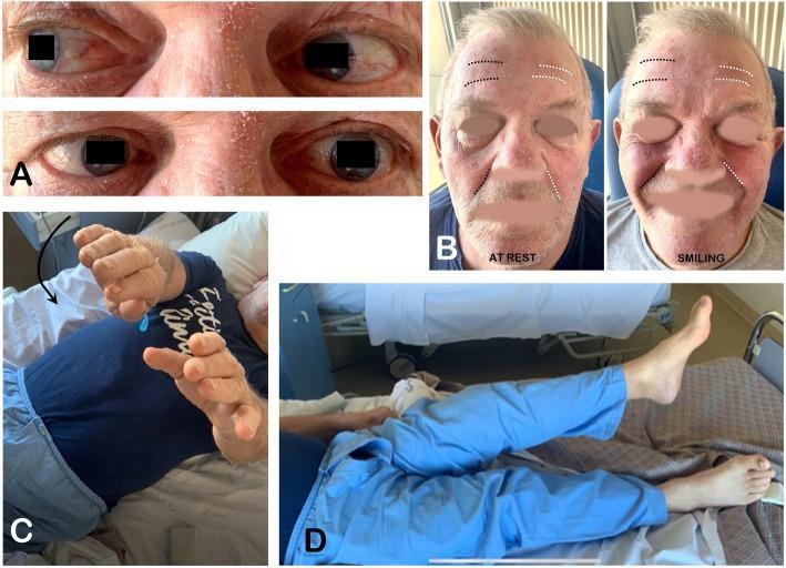
7. FOVILLE SYNDROME / RAYMOND-FOVILLE SYNDROME (Pontine Tegmentum)
Pathology
More dorsal involvement of the pons (tegmentum), involving:
- PPRF (Paramedian Pontine Reticular Formation) + CN VI nucleus → ipsilateral conjugate gaze palsy (eyes cannot look to side of lesion)
- CN VII nucleus/fascicle → ipsilateral LMN facial palsy
- Corticospinal tract
- Sometimes: Descending sympathetic fibres → ipsilateral Horner
Clinical Features
| Sign | Side |
|---|
| Ipsilateral conjugate gaze palsy (both eyes deviate away from lesion) | Ipsilateral |
| LMN facial palsy (all divisions) | Ipsilateral |
| Hemiplegia | Contralateral |
| Horner syndrome (ptosis, miosis, anhidrosis) | Ipsilateral (sometimes) |
Millard-Gubler vs Foville: Millard-Gubler → only CN VI palsy (one eye can't abduct). Foville → full conjugate gaze palsy (BOTH eyes can't look toward lesion, due to PPRF damage).
Fig 4 — Foville syndrome: (A) Left CN VI palsy — left eye esotropia, failed abduction. (B) Left LMN facial palsy — loss of forehead wrinkling, nasolabial fold asymmetry at rest and on smiling. (C,D) Right hemiparesis.
Management (Pontine Syndromes)
- Ischaemic pons: Same stroke protocol; basilar artery thrombectomy if basilar occlusion (door-to-recanalization within 6h, extended to 24h for basilar stroke with favourable imaging — ATTENTION trial)
- Pontine glioma (DIPG in children): See below
- MS: IV methylprednisolone 1g/day × 5 days for acute relapse
- MOA: Glucocorticoid → binds GR → inhibits NF-κB → reduces BBB permeability and inflammation
Surgical Approach: Brainstem Tumour (Pontine)
- Approach: Retrosigmoid / far lateral / suboccipital depending on location
- Most focal exophytic lesions: Safe to biopsy/resect via retrosigmoid craniotomy
- DIPG (Diffuse Intrinsic Pontine Glioma): Biopsy only; then focal RT (54 Gy) + ONC201 (H3K27M-mutant); NO radical surgery
- Safe entry zones for brainstem surgery:
- Lateral sulcus of pons (between CN V and VII)
- Supratrigeminal zone (above CN V)
- Peritrigeminal zone
- Median sulcus (floor of 4th ventricle)
🟢 SECTION C: MEDULLARY SYNDROMES
8. LATERAL MEDULLARY SYNDROME (Wallenberg Syndrome) — MOST COMMON BRAINSTEM SYNDROME
Demography
- Age: 40–70 years; younger with vertebral artery dissection (30–50 years)
- Sex: M > F
- Risk factors: HTN, DM, smoking, dyslipidaemia, vertebral artery dissection (trauma, neck manipulation, Marfan's)
- Incidence: Most common brainstem infarct syndrome
Aetiology
- Vertebral artery occlusion (50%) — large vessel atherosclerosis
- PICA (Posterior Inferior Cerebellar Artery) occlusion — less common than vert. artery
- Vertebral artery dissection (15%)
- Small vessel disease (13%)
- Cardioembolism (5%)
- Others: Cocaine, tumour, MS, neck manipulation, trauma
🧬 Anatomy/Pathology
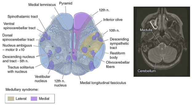
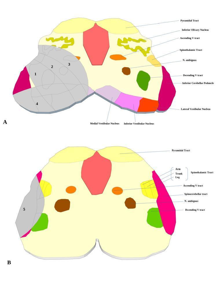
Structure damaged: Wedge-shaped lateral medulla (territory of PICA/vertebral artery), involving:
Fig 5 — Medullary cross-section showing lateral (yellow) and medial (blue) infarct zones. Key structures of lateral medullary syndrome include: spinothalamic tract, nucleus ambiguus, descending sympathetic tract, inferior cerebellar peduncle, vestibular nuclei, spinal trigeminal nucleus and tract. — Harrison's Principles of Internal Medicine, 21e
Fig 6 — Lateral medullary infarct subtypes: 1+2 = typical; 2+3 = ventral; 1+2+3 = large; 4 = dorsal; 5 = lateral. Rostral (A) vs caudal (B) levels differ in vestibular involvement.
🩺 Clinical Features (LATERAL mnemonic)
| # | Sign | Structure Damaged | Side |
|---|
| 1 | Loss of pain/temp face | Spinal nucleus + tract of CN V | Ipsilateral face |
| 2 | Ataxia (cerebellar) | Inferior cerebellar peduncle (restiform body) | Ipsilateral limbs + gait |
| 3 | Thermoanesthesia + pain/temp loss body | Spinothalamic tract | Contralateral limbs + trunk |
| 4 | Emetic symptoms (vertigo, N&V) | Vestibular nuclei | — |
| 5 | Regurgitation + dysphagia | Nucleus ambiguus (CN IX, X) | Ipsilateral |
| 6 | Anhidrosis + Horner | Descending sympathetics | Ipsilateral (ptosis, miosis, anhidrosis) |
| 7 | Locomotive difficulties (hiccups) | Dorsolateral middle medulla | — |
⚠️ Crossed sensory loss: Ipsilateral face pain/temp loss + Contralateral body pain/temp loss = PATHOGNOMONIC of lateral medullary syndrome. This is because the spinal trigeminal nucleus (face, ipsilateral) and spinothalamic tract (body, crosses in spinal cord) are both in the lateral medulla.
⚠️ Preserved: Motor function (pyramids = medial medulla), vibration/proprioception (dorsal columns = medial), tongue movements (XII = medial).
Additional signs (ocular):
- Skew deviation (ipsilateral hypotropia)
- Ocular tilt reaction
- Nystagmus (horizontal-torsional, beating away from lesion)
- Ipsipulsion (saccades and smooth pursuit pull toward lesion side)
- "Upside down" room sensation
Rostral vs Caudal lesion differences:
- Rostral (more severe): Dysphagia, hoarseness, facial paresis
- Caudal: Predominant vertigo, nystagmus, gait ataxia
🔬 Investigations
MRI Brain + Diffusion Weighted Imaging
- Indication: Acute brainstem syndrome; Wallenberg is a CLINICAL diagnosis — MRI confirms
- Finding: Hyperintense DWI + hypointense ADC lesion in lateral medulla (posterior to inferior olive, lateral to pyramid)
- Note: DWI may be initially negative in first 24h for lateral medullary infarcts — repeat MRI at 48–72h if clinically suspected
MRA (MR Angiography) of neck and posterior fossa
- Indication: All cases to identify vertebral artery stenosis/occlusion/dissection
- Finding: Vertebral artery narrowing, occlusion; dissection (double lumen, flap)
CT Angiogram neck
- Indication: If MRA not available; initial assessment
MRI neck (T1 fat-sat)
- Indication: Suspect vertebral artery dissection
- Finding: Crescentic hyperintense mural haematoma in vertebral artery wall
Echocardiography + Holter monitor
- Indication: Rule out cardioembolic source
- Finding: Left atrial thrombus, AF, patent foramen ovale
NIHSS score — document baseline and monitor
Barthel Index / mRS — functional outcome
💊 Management
Acute Phase
- ABC — airway is critical (dysphagia/dysphonia from nucleus ambiguus damage)
- NPO → NGT feeding if dysphagia
- Antiplatelet/anticoagulation as per stroke guidelines
- VTE prophylaxis (compression stockings → LMWH when safe)
- Head of bed 30°
Specific Drugs
- Aspirin 300 mg loading → 75 mg/day (all ischaemic strokes)
- Clopidogrel if aspirin intolerant
- LMWH/Heparin → Warfarin/NOAC if AF (cardioembolic source)
- Statin (Atorvastatin 80 mg) regardless of baseline LDL
Vertigo/Nausea:
- Prochlorperazine 12.5 mg IM/10 mg oral — D2 antagonist; vestibular suppression; SHORT-TERM only
- Betahistine (not evidence-based for central vertigo)
- NOT Betahistine for acute central vertigo
Hiccups (intractable):
- Chlorpromazine 25–50 mg TDS (FDA-approved) — D2 antagonist
- MOA: Dopamine D2 receptor antagonism → suppresses hiccup reflex arc (phrenic nerve + vagal + reticular formation)
- Baclofen 5–20 mg TDS — GABA-B agonist; inhibits phrenic nerve hyperexcitability
- Metoclopramide 10 mg TDS — D2 + 5HT3 antagonist
Dysphagia rehabilitation:
- SLT (speech and language therapy) assessment
- Modified diet; compensatory swallowing techniques
Intervention
- Vertebral artery stenting/angioplasty — for symptomatic high-grade stenosis resistant to medical therapy (intracranial atherosclerosis)
- Vertebral artery dissection: Anticoagulation (heparin → warfarin 3–6 months) or antiplatelet
- No surgical option for lateral medullary infarct itself
Follow-up
- 3 months: MRI brain + MRA, mRS/Barthel assessment, SLT review
- 6 months & 1 year: Functional rehabilitation, secondary prevention review
- Ongoing: Annual risk factor review (BP, lipids, HbA1c, AF screening)
- Dysphagia: Repeat videofluoroscopy at 3 months
9. MEDIAL MEDULLARY SYNDROME (Dejerine Syndrome)
Demography
- Age: 50–70 years
- Cause: Anterior spinal artery or vertebral artery occlusion (anteromedial branches)
- Less common than lateral medullary syndrome
Anatomy/Pathology
Lesion in medial medulla, involving:
- Pyramid (corticospinal tract) → contralateral hemiplegia (spares face — nuclei above)
- Medial lemniscus → contralateral loss of proprioception + vibration + discriminative touch (dorsal column modalities)
- CN XII fascicle → ipsilateral hypoglossal palsy (tongue deviates to side of lesion)
Clinical Features
| Sign | Side |
|---|
| Tongue deviation (LMN) — tongue to side of lesion | Ipsilateral |
| Hemiplegia (UMN) | Contralateral |
| Loss of vibration, proprioception, discriminative touch | Contralateral |
⚠️ Dejerine differs from Wallenberg: Pain/temperature and cerebellar signs are ABSENT (medial structure spared from these tracts). There is no Horner, no dysphagia, no crossed facial sensory loss.
⚠️ Bilateral medial medullary infarct = "heart-shaped" or "Y-shaped" DWI lesion — presents as quadriplegia + bilateral tongue palsy (devastating).
Investigations: Same as Wallenberg (MRI, MRA)
Positive DWI finding: Anteromedial medullary hyperintensity (anterior to inferior olive, medial to PICA territory)
Management: Same stroke protocol; focus on physio for motor recovery
10. AVELLIS SYNDROME
- Lesion: Dorsolateral tegmentum of medulla; CN X (nucleus ambiguus)
- Features:
- Ipsilateral palate + vocal cord paralysis (hoarseness, dysphagia)
- Contralateral spinothalamic loss (pain/temp body)
- Sometimes ipsilateral Horner (if descending sympathetics involved)
- No tongue palsy (XII spared)
11. JACKSON SYNDROME
- = Avellis + CN XII involvement
- Lesion: More extensive dorsolateral medullary tegmentum
- Features:
- Ipsilateral CN X: Palate, pharynx, vocal cord paralysis
- Ipsilateral CN XII: Tongue deviation toward lesion (LMN)
- Contralateral hemiplegia (corticospinal)
12. BABINSKI-NAGEOTTE SYNDROME
- Lesion: Extensive lateral medulla + inferior cerebellum (involves both dorsolateral AND some medial structures)
- Features: Combined Wallenberg features + contralateral hemiplegia (pyramid included)
- Rare; usually large infarcts
📐 CLASSIFICATIONS
Classification 1: By Level
MIDBRAIN:    Weber → Benedikt → Claude → Nothnagel → Parinaud
PONS:        Millard-Gubler → Foville/Raymond-Foville → One-and-a-half syndrome
MEDULLA:     Wallenberg (lateral) → Dejerine (medial) → Avellis → Jackson → Babinski-Nageotte
Classification 2: By Aetiology
| Cause | Syndromes |
|---|
| Ischaemia (most common) | Weber, Wallenberg, Dejerine, Millard-Gubler |
| Haemorrhage | Pontine haemorrhage (bilateral pinpoint pupils + quadriplegia!) |
| Tumour | Parinaud (pineal), DIPG (pons), brainstem glioma |
| Demyelination (MS) | Any syndrome, often incomplete, younger patients |
| Vertebral artery dissection | Wallenberg |
| Aneurysm (PCoA) | Weber |
| Infection/abscess | Any |
Classification 3: Adams & Victor Intramedullary Brainstem Syndrome Table (Viva Gold)
| Syndrome | Site | CN Involved | Tracts | Signs | Cause |
|---|
| Weber | Base midbrain | III | Corticospinal | CN III palsy + crossed hemiplegia | Vascular, tumour, aneurysm |
| Claude | Tegmentum midbrain | III | Red nucleus, sup. cerebellar peduncle | CN III + contralateral cerebellar ataxia + tremor | Vascular, tumour |
| Benedikt | Tegmentum midbrain | III | Red nucleus + corticospinal + sup. cerebellar peduncle | CN III + ataxia + tremor + corticospinal signs ± choreoathetosis | Infarct, haemorrhage, tuberculoma, tumour |
| Nothnagel | Tectum midbrain | III bilateral | Sup. cerebellar peduncles | CN III bilateral + gaze palsy + ataxia | Tumour |
| Parinaud | Dorsal midbrain | — | Supranuclear upgaze | Upgaze palsy + light-near dissociation + convergence retraction nystagmus | Pinealoma, hydrocephalus |
| Millard-Gubler + Raymond-Foville | Base of pons | VII + often VI | Corticospinal | CN VI/VII palsy + crossed hemiplegia | Infarct, tumour |
| Avellis | Tegmentum medulla | X | Spinothalamic | Palate + vocal cord palsy + contralateral hemianesthesia | Infarct, tumour |
| Jackson | Tegmentum medulla | X, XII | Corticospinal | Avellis + tongue palsy | Infarct, tumour |
| Dejerine (medial medullary) | Medial medulla | XII | Corticospinal + medial lemniscus | Tongue palsy + crossed hemiplegia + dorsal column loss | Infarct |
— Adams & Victor's Principles of Neurology, 12e, Table 33-5
🔑 KEY VIVA POINTS — QUICK FIRE
- Which syndrome has NO hemiplegia? → Wallenberg (pyramids spared — medial medulla)
- Which brainstem syndrome does NOT involve CN directly? → Parinaud (supranuclear)
- First sign of PCoA aneurysm? → Dilated pupil (parasympathetics on outside of CN III)
- Why is facial palsy LMN in pontine lesions? → Facial nucleus or intrapontine fascicle affected
- Millard-Gubler vs Foville? → MG = CN VI only; Foville = full conjugate gaze palsy (PPRF)
- Why crossed sensory in Wallenberg? → Spinothalamic (body) crosses at spinal cord level; trigeminal spinal nucleus (face) is ipsilateral in lateral medulla
- MRI timing: DWI positive immediately; FLAIR positive after 6–12h; lateral medullary DWI can be false negative initially → repeat at 48h
- Most common cause of Wallenberg under 50? → Vertebral artery dissection
- Bilateral pontine haemorrhage? → Bilateral pinpoint pupils (sympathetic damage) + quadriplegia + locked-in state; frequently fatal
- Best surgery for Parinaud + obstructive hydrocephalus? → ETV (endoscopic third ventriculostomy)
📊 SCORES USED IN BRAINSTEM SYNDROMES
| Score | Use | Range | Clinical Significance |
|---|
| NIHSS | Stroke severity | 0–42 | ≥6 → consider thrombectomy |
| GCS | Consciousness | 3–15 | <8 → consider intubation |
| mRS (modified Rankin Scale) | Functional outcome | 0–6 | Goal: mRS ≤2 at 3 months |
| Barthel Index | ADL function | 0–100 | Rehabilitation planning |
| Hunt & Hess | SAH severity (if aneurysm rupture) | 1–5 | Grade I–III → surgery feasible |
| Fisher Scale | SAH on CT (vasospasm risk) | 1–4 | Grade 3–4 → high vasospasm risk |
| ASPECTS | MCA/basilar territory infarct volume | 0–10 | <6 → poor thrombectomy outcome |
| pc-ASPECTS | Posterior circulation ASPECTS | 0–10 | For basilar artery occlusion decisions |
🧪 PATHOLOGY OF CLINICAL FEATURES
| Clinical Sign | Anatomical Structure Damaged | Pathway Explanation |
|---|
| Ipsilateral face pain/temp loss | Spinal nucleus/tract of CN V | Descends ipsilaterally in lateral medulla before crossing |
| Contralateral body pain/temp loss | Spinothalamic tract | Crosses in anterior white commissure of spinal cord; travels in lateral medulla |
| Ipsilateral Horner | Descending central sympathetics | Hypothalamospinal fibres descend through lateral tegmentum before synapsing at C8–T2 |
| Dysphagia/dysphonia | Nucleus ambiguus | Motor nucleus of CN IX, X; ipsilateral in lateral medulla |
| Ipsilateral cerebellar ataxia | Inferior cerebellar peduncle (restiform body) | Spinocerebellar + olivocerebellar fibres; no crossing |
| Vertigo + nystagmus | Vestibular nuclei (medial, inferior, lateral) | Vestibular nuclei in lateral medulla |
| Contralateral hemiplegia | Corticospinal (pyramidal) tract | Decussates at pyramidal decussation at craniocervical junction |
| Ipsilateral tongue deviation (LMN) | CN XII fascicle | Exits anteromedially; tongue tip points to weak side |
| Contralateral dorsal column loss | Medial lemniscus | Second-order neurones cross at medullary sensory decussation |
| Upgaze palsy | Rostral interstitial nucleus of MLF + posterior commissure | Supranuclear vertical gaze pathway |
| Light-near dissociation | Pretectal olivary nucleus (afferent limb of light reflex) | Bypasses pupillary constriction for light; near reflex preserved |
| Ipsilateral rubral tremor | Red nucleus + dentatorubrothalamic pathway | Cerebellar outflow interruption → Holmes tremor |
| Contralateral hemiataxia | Red nucleus / decussating superior cerebellar peduncle | Post-decussation brachium conjunctivum in midbrain tegmentum |
📋 FOLLOW-UP PROTOCOL
| Syndrome | Follow-up Modality | Frequency |
|---|
| Ischaemic brainstem stroke | MRI brain + MRA (neck + posterior fossa) | 3 months, 1 year; then annual if stable |
| Vertebral artery dissection | MRI neck (T1 fat-sat) + MRA | 3 months (healing confirmed) → 1 year |
| PCoA aneurysm (clipped) | CTA or DSA | 1 year post-clipping; then every 3–5 years |
| PCoA aneurysm (coiled) | MRA or DSA | 6 months → 1 year → then every 2–3 years (coil compaction) |
| Pineal tumour (post-op) | MRI brain + spine (if GCT) | 3 months post-op → then 6-monthly for 2 years → annually |
| Brainstem glioma (DIPG) | MRI brain | Every 8 weeks during treatment; 3-monthly after |
| MS (if aetiology) | MRI brain + cord | 3–6 monthly (first 2 years); annually if stable |
📝 SUMMARY TABLE
| Syndrome | Location | Key CN | Ipsilateral | Contralateral | Cause | Memory Hook |
|---|
| Weber | Midbrain ventral | III | CN III palsy | Hemiplegia | PCA/basilar perforators, PCoA aneurysm | "W3" — Weber + CN 3 + Weakness |
| Benedikt | Midbrain tegmentum | III | CN III palsy | Tremor + ataxia + mild hemiparesis | PCA infarct | "B = Both (tremor + weakness)" |
| Claude | Midbrain tegmentum dorsal | III | CN III palsy | Cerebellar ataxia only | Vascular, tumour | "Claude = Cerebellum" |
| Nothnagel | Midbrain tectum | III bilateral | Bilateral CN III | Ataxia | Tumour | "N = No hemiplegia, Numerous CN IIIs" |
| Parinaud | Dorsal midbrain | None (supra) | Upgaze palsy, convergence retraction nystagmus, lid retraction, light-near dissociation | Hydrocephalus | Pinealoma, hydrocephalus | "P = Pineal + Pupils near but not light" |
| Millard-Gubler | Ventral pons | VI + VII | CN VI + CN VII palsy | Hemiplegia | Basilar perforators | "MG = Motor Gone (one eye + face)" |
| Foville | Pontine tegmentum | VI + VII + PPRF | Conjugate gaze palsy + CN VII | Hemiplegia | Pontine infarct | "F = Full gaze palsy + Face" |
| Wallenberg | Lateral medulla | IX, X, V spinal | Horner, ataxia, dysphagla, ipsilat face pain/temp | Body pain/temp | PICA/vertebral artery | "LATERAL = 7 signs, no hemiplegia" |
| Dejerine | Medial medulla | XII | Tongue deviation | Hemiplegia + dorsal column loss | ASA/vertebral art. | "D = Dorsal columns + deviation of tongue" |
| Avellis | Dorsolateral medulla | X | Palate + vocal cord | Pain/temp | Infarct, tumour | "A = Ambiguus (X) + spinothalamic" |
| Jackson | Dorsolateral medulla | X + XII | Palate + vocal cord + tongue | Hemiplegia | Infarct | "J = Jackson = XII added to Avellis" |
*Primary References: Brazis PW, Localization in Clinical Neurology 8e; Adams & Victor, Principles of Neurology 12e; Harrison's Principles of Internal Medicine 21e; Bradley & Daroff's Neurology in Clinical Practice.*Here is your complete viva guide on Brainstem Syndromes. Let me give you the key takeaways to drill:
🎯 Top Viva Traps — Don't Get Caught
- "Which brainstem syndrome has NO hemiplegia?" → Wallenberg — pyramids (medial medulla) are completely spared
- "Why does the tongue point toward the lesion in medial medullary syndrome?" → LMN CN XII palsy — weak side is ipsilateral; tongue tip deviates to the paralysed side
- "Which CN III palsy hits parasympathetics first?" → Compressive (PCoA aneurysm, uncal herniation) — parasympathetics are on the outside of CN III. Ischaemic CN III palsy typically SPARES the pupil (vasa nervorum supply the core)
- "Millard-Gubler vs Foville?" → MG = CN VI alone (one eye can't look out); Foville = PPRF damaged = full conjugate gaze palsy (both eyes look away from lesion)
- "Best initial investigation for acute Wallenberg?" → MRI DWI — but warn that it can be false-negative in first 24h for lateral medullary infarcts; repeat at 48–72h
The guide covers: demography, pathological anatomy of each sign, all 12 classic syndromes, classification tables (Adams & Victor complete table), drug MOAs and doses, surgical steps with indications/contraindications, ETV steps for Parinaud, thrombectomy for basilar occlusion, clipping vs coiling for PCoA aneurysm, scoring systems, and follow-up modalities with frequency. All images are sourced from Localization in Clinical Neurology 8e and Harrison's 21e.