
waste product from amino acid
The main waste product from amino acid catabolism is ammonia (NH3), which is toxic and must be eliminated. In humans, ammonia is converted to urea in the liver (via the urea cycle) and excreted in urine. Other waste products include CO2, water, and carbon skeletons that feed into metabolic pathways (TCA cycle, gluconeogenesis, ketogenesis).
Searching sources...
Reading File
Finding Sources
Finding Sources
Reading File
Reading File
Finding Sources
Reading File
Reading File
Searching Images
urea cycle amino acid catabolism ammonia diagram
Loading Image
Here is a thorough breakdown of the waste products generated from amino acid catabolism, drawn from authoritative medical biochemistry textbooks.
Waste Products from Amino Acid Catabolism
When amino acids are broken down (catabolized) for energy or to remove excess nitrogen, three main categories of waste are produced:
1. Nitrogenous Waste - The Primary Issue
The alpha-amino group (-NH2) removed from amino acids is the main metabolic challenge because free ammonia (NH3) is highly toxic to the brain and other tissues.
How ammonia is generated:
Step 1 - Transamination:
Most amino acids first transfer their amino group to alpha-ketoglutarate (via aminotransferases), producing glutamate and the corresponding alpha-keto acid.
Step 2 - Oxidative Deamination:
Glutamate is then deaminated by glutamate dehydrogenase (GDH) back to alpha-ketoglutarate, releasing free NH3. GDH is unusual in that it can use either NAD+ or NADP+ as coenzyme.
"Degradation involves transamination by pyridoxal phosphate-requiring aminotransferases... The glutamate undergoes oxidative deamination to alpha-ketoglutarate and ammonia (NH3) by glutamate dehydrogenase."
- Biochemistry, Lippincott Illustrated Reviews, 8th ed.
Transport of ammonia to the liver:
Because free NH3 is toxic in blood, tissues package it safely:
- Glutamine - formed by glutamine synthetase (ATP-requiring) from glutamate + NH3; the main carrier in blood
- Alanine - formed by transamination of pyruvate; carries amino groups from muscle to liver (glucose-alanine cycle)
2. Urea - The Main Excretory Waste Product
Once ammonia reaches the liver, it is detoxified in the urea cycle (also called the ornithine cycle). Urea is the final, non-toxic nitrogen waste excreted in urine.
Overall urea cycle equation:
NH3 + CO2 + aspartate + 3 ATP + 2 H2O → urea + fumarate + 2 ADP + 2 Pi + AMP + PPi
- Mulholland and Greenfield's Surgery, 7th ed.
The 5 key enzymes and their locations:
| Step | Enzyme | Location | Product |
|---|---|---|---|
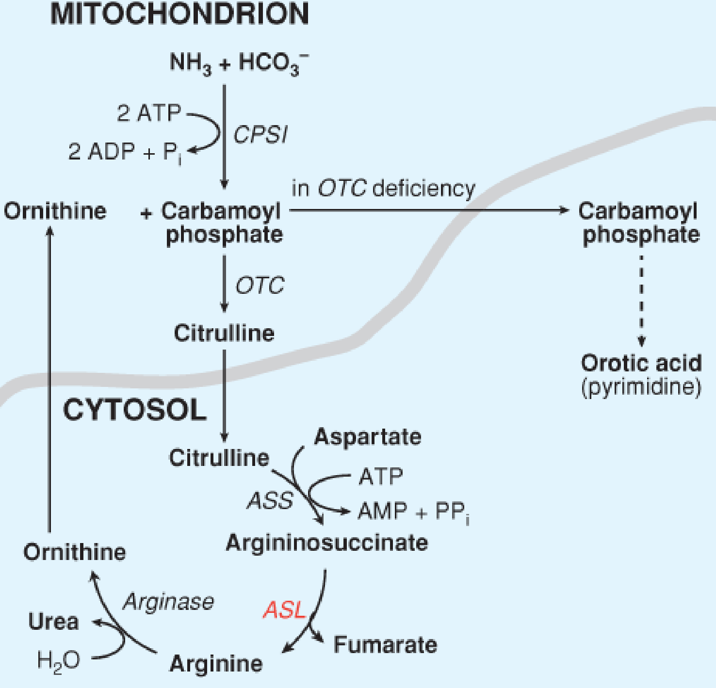
| 1 | Carbamoyl phosphate synthetase I (CPS I) | Mitochondria | Carbamoyl phosphate |
| 2 | Ornithine transcarbamoylase (OTC) | Mitochondria | Citrulline |
| 3 | Argininosuccinate synthetase (ASS) | Cytosol | Argininosuccinate |
| 4 | Argininosuccinate lyase (ASL) | Cytosol | Arginine + Fumarate |
| 5 | Arginase | Cytosol | Urea + Ornithine |
The urea cycle diagram below (from Lippincott Biochemistry) shows the full pathway:
Each urea molecule contains 2 nitrogen atoms:
- One from NH3 (via carbamoyl phosphate)
- One from aspartate
The urea is then excreted in the urine. Normal blood urea nitrogen (BUN) reflects this ongoing excretion.
3. Carbon Skeleton Waste Products
After deamination, the remaining alpha-keto acid (carbon skeleton) enters central metabolic pathways. These are not "waste" in the traditional sense but are processed or expelled:
| Type | Fate | Examples |
|---|---|---|
| Glucogenic amino acids | Carbon skeleton → pyruvate or TCA cycle intermediates → glucose or CO2 | Alanine, Glycine, Serine, Aspartate, Glutamate |
| Ketogenic amino acids | Carbon skeleton → acetyl-CoA or acetoacetyl-CoA → ketone bodies | Leucine, Lysine |
| Both (mixed) | Yield both glucose and ketone precursors | Isoleucine, Phenylalanine, Tyrosine, Tryptophan |
Final carbon waste: CO2 + H2O (excreted via lungs and kidneys).
4. Summary Table
| Waste Product | Origin | Elimination Route |
|---|---|---|
| Ammonia (NH3) | Oxidative deamination of glutamate | Converted to urea in liver |
| Urea | Urea cycle in liver | Excreted in urine (kidneys) |
| CO2 | TCA cycle oxidation of carbon skeletons | Exhaled by lungs |
| Water (H2O) | Urea cycle + TCA cycle | Urine, sweat, breath |
| Fumarate | Released by ASL in urea cycle | Enters TCA cycle |
| Creatinine | Breakdown of creatine phosphate (muscle) | Excreted in urine |
Clinical Relevance
- Hyperammonemia occurs when the urea cycle fails (enzyme defects such as OTC deficiency, ASL deficiency, CPS I deficiency). Even a 40-hour-old neonate can present with cerebral edema, seizures, and respiratory alkalosis from ammonia buildup - Lippincott Biochemistry, 8th ed. (Case 9).
- Elevated BUN reflects increased protein catabolism (starvation, sepsis, GI bleed) or reduced renal excretion.
- Liver failure impairs the urea cycle, causing ammonia accumulation and hepatic encephalopathy.
- Branched-chain amino acids (valine, leucine, isoleucine) are an exception - they cannot be catabolized in the liver and are handled mainly by muscle.
"The liver is central and critical to the body's nitrogen balance and amino acid metabolism."
- Sabiston Textbook of Surgery, Biological Basis of Modern Surgical Practice
why shouldn't we take excess protein
Excess dietary protein creates more ammonia than the liver's urea cycle can efficiently handle, stressing the kidneys (which excrete urea), raising blood urea nitrogen, and over time contributing to kidney damage, bone calcium loss, and cardiovascular risk from high-protein animal food sources.
Searching sources...
Finding Sources
Finding Sources
Searching PubMed
high protein diet adverse effects kidney health
Finding Sources
Finding Sources
Finding Sources
Reading File
Finding Sources
Reading File
Finding Sources
Here is a complete answer to why excess protein is harmful, drawing directly from medical biochemistry and clinical textbooks.
Why You Should NOT Consume Excess Protein
The short answer: the body cannot store protein like it stores fat or glycogen. Any protein eaten beyond your needs must be broken down, and the byproducts of that breakdown create a cascade of problems.
1. No Physiologic Benefit Beyond the RDA
"There is no physiologic advantage to the consumption of more protein than the RDA."
- Biochemistry, Lippincott Illustrated Reviews, 8th ed.
The recommended daily allowance (RDA) for a normal adult is 0.8 g/kg body weight (~56 g/day for a 70 kg person). Athletes may benefit from up to ~1 g/kg, but beyond that the benefits plateau and harms begin.
2. Excess Protein is Converted to Fat
The body cannot stockpile amino acids. When you eat more protein than you need:
- The excess amino acids are deaminated (amino group stripped off)
- The carbon skeletons are converted to acetyl-CoA and channeled into fatty acid synthesis
- Result: the extra protein ends up stored as body fat, not muscle
3. Increased Ammonia and Urea Load (Liver and Kidney Stress)
This is the biochemical core of the problem (directly linked to the previous topic):
- More protein → more amino acid catabolism → more ammonia (NH3) produced
- The liver must run the urea cycle harder to convert NH3 → urea
- The kidneys must filter and excrete the increased urea load
- Over time, this raises blood urea nitrogen (BUN) and places chronic stress on both organs
In patients with chronic kidney disease (CKD), this is so harmful that protein intake is actively restricted to 0.6-1.0 g/kg/day to slow disease progression. As the 2025 umbrella review (PMID 39657217) confirms, low-protein diets improve kidney function markers in CKD.
4. Kidney Stones and Osteoporosis
This is a direct, well-documented consequence:
"When excess protein is eliminated from the body as urinary nitrogen, it is often accompanied by increased urinary calcium, thereby increasing the risk of nephrolithiasis (kidney stones) and osteoporosis."
- Biochemistry, Lippincott Illustrated Reviews, 8th ed.
Mechanism:
- High protein intake generates an acid load (from sulfur-containing amino acids like methionine and cysteine)
- The body buffers this acid by releasing calcium carbonate from bone → bone resorption and calcium in urine
- High urinary calcium = calcium oxalate kidney stones + reduced bone mineral density over time
5. Cardiovascular Risk
- High animal protein diets are commonly high in saturated fat and red meat, which raise LDL cholesterol
- Research cited in Fuster and Hurst's The Heart, 15th ed. links low-carbohydrate/high-protein diets to increased incidence of cardiovascular disease and mortality
- The concern is greater with red and processed meat as the protein source, not plant proteins
6. Liver Stress
The liver is the primary site of amino acid metabolism and urea synthesis. A chronically high protein diet forces the liver to:
- Work harder to deaminate excess amino acids
- Run the urea cycle continuously at near-maximum capacity
- The systematic review on whey protein supplements (PMID 32702243) specifically flags liver and kidney disease as potential adverse effects of excessive protein supplementation.
7. Gut Microbiome Disruption
High protein diets can shift the gut microbiome toward putrefactive bacteria that ferment undigested protein in the colon, producing toxic byproducts like ammonia, phenols, and hydrogen sulfide - contributing to gut inflammation and dysbiosis.
Summary Table
| Problem | Mechanism |
|---|---|
| No muscle gain beyond needs | Excess protein → fat, not more muscle |
| Liver stress | More deamination → urea cycle overload |
| Kidney stress | Higher urea filtration load |
| Kidney stones | Increased urinary calcium excretion |
| Osteoporosis | Bone calcium mobilized to buffer acid load |
| Cardiovascular risk | High animal protein often paired with saturated fat |
| Gut dysbiosis | Undigested protein fermented by colonic bacteria |
| Weight gain | Excess carbon skeletons → fatty acid synthesis |
The Right Amount
| Population | Protein (g/kg/day) |
|---|---|
| Normal adult | 0.8 |
| Athletes | ~1.0-1.2 |
| Pregnant/lactating | +30 g/day extra |
| Infants | ~2.0 |
| CKD (not on dialysis) | 0.6-1.0 |
The bottom line: protein is essential, but more is not better. The body is not built to store it, and dismantling the excess creates a nitrogen waste burden that stresses the liver, kidneys, and bones.
gout
Gout is a form of inflammatory arthritis caused by deposition of monosodium urate crystals in joints and soft tissues, resulting from hyperuricemia (elevated serum uric acid). The classic presentation is sudden, severe pain in the big toe (podagra). It is directly related to purine metabolism - uric acid is the end product of purine breakdown in humans.
Searching sources...
Reading File
Finding Sources
Finding Sources
Searching PubMed
gout management treatment guidelines
Reading File
Reading File
Reading File
Reading File
Reading File
Loading Image
Loading Image
Loading Image
Here is a complete, textbook-sourced overview of gout.
Gout
Definition
Gout is a metabolic and inflammatory disease marked by transient attacks of acute arthritis initiated by deposition of monosodium urate (MSU) crystals in and around joints, resulting from sustained hyperuricemia (serum uric acid > 6.8 mg/dL - the limit of urate solubility in serum).
"Gout is a disease of both metabolism and inflammation, and its pathogenesis requires the intersection of two distinct processes: (1) the intrinsic accumulation of urate, at levels sufficient to drive the precipitation of MSU crystals, and (2) an inflammatory response to the crystals so formed."
- Firestein & Kelley's Textbook of Rheumatology
Biochemical Basis - Connection to Purine Metabolism
Uric acid is the end product of purine catabolism in humans. Purines (adenine, guanine) from DNA/RNA breakdown and dietary sources are ultimately degraded to uric acid via xanthine oxidase (XO).
Key steps:
- Purines → Hypoxanthine → Xanthine → Uric acid (via Xanthine oxidase)
- Humans lack uricase (present in most mammals), so uric acid is the terminal product rather than the more soluble allantoin
Classification of Gout
| Type | Mechanism | Examples |
|---|---|---|
| Primary gout (90%) | Reduced uric acid excretion (unknown cause) | Polymorphisms in URAT1, GLUT9, KCNQ1 transporters |
| Primary - enzyme defect | Overproduction | Partial HGPRT deficiency, PRPP synthetase superactivity |
| Secondary - overproduction | High cell turnover → excess purines | Leukemia, chemotherapy, Lesch-Nyhan syndrome |
| Secondary - underexcretion | Reduced renal clearance | Chronic kidney disease, thiazide diuretics, lead poisoning |
"In >90% of individuals with hyperuricemia, the cause is underexcretion of uric acid."
- Lippincott Biochemistry, 8th ed.
Risk factors:
- Male sex (90% of cases)
- Age (usually develops after 20-30 years of hyperuricemia)
- Alcohol consumption
- Obesity
- Diet rich in meat, seafood (shellfish), fructose
- Drugs: thiazide diuretics, low-dose aspirin, cyclosporine
- Genetic predisposition
Pathogenesis - The Inflammatory Cascade
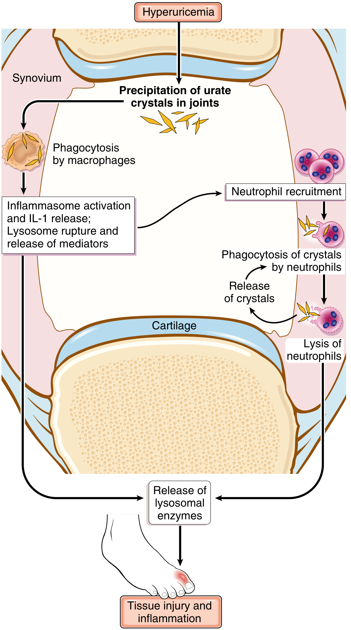
The sequence of events (Robbins & Kumar Pathologic Basis of Disease):
- Hyperuricemia → MSU crystals precipitate in the joint space (cooler peripheral joints favor crystal formation)
- Resident synovial macrophages phagocytose MSU crystals
- Crystals activate the NLRP3 inflammasome → caspase-1 activation → release of IL-1β (the master cytokine)
- IL-1β triggers massive neutrophil recruitment into the joint
- Neutrophils phagocytose crystals → phagolysosomes rupture → release of proteases, free radicals, arachidonic acid metabolites
- Vicious cycle: inflammation → more neutrophils → more crystal phagocytosis → more tissue damage
- Acute attack typically remits in days to weeks as crystals are solubilized
Clinical Stages
| Stage | Features |
|---|---|
| Asymptomatic hyperuricemia | Elevated urate, no symptoms. Only ~10% ever develop gout |
| Acute gouty arthritis | Sudden severe joint pain, redness, warmth, swelling. Classic: 1st metatarsophalangeal joint (podagra). Starts at night, peaks in 24 hrs |
| Intercritical gout | Symptom-free intervals between attacks |
| Chronic tophaceous gout | Persistent inflammation, tophi deposits, joint destruction |
Morphology / Pathology
Acute phase: Dense neutrophilic infiltrate in synovium and synovial fluid. Slender needle-shaped urate crystals inside neutrophils.
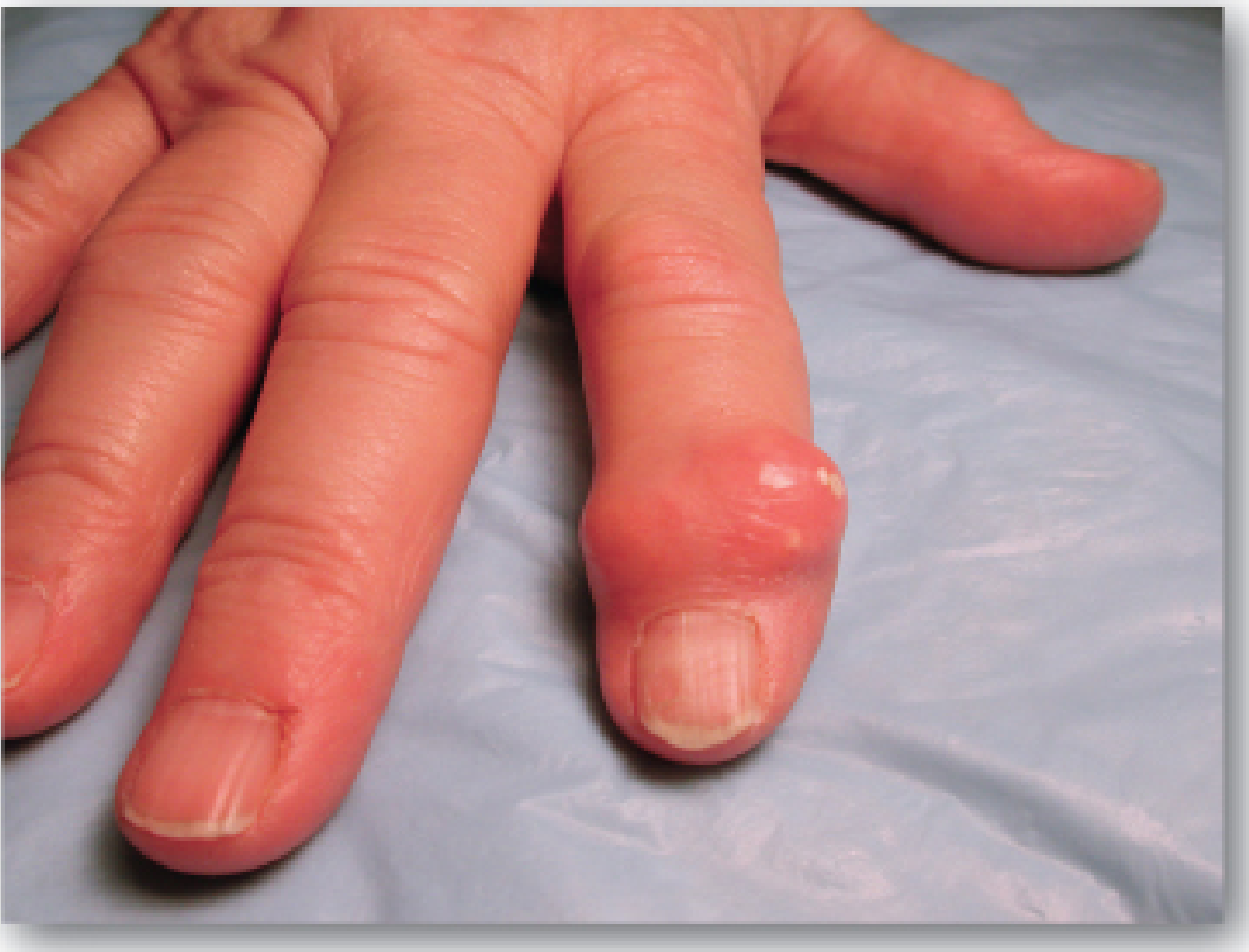
Chronic tophaceous arthritis: Tophi - nodular aggregates of MSU crystals + inflammatory tissue in synovium and periarticular tissue → severe cartilage damage.
Clinical photo showing a tophus on the finger (monosodium urate crystal deposit):
Diagnosis
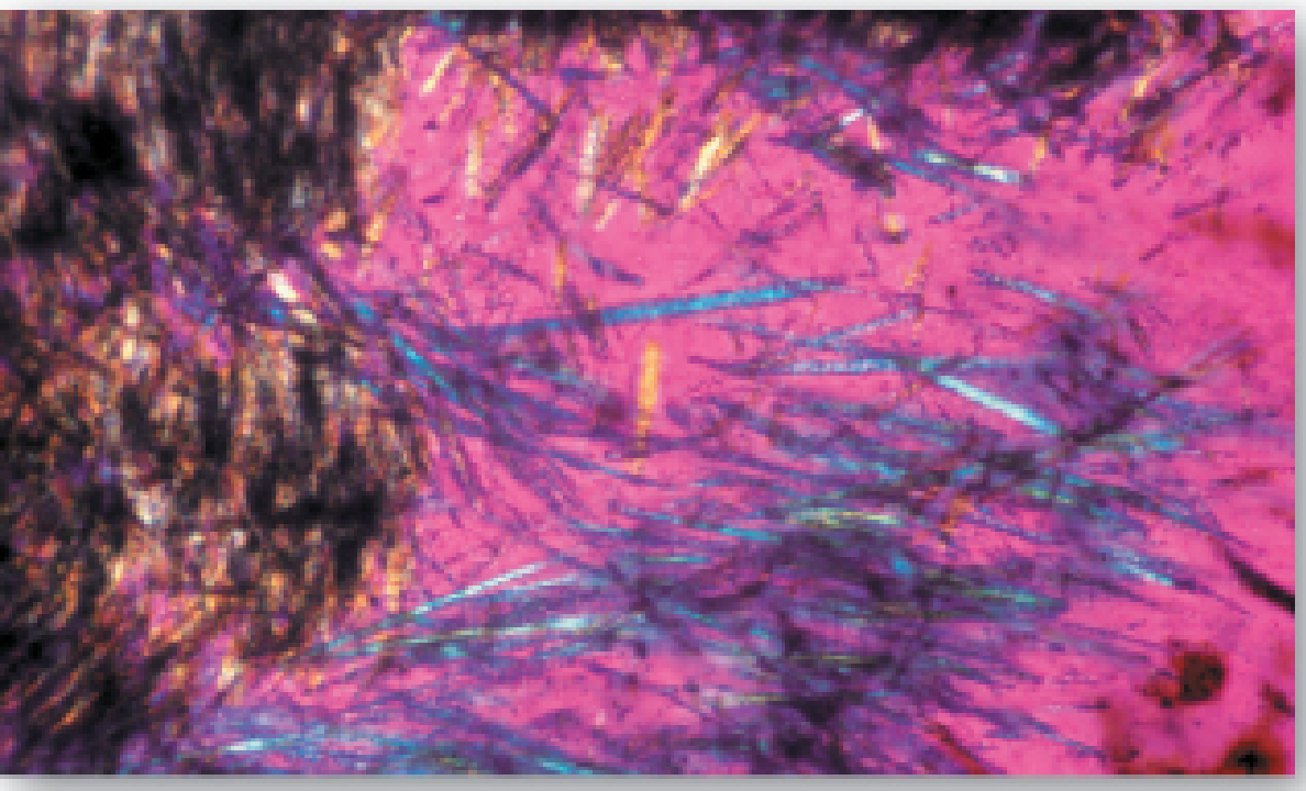
The gold standard is synovial fluid aspiration with polarized light microscopy:
"Demonstration of uric acid crystals in joint aspirate fluid is pathognomonic for gout."
- Tietz Textbook of Laboratory Medicine, 7th ed.
Key microscopy finding: Needle-shaped MSU crystals showing negative birefringence under compensated polarized light - crystals appear yellow when parallel to the polarizer axis.
Important: Serum uric acid is often normal during an acute attack and is NOT a component of the diagnostic criteria alone.
Treatment
Acute Attack
| Drug | Mechanism | Notes |
|---|---|---|
| Colchicine | Inhibits microtubule formation → limits neutrophil migration into joint | No effect on uric acid levels |
| NSAIDs (e.g., indomethacin) | Inhibit prostaglandin synthesis → reduce inflammation | First choice if no contraindications |
| Corticosteroids (e.g., prednisone) | Broad anti-inflammatory | Used when NSAIDs/colchicine contraindicated |
Long-Term Urate-Lowering Therapy (ULT)
Target: serum uric acid < 6.5 mg/dL (below saturation point)
| Drug | Mechanism | Use in |
|---|---|---|
| Allopurinol | Structural analog of hypoxanthine; inhibits xanthine oxidase (XO) → reduces uric acid synthesis | Overproducers; most common first-line ULT |
| Febuxostat | Non-purine XO inhibitor | Allopurinol-intolerant patients |
| Probenecid / Sulfinpyrazone | Uricosuric - increase renal excretion of uric acid | Underexcretors with normal renal function |
Allopurinol is oxidized to oxypurinol, a long-lived inhibitor of XO. This causes accumulation of hypoxanthine and xanthine - both more soluble than uric acid and less likely to form crystals.
- Lippincott Biochemistry, 8th ed.
Complications
- Urate nephropathy - MSU crystal deposition in renal interstitium → chronic kidney disease
- Uric acid urolithiasis - kidney stones (radiolucent on X-ray)
- Joint destruction - from chronic tophaceous arthritis
- Cardiovascular disease - hyperuricemia is an independent risk factor
Diet and Lifestyle
Foods that increase uric acid: red meat, organ meats, shellfish, beer, fructose-sweetened drinks, alcohol.
Foods that decrease risk: low-fat dairy products, cherries, vitamin C, adequate hydration.
The 2024 Chinese Guidelines for Hyperuricemia and Gout (PMID 40692263) reinforce that dietary modification combined with pharmacological ULT is the standard of care, with special attention to comorbidities like CKD and cardiovascular disease.
This is a shared conversation. Sign in to Orris to start your own chat.