
again
toacs imm fcps exam radiology
What do you need for FCPS Radiology TOACS/IMM exam?
interpretation of cases, anatomy and physics
How would you like to proceed?
can you help me pass imm fcps toacs radiology exam
- Case Interpretation - Show you real medical images (X-rays, CT, MRI, ultrasound) and walk through systematic reporting, findings, and differentials
- Radiological Anatomy - Spotters on plain films, CT cross-sections, MRI anatomy, angiograms
- Physics & Principles - X-ray physics, MRI physics, ultrasound principles, radiation protection, CT parameters - all commonly tested in IMM
- High-Yield Topic Revision - Chest, abdomen, MSK, neuro, paeds, breast, interventional
- Viva-style Q&A - I ask, you answer, I give feedback
- Which area do you want to begin with? (Chest / Abdomen / Neuro / MSK / Physics / Anatomy)
- Do you want me to quiz you (I show a case, you interpret), or teach you a topic systematically?
- Are there any weak areas you want to focus on?
im doing chest right now. i woild like you do all these suggestiosns
- 🫁 Case - I show you a chest image, you interpret it (findings → diagnosis → management)
- 🦴 Anatomy spotter - related to the case (e.g., identify structures on the CXR)
- ⚡ Physics pearl - one high-yield physics point tied to the modality used
chest X-ray pneumothorax
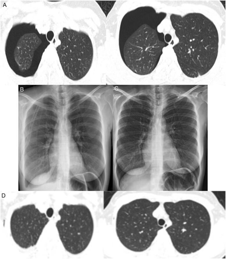
A multi-panel medical image sequence documenting the progression and resolution of a right-sided pneumothorax. Panel A presents axial Thorax Computed Tomography (TCT) lung window slices showing a significant right pneumothorax, characterized by a large, dark, air-filled pleural space (radiolucency) causing collapse of the right lung toward the mediastinum; the left lung remains fully expanded with normal density. Panel B is a Posteroanterior (PA) chest X-ray taken after tube thoracostomy, showing the right lung successfully re-expanded with the chest tube in situ. Panel C displays a follow-up chest X-ray after removal of the chest tube on the fifth day, maintaining full lung expansion and clear costophrenic angles. Panel D shows a follow-up axial TCT image two weeks post-discharge, confirming complete resolution of the pneumothorax with bilateral symmetric lung expansion and normal lung parenchyma. This sequence illustrates the clinical management of a spontaneous pneumothorax in the context of COVID-19 infection, from acute presentation to complete recovery.
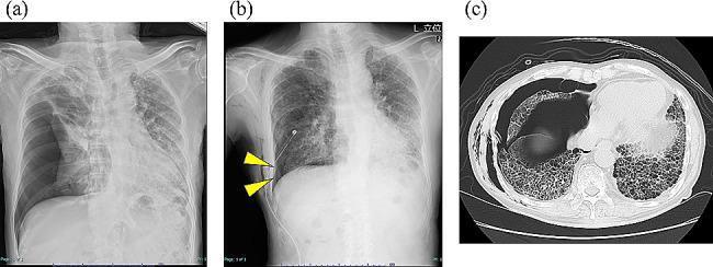
Educational clinical image set documenting a secondary pneumothorax in a patient with chronic hypersensitivity pneumonitis. Panel (a) is a posterior-anterior (PA) chest X-ray showing a large right-sided tension pneumothorax with significant mediastinal shift to the left and complete collapse of the right lung. Panel (b) is a follow-up chest X-ray post-thoracic drainage showing a visible chest tube and a persistent, mild residual pneumothorax cavity (indicated by yellow arrowheads) along the right lateral chest wall. Both radiographs reveal underlying diffuse reticular opacities consistent with interstitial lung disease. Panel (c) is an axial non-contrast chest CT scan (lung window) demonstrating extensive bilateral subpleural honeycombing and ground-glass opacities characteristic of advanced pulmonary fibrosis. The CT also captures the collapse of the right middle and lower lobes and an associated pleural air space. The series demonstrates the complication of intractable air leak in the context of advanced fibrotic lung disease.
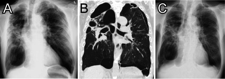
This composite figure demonstrates diagnostic imaging findings of bullous lung disease and pneumothorax in a patient with COPD. Image A is a frontal chest X-ray on presentation showing multiple large, thin-walled, hyperlucent areas consistent with giant bullae, particularly prominent in the lower zones, and a concomitant mild left-sided pneumothorax identified by a peripheral lucency and lack of lung markings. Image B is a coronal reformatted CT scan of the chest, providing high-resolution visualization of the lung parenchyma. It reveals extensive emphysematous changes and multiple, well-defined, thin-walled air-filled bullae of varying sizes occupying significant portions of both hemithoraces, with clear demarcation between the bullae and remaining compressed lung tissue. Image C is a follow-up chest X-ray taken 10 days later, showing stable appearance of the giant bullae but interval improvement and resolution of the previously seen left-sided pneumothorax, with better expansion of the underlying lung. The series highlights the importance of distinguishing between giant bullae and pneumothorax in patients with advanced obstructive lung disease.
🫁 STATION 1 - CHEST X-RAY
its a tension pneumothorax
✅ FEEDBACK - Station 1
🔍 Full Systematic Description (how to say it in the exam)
- Complete collapse of the right lung
- Right hemithorax shows no lung markings peripherally
- A visible pleural white line separates lung from air
- Underlying diffuse reticular opacities in both lungs (this patient has interstitial lung disease - the background cause)
⚠️ What makes it TENSION (not simple) pneumothorax?
| Feature | Simple PTX | Tension PTX |
|---|---|---|
| Mediastinal shift | No | Yes - AWAY from side |
| Tracheal deviation | No | Yes |
| Diaphragm | Normal | Depressed ipsilaterally |
| Haemodynamic compromise | No | Yes (clinically) |
Key TOACS point: Tension PTX is a clinical diagnosis - do NOT wait for X-ray if the patient is haemodynamically unstable. Immediate needle decompression at 2nd intercostal space, midclavicular line.
🔴 Immediate Management
- Do NOT wait for imaging if clinically suspected
- Needle decompression - 2nd ICS, MCL (or 4th/5th ICS, anterior axillary line - ATLS now accepts both)
- Followed by chest drain - 5th ICS, anterior axillary line (triangle of safety)
- High-flow oxygen
🦴 ANATOMY SPOTTER
- Anterior border: Lateral edge of pectoralis major
- Posterior border: Lateral edge of latissimus dorsi
- Inferior border: 5th ICS / horizontal line at nipple level
- Apex: Axilla
⚡ PHYSICS PEARL #1 - Chest X-Ray
| Density (Hounsfield / opacity) | Appears on X-ray |
|---|---|
| Air (-1000 HU) | Black (radiolucent) |
| Fat (-100 HU) | Dark grey |
| Soft tissue (0-80 HU) | Grey |
| Bone (400-1000 HU) | White |
| Metal (>1000 HU) | Bright white |
- Air attenuates very few X-ray photons → more photons hit the film/detector → black
- That's why a pneumothorax, pneumoperitoneum, and emphysema all appear hyperlucent (black)
if i share imm pastpaper files can you test me on them
- Read through all the questions
- Quiz you one by one in TOACS station format
- Give full feedback on each answer (what you got right, what to add, exam tips)
- Link related anatomy/physics where relevant
- 📝 Word (.docx)
- 🖼️ Images (JPG, PNG) - if the paper is scanned
- 📊 Excel/PowerPoint
its not very clear and its not easy to understant
- Blurry or low quality scan - I can still read most text and describe images
- Handwritten notes - I can interpret these
- Mixed language (Urdu + English) - I can handle that too
- Poor photo of a printed paper - still worth trying
Toacs past papers , that i downloaded from a link shared by senior... https://drive.google.com/drive/folders/1ewjxl9LAfBulIDvTiYSXJXQfmQGQlk-U
- Open the Drive folder
- Download the files to your phone/computer
- Attach them here in the chat (drag & drop or paperclip icon)
- Right-click a file in Drive → "Get link" → set to "Anyone with the link"
- Paste the direct file link here (not the folder link)
- I already know the high-yield TOACS radiology topics
- I can generate realistic exam-style questions based on what commonly appears in FCPS IMM radiology
- Many candidates report the same recurring cases (lobar collapse, pleural effusion, mediastinal masses, pneumonia patterns, etc.)
https://drive.google.com/drive/folders/1GlqNiGSY8knr4wTcW0t1E0t7NeNPSHLk
- Open that Drive folder on your phone/computer
- Select all files → Download (it'll zip them)
- Upload the zip or individual files here in the chat
- Lobar collapse (all 5 lobes)
- Pleural effusion
- Consolidation vs collapse
- Mediastinal masses (3 zones)
- Interstitial patterns (reticular, nodular, honeycomb)
- Hilum - anatomy + pathology
- Chest CT basics
let do more
Because the neurovascular bundle (intercostal nerve, artery, vein) runs in the subcostal groove - under the lower rib. Going over the upper border avoids injuring it. ✅
lobar collapse chest X-ray left lower lobe
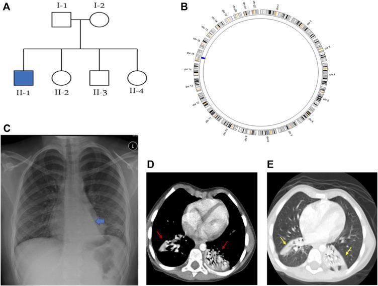
This composite educational image illustrates the genetic and radiological manifestations of Primary Ciliary Dyskinesia (PCD) in a pediatric patient. Panel (A) presents a family pedigree showing an autosomal recessive inheritance pattern. Panel (B) is an AgileMultiIdeogram showing a region of homozygosity (ROH) on chromosome 15. Panel (C) is a posteroanterior (PA) chest X-ray demonstrating a 'flat-waist sign' and a triangular retrocardiac opacity, classic indicators of left lower lobe (LLL) collapse. Panels (D) and (E) are axial CT chest scans in soft-tissue and lung windows, respectively. These diagnostic images reveal bilateral pathology, specifically highlighting subsegmental collapse in both the right and left lower lobes (indicated by red and yellow arrows). The images collectively demonstrate the secondary pulmonary complications of PCD, such as chronic atelectasis and lobar collapse, essential for clinical diagnosis and management in pediatric pulmonology and medical genetics.
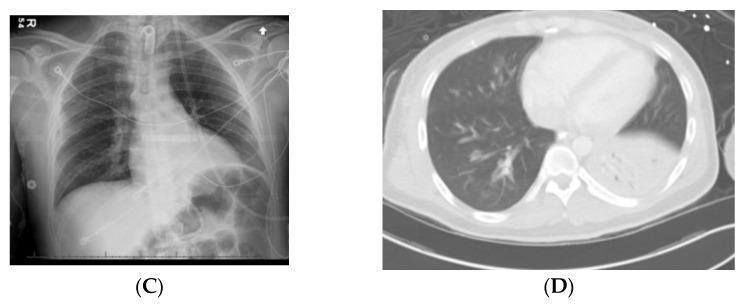
This composite figure displays diagnostic imaging of chest pathologies. Image C is an anteroposterior chest X-ray demonstrating a significantly elevated left hemidiaphragm caused by multiple dilated, gas-filled bowel loops within the upper abdomen, consistent with an opioid-induced ileus. Monitoring leads and a central line are visible. Image D is an axial CT scan of the chest (lung window) showing a dense area of consolidation and volume loss in the left lower lobe, indicative of collapse and persistent pneumonia or aspiration. The right lung remains clear. These images illustrate common secondary pulmonary and gastrointestinal complications in trauma patients with spinal cord injuries, emphasizing the impact of abdominal distention on diaphragmatic position and the radiological presentation of lobar consolidation.
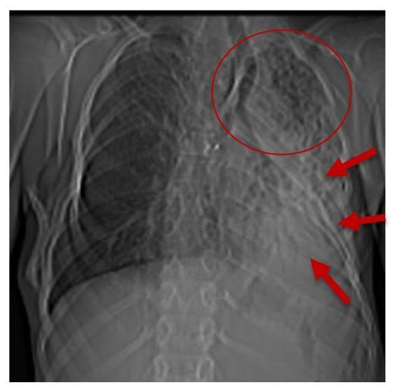
Frontal chest x-ray demonstrating multi-lobar pathology in the left lung with associated mediastinal changes. Key findings include a dense opacity in the left lower lobe consistent with atelectasis (indicated by red arrows) and reticular, net-like infiltrates throughout the left upper lobe (circled in red), suggesting an alveolo-interstitial pneumopathy. There is a noticeable ipsilateral shift of the mediastinum towards the left, signifying volume loss. The right lung appears hyperinflated, providing a compensatory response to the left-sided collapse. Additionally, the image shows evidence of extraskeletal calcification near the tracheal bifurcation and possible heterotopic bone formation in the right laterodorsal region. These findings are highly relevant for diagnosing complex systemic conditions manifesting with respiratory impairment and progressive ossification, such as fibrodysplasia ossificans progressiva (FOP).
🫁 STATION 2 - LOBAR COLLAPSE