


Summarise different parts of upper respiratory tract and lower respiratory tract important of them and functions of respiratory system in detail in understanding way with full knowledge to attempt them in exam point of view
respiratory system diagram upper lower tract anatomy
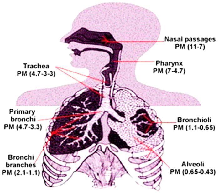
This medical illustration is a pathophysiology diagram depicting the human respiratory system and the deposition patterns of particulate matter (PM) based on size. The diagram utilizes a human silhouette to map the upper and lower respiratory tracts. Labeled anatomical regions include the nasal passages, pharynx, trachea, primary bronchi, bronchi branches, bronchioli, and alveoli. Each region is annotated with a specific PM size range (presumably in micrometers) that typically deposits there. The data shows a progressive decrease in particle size relative to the depth of penetration: the upper tract (nasal passages) captures larger particles (11–7 µm), the conductive airways (trachea and bronchi) capture mid-sized particles (4.7–1.1 µm), and the deepest acinar regions (bronchioli and alveoli) receive the smallest, ultrafine particles (1.1–0.43 µm). This visual serves as an educational tool for environmental health and pulmonology, illustrating how different particle diameters interact with respiratory anatomy to impact lung health.
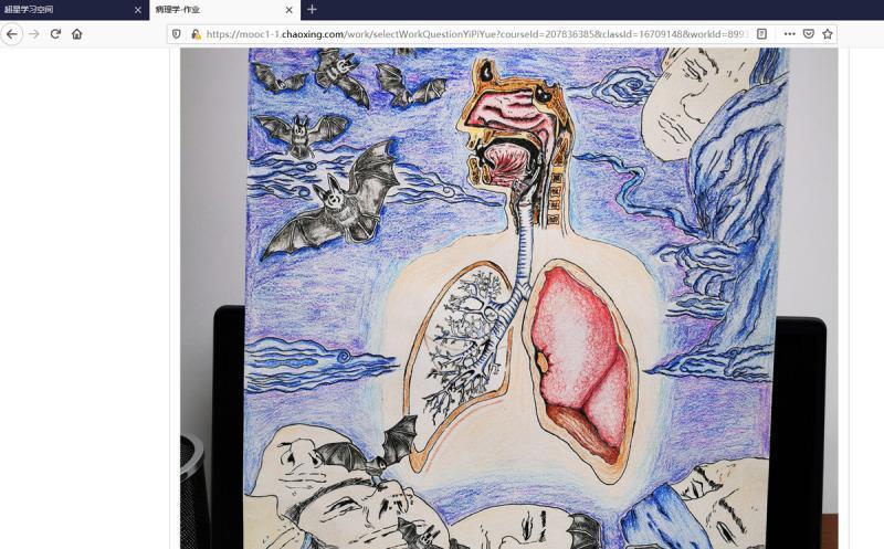
This educational anatomical illustration combines a sagittal section of the upper respiratory tract with a coronal view of the lower respiratory system, created as a student project. The superior portion depicts the nasal cavity, oral cavity, pharynx, and larynx in cross-section. Below, the trachea descends and bifurcates into the primary bronchi. The lungs are shown with asymmetrical detailing: the right lung is rendered in a solid pink/red hue indicating tissue parenchyma, while the left lung features a detailed schematic of the bronchial tree and alveolar branching patterns. The central anatomical figure is set within a light-colored torso silhouette. Surrounding the anatomical drawing are symbolic elements including bats and several human faces in states of rest or mask-wearing, set against a blue, wavy background. This visual serves as a medical student's creative synthesis of respiratory anatomy and pathology, likely contextualizing the COVID-19 pandemic and zoonotic disease transmission. The medical focus is on the structural continuity between the upper and lower respiratory tracts.
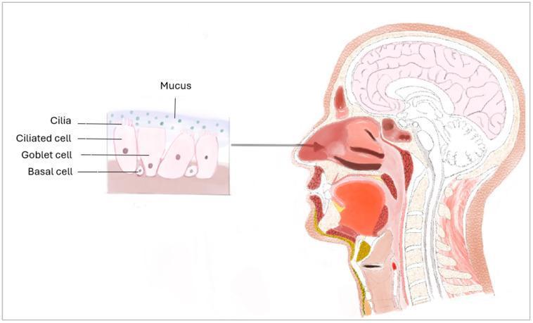
This medical illustration presents a sagittal anatomical diagram of the human head and neck, coupled with a histological inset of the nasal mucosa. The main diagram depicts the upper respiratory tract including the nasal cavity, hard and soft palate, tongue, pharynx, larynx, and trachea, alongside the brain and cervical spine. An arrow points from the nasal conchae to a detailed histological cross-section of the respiratory epithelium. This inset illustrates a pseudostratified ciliated columnar epithelium, highlighting its cellular composition: basal cells positioned along the basement membrane; secretory goblet cells; and ciliated cells featuring hair-like cilia at the apical surface. A superficial layer of mucus is shown above the cilia, representing the mucociliary clearance mechanism. This educational visual is designed to demonstrate the relationship between gross anatomy and microscopic tissue structure in the context of respiratory physiology and physical defense barriers against pathogens.
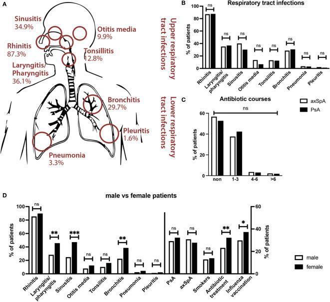
This medical infographic displays data on respiratory tract infections (RTIs) in patients with spondyloarthritis (SpA). Panel A utilizes an anatomical diagram of the human respiratory system to map the frequency of specific infections. Upper respiratory tract infections (URTIs) shown include rhinitis (87.3%), laryngitis/pharyngitis (36.1%), sinusitis (34.9%), tonsillitis (12.8%), and otitis media (9.9%). Lower respiratory tract infections (LRTIs) include bronchitis (29.7%), pneumonia (3.3%), and pleuritis (1.6%). Panel B provides a bar chart comparing RTI frequencies between axial spondyloarthritis (axSpA) and psoriatic arthritis (PsA) cohorts, showing no significant differences. Panel C illustrates the distribution of antibiotic courses prescribed, categorized by patient subgroup. Panel D presents a sex-disaggregated analysis, revealing that female patients (black bars) report significantly higher rates of laryngitis/pharyngitis, sinusitis, and bronchitis compared to male patients (white bars), alongside higher rates of antibiotic treatment and influenza vaccination. The graphic effectively summarizes the epidemiological burden of RTIs within these chronic inflammatory disease populations, highlighting gender-specific variations and clinical management patterns.
alveoli gas exchange conducting zone respiratory bronchiole diagram
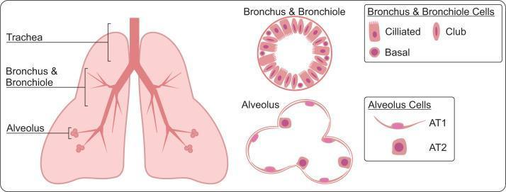
This medical illustration depicts the human lung architecture from a macroscopic to a microscopic cellular level. The left side features an anatomical diagram showing the hierarchy of the respiratory system: the central trachea bifurcates into the primary bronchi and smaller bronchioles, eventually terminating in alveolar sacs. To the right, high-magnification cross-sectional diagrams detail the micro-architecture and cellular composition of these regions. The Bronchus & Bronchiole section shows a circular lumen lined with three distinct cell types: elongated ciliated cells with hair-like projections, oval-shaped club cells, and circular basal cells. The Alveolus section illustrates the thin, multi-lobed structure of alveolar sacs, primarily composed of Alveolar Type 1 (AT1) cells, which are characterized as thin, flat squamous cells optimized for gas exchange, and Alveolar Type 2 (AT2) cells, which are larger, cuboidal cells responsible for surfactant production. This diagram serves as a physiological and histological reference for the organization of the lower respiratory tract.
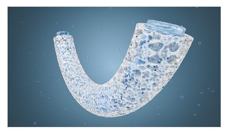
A schematic diagram representing a tubular, U-shaped porous sponge designed for gas-liquid exchange, modeled after physiological systems like lung alveoli or blood vessels. The illustration highlights a high-porosity structural wall with a heterogeneous network of interconnected pores of varying sizes. The sponge material is shown in a light, fiber-like texture, with visible blue-tinted gas bubbles or fluid droplets penetrating and diffusing through the porous walls. Both ends of the tubular structure are sealed with a translucent blue material, suggesting the containment of an inner aqueous solution or reaction channel. The educational focus of this visual is the demonstration of gas diffusion mechanisms across semi-permeable, virtually wall-less containers, applicable in biomedical engineering for respiratory support systems or controlled mineralization studies. The diagram illustrates how the hierarchical pore structure increases surface area for efficient gas-liquid interface reactions while maintaining mechanical stability.
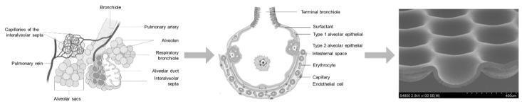
This figure illustrates the multiscale engineering approach to mimicking human lung anatomy for tissue engineering. The progression moves from macroscale anatomy to microscale fabrication in three panels. The left panel is a schematic diagram of the pulmonary parenchyma, labeling the bronchiole, pulmonary artery and vein, respiratory bronchioles, alveolar ducts, alveolar sacs, and the capillaries of the interalveolar septa. The middle panel provides a detailed cross-sectional illustration of a single alveolus, identifying the blood-air barrier components: surfactant layer, Type 1 and Type 2 alveolar epithelial cells, interstitial space, endothelial cells, and erythrocytes within the capillary. The right panel is a scanning electron microscopy (SEM) image of a bioengineered 'Lung-MatriGrid®' scaffold. This microfabricated structure features an array of concave, dome-shaped micro-cavities designed to replicate the three-dimensional geometry and dimensions of human alveoli. The scaffold, likely made of polycarbonate, provides a biomimetic surface for co-culturing epithelial and endothelial cells to study the blood-air barrier in vitro.
Functional Organization of the Respiratory System
Complete Exam-Focused Guide (GK Pal Physiology / Jaypee Physiology)
Overview: What is Respiration?
| Stage | What Happens |
|---|---|
| Ventilation | Exchange of gases between atmosphere and pulmonary capillaries |
| Transport | O₂ and CO₂ carried in blood between lungs and tissues |
| Tissue/Exchange | O₂ used by mitochondria; CO₂ produced (cellular respiration) |
Components of the Respiratory System
- Upper Respiratory Tract (URT) - nose to larynx
- Lower Respiratory Tract (LRT) - trachea, bronchi, lungs
UPPER RESPIRATORY TRACT (URT)
1. NOSE
- Lined with olfactory epithelium (receives smell)
- Nasal filters: Nasal hairs filter particles >10 µm
- Secretions contain immunoglobulins and interferons
- Nose cross-sectional area is greatly increased by nasal turbinates (conchae)
- Nose offers about 50% of resistance to airflow in the respiratory system
- Resistance increases during viral infections (e.g., common cold)
2. PARANASAL SINUSES
- They act as resonance chambers for voice
- They lighten the skull for upright posture
- They protect the brain during facial trauma
Clinical Box: The sinuses open onto the nasal turbinates, so nasal infection can spread to sinuses (sinusitis), blocking drainage and causing severe pain.
3. PHARYNX
- Nasopharynx - behind the nose; contains Eustachian tube openings and posterior nares open here
- Oropharynx - located behind the mouth; soft palate above
- Laryngopharynx - starts from level of hyoid bone below; continues as esophagus
4. LARYNX AND GLOTTIS
- Epiglottis, arytenoids, and vocal cords
- Glottis = the vocal cords + rima glottidis
- Phonation - production of sound via vocal cords
- Epiglottis closes during swallowing to prevent aspiration of food into the respiratory tract
- Arytenoids enlarge during infections (especially in children), which increases resistance to air flow (croup)
- During swallowing, abductor muscles contract and pull vocal cords apart; incomplete closure of glottis during unconscious states causes food and fluid to enter the lungs, leading to aspiration pneumonia
- Cervical vagotomy in animals produces laryngeal congestion and edema (partly due to aspiration)
Laryngeal paralysis = arytenoid muscles paralyzed = "respiratory stridor"
LOWER RESPIRATORY TRACT (LRT)
I. CONDUCTING ZONE
Functions of Conducting Zone:
- Warm and humidify inspired air
- Distribute air evenly to deeper parts of the lungs
- Non-specific defense: Removes dust, bacteria, and noxious agents from the respiratory tract
Structural Specializations:
- Cartilage is present in the trachea and first 16 generations, preventing airway collapse; it is absent in bronchioles; it is absent totally in the bronchioles (the smallest airways use smooth muscle tone instead)
- Airways are lined by ciliated pseudostratified columnar epithelium with goblet cells that secrete mucus
- Mucociliary escalator: Mucus traps particles; cilia beat upward (toward the pharynx) where they are swallowed or expectorated
- Walls contain smooth muscle innervated by sympathetic (β₂ - bronchodilation) and parasympathetic (muscarinic - bronchoconstriction) fibers
TRACHEA - Key Points:
- Bifurcates at carina (T4-T5 level) into right and left main bronchi
- Right main bronchus is more vertical → aspirated foreign bodies usually go to the right
- Blood supply: Inferior thyroid artery (bronchial circulation)
- Nerve supply: Autonomic nervous system (ANS)
BRONCHIAL TONE:
- Sympathetic stimulation via β₂ receptors → bronchodilation (e.g., salbutamol/albuterol in asthma)
- Parasympathetic stimulation (vagal, muscarinic) → bronchoconstriction (e.g., in asthma, methacholine challenge)
- Bronchoconstriction is also produced by inflammatory mediators, cytokines, adenosine, histamine, and leukotrienes
CHANGES IN CONDUCTING ZONE AS AIRWAYS BRANCH:
- Airway generations increase (0 → 16)
- Individual airway diameter gets smaller
- Number of airways increases dramatically (in parallel) → total cross-sectional area increases enormously
- Cartilage decreases (absent in bronchioles)
- Mucus-secreting glands decrease
- Amount of cartilage in airway wall decreases
- Cilia present throughout (up to terminal bronchioles)
Dead Space (exam important): Gas exchange does not occur in the conducting zone. This volume is called anatomic dead space = approximately 150 mL (generation 0-16).
II. RESPIRATORY ZONE
- Respiratory bronchioles (17-19) - transitional; have occasional alveoli budding off their walls; still have cilia and smooth muscle
- Alveolar ducts (20-22) - completely lined with alveoli; no cilia, little smooth muscle
- Alveolar sacs (23) - terminal clusters of alveoli
ALVEOLI - Must Know for Exams:
- Each lung contains approximately 300 million alveoli
- Diameter of each alveolus ≈ 200 µm
- Total surface area ≈ 70-100 m² (size of a tennis court)
- This large surface area enables rapid and efficient gas exchange
| Cell | Name | Function |
|---|---|---|
| Type I pneumocyte | Alveolar Type 1 (AT1) | Thin, flat squamous cells; allow gas diffusion across the air-blood barrier |
| Type II pneumocyte | Alveolar Type 2 (AT2) | Cuboidal cells; produce surfactant; have regenerative capacity for both Type I and Type II cells |
| Alveolar macrophages | "Dust cells" | Phagocytosis of foreign particles; since alveoli have no cilia, macrophages migrate to bronchioles where cilia take over |
- Reduces surface tension of alveoli
- Prevents alveolar collapse (atelectasis) during expiration
- Deficiency in premature babies → Neonatal Respiratory Distress Syndrome (NRDS)
PLEURA:
- Parietal pleura = outer layer lining the chest wall
- Visceral pleura = inner layer covering the lungs
- Pleural fluid (15-20 mL) acts as a lubricant allowing lung sliding, facilitates changes in lung size and shape, and keeps the lungs expanded against the chest wall
- Pleural fluid is produced mainly by the parietal pleura and removed by lymphatics
Pleural effusion = accumulation of excess fluid in the pleural space; can compress the lung.
FUNCTIONS OF THE RESPIRATORY SYSTEM
A. Respiratory Functions (Primary)
1. Gas Exchange (Most Important)
- O₂ moves from alveoli → blood by simple diffusion down a pressure gradient
- CO₂ moves from blood → alveoli in the same way
- Governed by Fick's law: Rate of diffusion ∝ (Surface area × pressure gradient) / thickness of membrane
2. Regulation of Blood pH
- By controlling CO₂ levels, the respiratory system regulates blood pH (via the carbonic acid-bicarbonate buffer system)
- ↑ ventilation → ↓ CO₂ → ↑ pH (alkalosis)
- ↓ ventilation → ↑ CO₂ → ↓ pH (acidosis)
B. Non-Respiratory Functions (High-Yield Exam Topic)
| Function | Details |
|---|---|
| Left ventricle reservoir | Due to high compliance, pulmonary vessels hold large volumes - serves as a blood reservoir for the left ventricle |
| Filtering small emboli | Lung filters venous blood clots, air bubbles, and aggregates before they enter systemic circulation |
| ACE production | Pulmonary endothelium contains Angiotensin Converting Enzyme (ACE) which converts angiotensin I → angiotensin II (important in blood pressure regulation) |
| Synthesis and release | Synthesizes and releases bradykinin, histamine, serotonin, and prostaglandins into circulation |
| Removal from blood | Lungs inactivate bradykinin, serotonin, prostaglandins (PGE₂, PGF₂, leukotrienes) |
| Metabolic | Synthesis of surfactant (phospholipids) |
| Speech | Controlled expiratory flow over vocal cords produces phonation |
| Defense functions | Mucociliary clearance, alveolar macrophages, IgA secretion in respiratory mucosa |
| Warming/humidifying | Air reaches near body temperature and 100% humidity by the time it reaches the trachea |
QUICK SUMMARY TABLE (Exam Blueprint)
| Feature | Conducting Zone | Respiratory Zone |
|---|---|---|
| Generations | 0-16 | 17-23 |
| Structures | Trachea, bronchi, bronchioles, terminal bronchioles | Respiratory bronchioles, alveolar ducts, alveolar sacs |
| Gas exchange | NO | YES |
| Cilia | YES | Only in respiratory bronchioles |
| Cartilage | YES (trachea-bronchi); NO in bronchioles | Absent |
| Smooth muscle | YES | Present (respiratory bronchioles) |
| Function | Warm, humidify, filter, conduct air | Gas exchange (O₂/CO₂) |
| Dead space | YES (anatomic dead space ~150 mL) | NO (participates in exchange) |
IMPORTANT TO KNOW (Exam Must-Read)
- Anatomic dead space = 150 mL = volume of conducting zone (no gas exchange here)
- Physiologic dead space = anatomic + alveolar dead space (alveoli ventilated but not perfused)
- Surfactant is produced by Type II pneumocytes; deficiency = NRDS in neonates
- Right bronchus is more vertical, shorter, wider → foreign body aspiration goes to right lung preferentially
- β₂ agonists (salbutamol, albuterol) = bronchodilators used in asthma
- Parasympathetic (vagal/muscarinic) = bronchoconstriction
- ACE is produced by pulmonary capillary endothelium
- Paranasal sinuses functions: resonance + lighten skull + protect brain
- Epiglottis prevents aspiration during swallowing
- Alveolar macrophages provide defense since alveoli have NO cilia
- Type I pneumocytes = gas diffusion; Type II = surfactant + regeneration
- Each lung contains ~300 million alveoli with total surface area ~70-100 m²