
Atrial septal defect
"atrial septal defect"
atrial septal defect adults diagnosis management guidelines AHA
diagram showing types of atrial septal defect secundum primum sinus venosus
https://www.acc.org/latest-in-cardiology/articles/2022/03/08…
atrial septal defect color Doppler echocardiogram
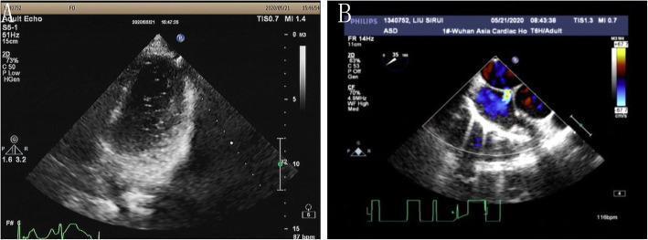
This composite diagnostic image displays echocardiographic findings of an atrial septal defect (ASD) in a 16-year-old patient. Panel A shows a transthoracic echocardiogram (TTE) in a grayscale 2D view. It illustrates the right heart morphology where varied echogenicity within the atrial chambers suggests right-to-left shunting. Panel B presents a transesophageal echocardiogram (TEE) utilizing color Doppler mapping. This panel clearly demonstrates a bidirectional shunt across the atrial septum. A physical interruption in the septal wall is visible, accompanied by a high-velocity color signal. The color Doppler displays a shift from blue to red, indicating the flow direction from the left atrium to the right atrium (left-to-right shunt) at the site of the ASD. The images are essential for delineating ASD morphology, measuring the defect size (approximately 0.6 cm in this case), and assessing the surrounding septal rims before transcatheter closure interventions. These views are typical in cardiology for diagnosing congenital heart defects and planning septal occluder placement.
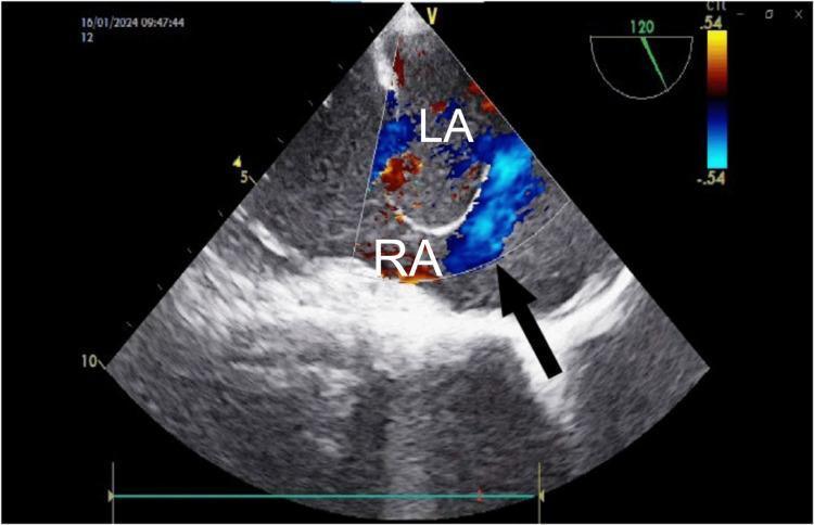
This diagnostic ultrasound image is a mid-esophageal transesophageal echocardiogram (TEE) with color Doppler, focused on the interatrial septum. The image demonstrates a significant atrial septal defect (ASD) associated with an atrial septal aneurysm (ASA). The right atrium (RA) and left atrium (LA) are clearly labeled. A large jet of color flow, predominantly represented in shades of blue, indicates shunting across the septal defect. A black arrow points specifically to the region of the defect where the septal wall is discontinuous, allowing for the passage of blood between the chambers. The color Doppler scale on the right indicates velocity and direction, with the blue flow signifying movement away from the probe (from LA to RA). The interatrial septum displays an aneurysmal bulge toward the right atrium, consistent with pressure differentials often seen in such congenital heart defects. This image serves as a clinical example of using TEE for the evaluation and intraoperative monitoring of septal anomalies and hemodynamics.
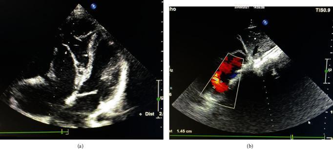
Transthoracic echocardiogram (TTE) images in a patient with Ebstein's anomaly and Atrial Septal Defect (ASD). Panel (a) shows a two-dimensional apical four-chamber view demonstrating cardiac anatomy in situs solitus and levocardia. There is visual evidence of Ebstein anomaly (types B-C), characterized by the apical displacement of the septal tricuspid valve leaflet and subsequent atrialization of the right ventricle. Panel (b) utilizes Color Doppler flow mapping to visualize an atrial septal defect. The Doppler overlay shows a mosaic flow pattern across the interatrial septum, indicating a left-to-right shunt. Linear measurements in panel (b) mark the ASD diameter, which correlates with a 8.3 mm defect. These diagnostic images are essential for evaluating congenital heart disease, assessing valvular displacement, and quantifying shunting in adult congenital cardiology.
Atrial septal defect (ASD)
Main types
- Ostium secundum ASD: Most common. Located at the fossa ovalis in the central atrial septum. Often suitable for transcatheter device closure.
- Ostium primum ASD: Low in the septum and commonly associated with atrioventricular valve abnormalities. Usually requires surgery.
- Sinus venosus ASD: Near the superior or inferior vena cava; may occur with partial anomalous pulmonary venous return. Usually surgical.
- Coronary sinus defect: Rare.
Symptoms and signs
- Breathlessness on exertion, reduced exercise tolerance, fatigue
- Palpitations or atrial fibrillation/flutter
- Frequent respiratory infections in children
- Fixed wide splitting of the second heart sound, sometimes with a systolic flow murmur
- Right-heart enlargement and pulmonary hypertension in untreated significant defects
Diagnosis
- Transthoracic echocardiography with color Doppler is the main test.
- Transesophageal echocardiography, cardiac MRI, or CT may define anatomy and associated abnormal pulmonary veins.
- ECG may show right-sided conduction or chamber changes.
- Cardiac catheterization is used selectively, particularly when pulmonary hypertension or closure suitability is uncertain.
Treatment
- Right atrial/right ventricular enlargement from volume overload, and/or
- A significant shunt, commonly Qp:Qs >1.5:1.
Follow-up


Interpret and treatment options
"atrial septal defect" AND closure
2025 ACC AHA adult congenital heart disease guideline atrial septal defect closure pulmonary vascular resistance
Interpretation of the echocardiogram
| Finding | Meaning |
|---|---|
| 18 mm ostium secundum ASD | An opening in the central part of the atrial septum, the commonest ASD type |
| Left-to-right shunt | Oxygenated blood is passing from the left atrium into the right atrium, causing extra blood flow to the right heart and lungs |
| Right atrium and right ventricle dilated | The ASD is already causing significant right-heart volume overload |
| LVEF 59%, normal biventricular function | The pumping function of the left ventricle is normal |
| All valves normal | No important valve disease was reported |
| No clot/effusion, no LV dysfunction | Reassuring findings |
Bottom line
Most likely treatment option: transcatheter device closure
- No chest incision or sternotomy
- Usually 1 to 2 nights in hospital
- Faster recovery than surgery
- High closure success when the rims and anatomy are suitable
When surgery is used
- The defect has inadequate rims for a device
- The ASD is too large or has unusual anatomy
- It is actually a sinus venosus or primum defect rather than a secundum ASD
- There is associated anomalous pulmonary venous drainage or another cardiac condition needing repair
Tests to arrange before choosing treatment
-
TEE, preferably 3D TEE
To confirm ASD type, exact diameter, number of defects, and all device-supporting rims. -
ECG and rhythm assessment
To check for atrial arrhythmias, particularly atrial fibrillation or flutter. -
Assessment of pulmonary pressures and shunt size
The report does not give pulmonary artery pressure, estimated right-ventricular systolic pressure, Qp:Qs ratio, or pulmonary vascular resistance. -
Cardiac catheterization, if pulmonary hypertension is suspected or echo measurements are uncertain
This measures the shunt and pulmonary vascular resistance directly. Closure should be avoided if there is severe fixed pulmonary vascular disease. The ACC adult ASD review notes that ASD closure should be avoided when pulmonary vascular resistance remains high despite pulmonary-hypertension treatment. -
Cardiac MRI or CT, sometimes
Especially if there is concern for anomalous pulmonary veins or if echo images are incomplete.
Medicines
- Diuretics for fluid retention or breathlessness due to heart failure
- Medicines for atrial fibrillation/flutter if present
- Anticoagulation only if an arrhythmia, clot risk, or another indication is confirmed
After closure
Practical recommendation
Explain what TEE and device closure involve
"atrial septal defect" AND transcatheter closure
American College Cardiology atrial septal defect transesophageal echocardiography device closure procedure patient
transesophageal echocardiography guided transcatheter device closure secundum atrial septal defect diagram
1. What is a TEE?
What TEE checks in your case
- The exact size and shape of the hole
- Whether there is one ASD or more than one
- The tissue margins around it, called rims. These are needed to anchor a closure device safely.
- Distance from the aorta, vena cavae, mitral valve, and pulmonary veins
- Direction and size of the shunt
- Right-heart enlargement and estimated lung pressures
- Any unexpected abnormal pulmonary vein drainage or clot
What happens during a TEE
- You usually fast for about 6 hours beforehand. The hospital will give exact instructions.
- An IV line is placed, and the throat is numbed with spray.
- Sedation is usually given, so most people are sleepy and recall little.
- A mouth guard is placed and the probe is gently passed into the esophagus.
- Images are taken for about 15 to 30 minutes.
- You are observed until the sedation wears off, then need someone else to take you home. Avoid driving that day.
TEE risks
2. What is ASD device closure?
Steps in the procedure
-
Preparation and anesthesia
Usually general anesthesia or deep sedation is used. You do not feel the procedure. -
Groin-vein access
The cardiologist makes a small puncture in the femoral vein, usually in the right groin. -
Catheter goes to the heart
A thin tube is moved through the vein into the right atrium and across the ASD. No chest incision is needed. -
Measurements and imaging
TEE, or sometimes intracardiac echocardiography, helps measure the ASD and confirm the rim anatomy. In some patients, balloon sizing is performed. -
Device placement
The left-sided disc is opened in the left atrium, gently pulled against the septum, then the right-sided disc is opened in the right atrium. -
Safety check before release
The team confirms that the device is stable, does not interfere with valves or nearby vessels, and that there is little or no residual shunt. -
Release and recovery
The device is detached from the delivery cable. The groin puncture is compressed or closed. The procedure often takes about 1 to 2 hours, but total time in the procedure room may be longer.
Is it likely to be an option for this report?
Benefits expected from closure
- Stops the abnormal left-to-right shunt
- Reduces excess blood flow to the lungs
- Allows the enlarged right atrium and right ventricle to remodel over time
- May improve breathlessness, fatigue, or exercise tolerance if present
- Helps prevent later complications such as atrial arrhythmias, right-heart failure, and pulmonary vascular disease
Risks of device closure
- Bruising or bleeding at the groin
- Temporary palpitations or arrhythmias
- A small residual leak around the device
- Device movement or embolization, rarely requiring catheter retrieval or surgery
- Clot formation on the device
- Pericardial effusion or cardiac erosion, very rare
- Allergy or kidney issues related to contrast, if contrast is used
- Need for surgery if device deployment is not safe or successful
After the procedure
- Observation overnight, occasionally discharge the same day depending on the center and recovery
- Echocardiogram before discharge or at early follow-up
- Antiplatelet medication, often aspirin with or without another agent for a limited time. Your cardiologist must choose the medicines and duration for you.
- Avoid strenuous exercise and heavy lifting for roughly a week, subject to the treating team's advice.
- Follow-up echocardiography to confirm device position, no significant leak, and improvement in right-heart size.
Questions to ask at the TEE/consultation
- Is this definitely a secundum ASD?
- What are the ASD dimensions and all rim measurements?
- Are the rims adequate for device closure?
- What are the pulmonary artery pressure and Qp:Qs shunt ratio?
- Do I need cardiac catheterization before closure?
- Which device is proposed, and why?
- What antiplatelet treatment and follow-up schedule will I need?