
Inner ear and middle ear anatomy with images
Reading File
Now let me search the library for ear anatomy content and images in parallel.
Finding Sources
Finding Sources
Finding Sources
The DB is temporarily overloaded. Let me retry and also search the web for high-quality anatomical images in parallel.
Finding Sources
Searching the Web
inner ear middle ear anatomy labeled diagram
Now let me retry the DB and also extract detailed content from TeachMeAnatomy:
Finding Sources
Reading Web Page
https://teachmeanatomy.info/head/organs/ear/middle-ear
Loading Image
Excellent! I have textbook matches. Now let me read the key ear anatomy sections from the medical library - Gray's Anatomy for Students and Scott-Brown's Otorhinolaryngology and Junqueira's are ideal sources. Let me read them in parallel.
Reading File
Reading File
Reading File
Now let me get more of the Gray's Anatomy content (middle ear and inner ear sections) and fetch the textbook diagram image in parallel:
Reading File
Loading Image
Loading Image
Excellent! I have rich content now. Let me fetch the textbook images for middle ear and tympanic membrane, and also read the Gray's inner ear section:
Loading Image
Loading Image
Reading File
I have excellent textbook images and content. Let me now fetch the inner ear diagram from Gray's and read the inner ear section:
Reading File
Loading Image
Now let me get the inner ear section from Gray's:
Reading File
Let me fetch the inner ear diagrams:
Loading Image
Loading Image
I now have all the content and images I need. Let me compose the comprehensive answer.
Ear Anatomy: Middle Ear and Inner Ear
The ear is organized into three regions: the external ear, the middle ear, and the inner (internal) ear. All three work in sequence to capture sound and convert it into neural signals. Below is a detailed breakdown of the middle and inner ear with textbook-quality images.
Overview
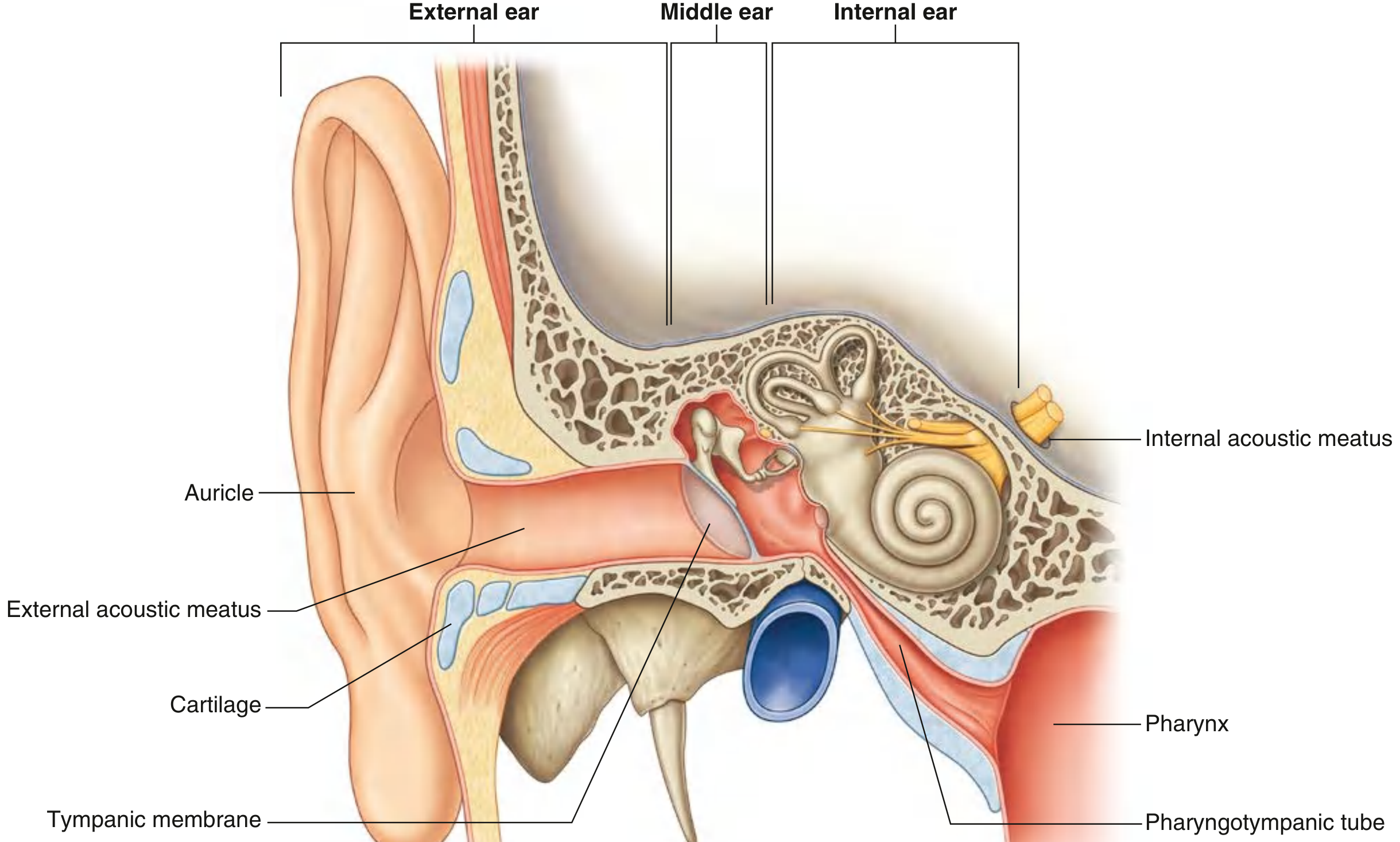
Fig. 8.116 Right Ear - Gray's Anatomy for Students. Three divisions are labeled: external ear (auricle + external acoustic meatus), middle ear (tympanic cavity), and internal ear (cochlea + labyrinth).
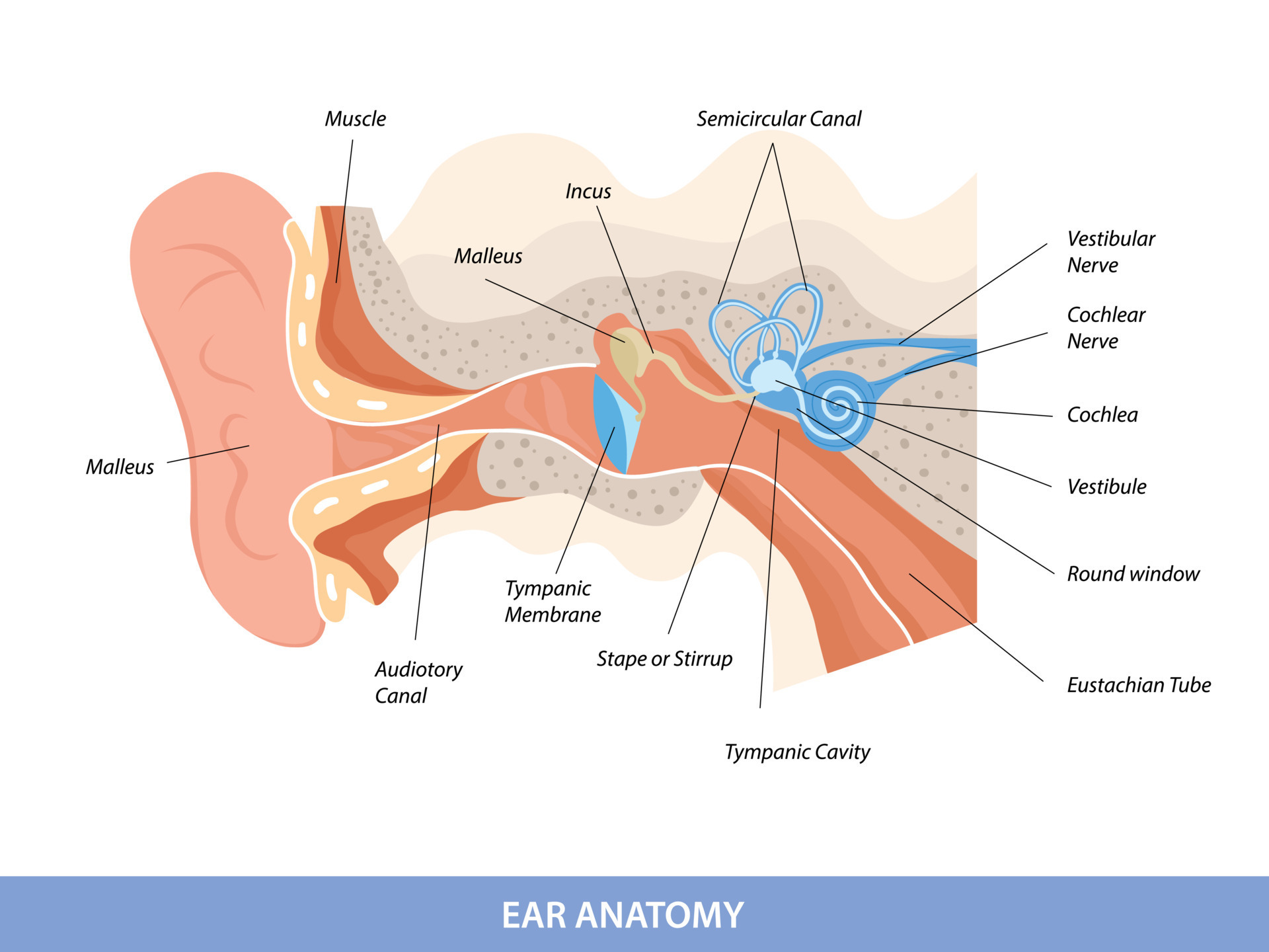
MIDDLE EAR
The middle ear lies within the petrous part of the temporal bone. It extends from the tympanic membrane (laterally) to the lateral wall of the inner ear (medially). Its primary role is to mechanically amplify and transmit sound vibrations from the tympanic membrane to the oval window of the inner ear.
Divisions of the Tympanic Cavity
The middle ear consists of two parts:
- Tympanic cavity proper - the main air-filled space
- Epitympanic recess - the superior extension above the level of the tympanic membrane, housing the head of the malleus and body of the incus
Boundaries (Six Walls)
| Wall | Name | Key Relationship |
|---|---|---|
| Roof | Tegmental wall (tegmen tympani) | Separates middle ear from middle cranial fossa |
| Floor | Jugular wall | Overlies the internal jugular vein |
| Lateral | Membranous wall | Largely formed by the tympanic membrane |
| Medial | Labyrinthine wall | Contains the oval window, round window, and promontory |
| Posterior | Mastoid wall | Communicates via aditus to mastoid antrum |
| Anterior | Carotid wall | Contains opening of pharyngotympanic tube; internal carotid artery is anterior |
Middle Ear - Detailed Anatomy
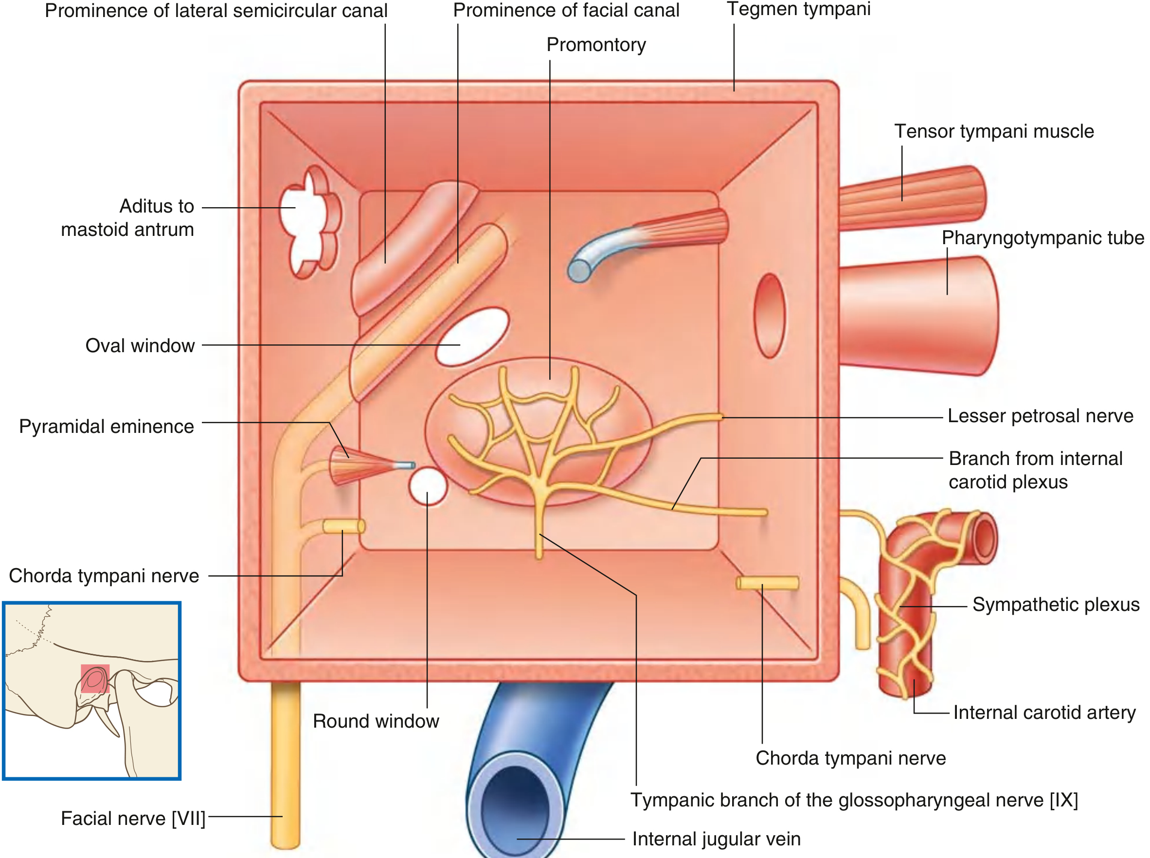
Fig. 8.123 Middle Ear Boundaries - Gray's Anatomy for Students. The medial (labyrinthine) wall shows the oval window, round window, promontory, facial canal prominence, chorda tympani nerve, and aditus to mastoid antrum.
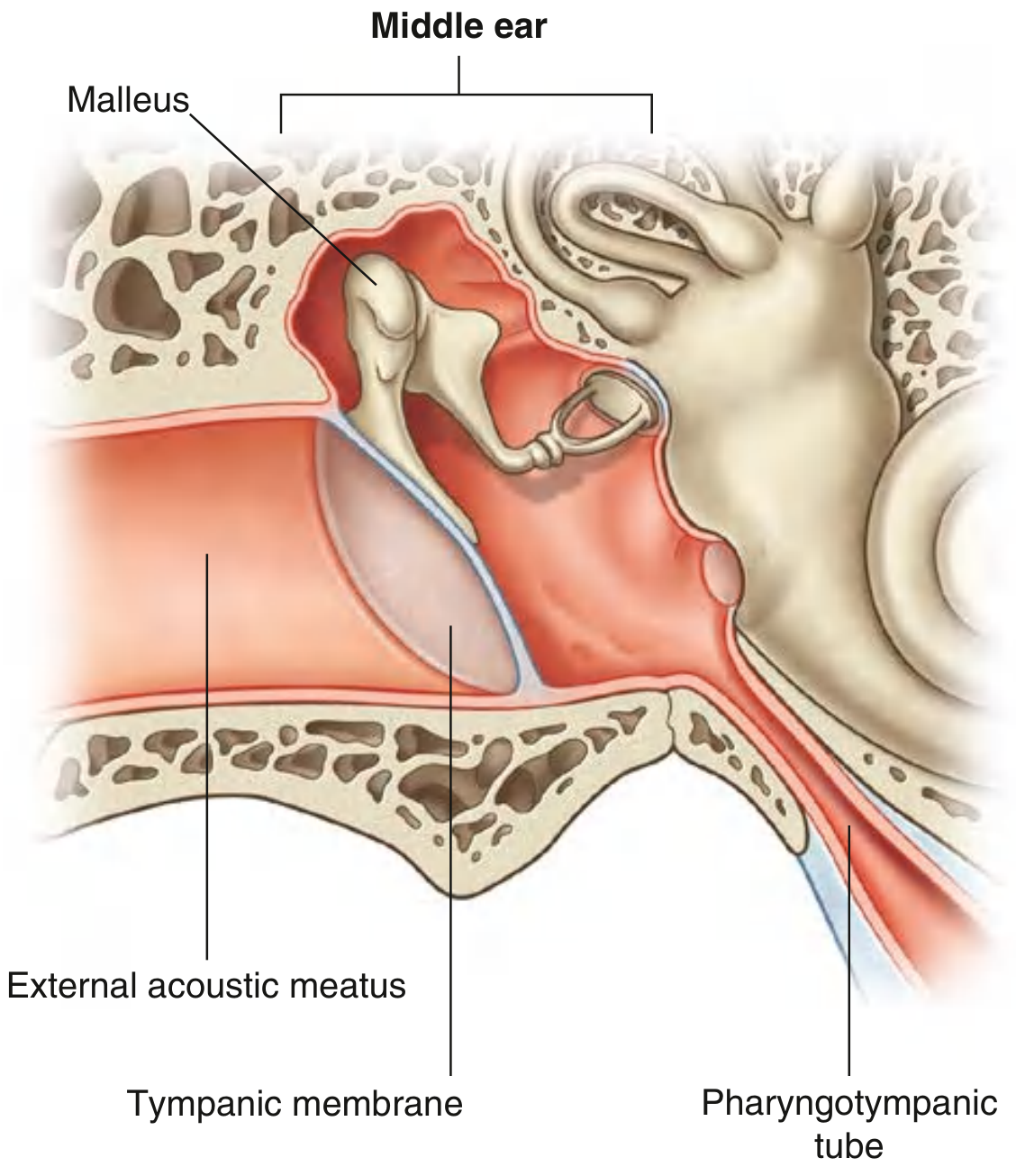
Fig. 8.120 Middle Ear - Gray's Anatomy for Students.
Tympanic Membrane
The tympanic membrane is an oblique, cone-shaped membrane separating the external acoustic meatus from the middle ear. It has three layers:
- Outer layer: skin (continuous with the canal)
- Middle: fibrous connective tissue core
- Inner layer: mucous membrane
Key landmarks on the tympanic membrane:
- Umbo - the deepest point, where the handle of the malleus attaches
- Handle (manubrium) of malleus - runs superiorly from the umbo
- Lateral process of malleus - produces a small bulge superiorly
- Cone of light - a bright anteroinferior reflection seen on otoscopy
- Pars tensa - the large, taut inferior portion
- Pars flaccida (Shrapnell's membrane) - the small, slack superior portion above the malleolar folds
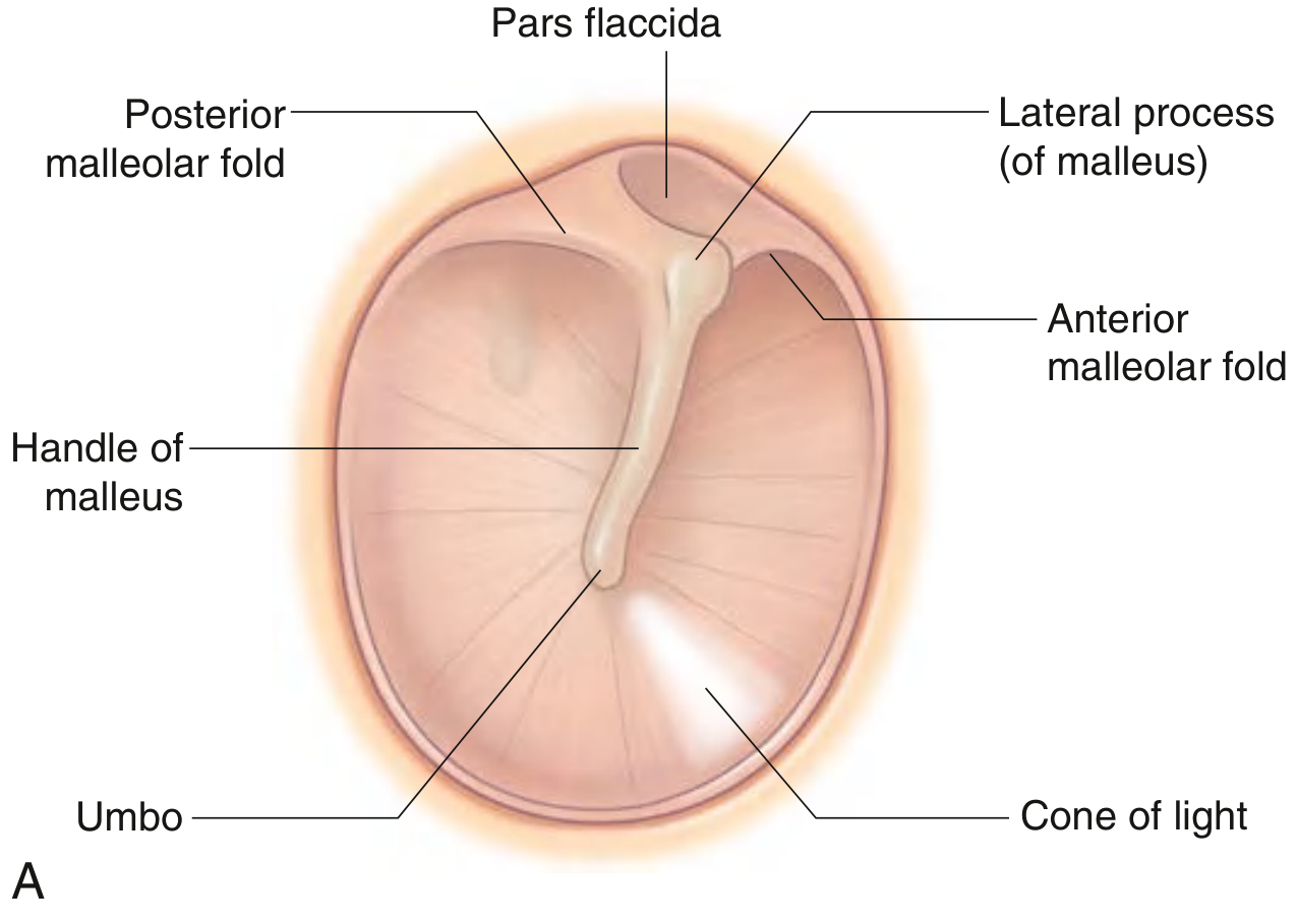
Fig. 8.121 Tympanic Membrane (Right Ear) - Gray's Anatomy for Students. The pars flaccida, pars tensa, handle of malleus, umbo, and cone of light are all labeled.
Innervation of the tympanic membrane:
- Outer surface: auriculotemporal nerve (CN V3), auricular branch of vagus (CN X)
- Inner surface: tympanic branch of glossopharyngeal nerve (CN IX)
Auditory Ossicles (The Three Bones)
Three small bones form a chain bridging the tympanic membrane to the oval window:
1. Malleus (Hammer)
The largest ossicle. Parts include:
- Head - in the epitympanic recess; articulates with the incus
- Neck - constriction below the head
- Anterior process - attached by ligament to the anterior wall
- Lateral process - attached to malleolar folds of the tympanic membrane
- Handle (manubrium) - embedded in the tympanic membrane
2. Incus (Anvil)
The middle ossicle. Parts include:
- Body - articulates with the head of the malleus; sits in the epitympanic recess
- Short limb - projects posteriorly, attached to the posterior wall by a ligament
- Long limb - descends parallel to the malleus handle, then bends medially to articulate with the stapes
3. Stapes (Stirrup)
The smallest bone in the human body. Parts include:
- Head - articulates with the long limb of the incus
- Anterior and posterior limbs (crura) - arch over to the oval base
- Base (footplate) - seated in the oval window on the labyrinthine wall
The ossicles are connected by synovial joints and amplify sound pressure ~22-fold as vibrations pass from the large tympanic membrane to the small oval window (impedance matching).
Muscles of the Middle Ear
Two small muscles protect against very loud sounds (acoustic reflex):
| Muscle | Origin | Insertion | Innervation | Action |
|---|---|---|---|---|
| Tensor tympani | Cartilaginous part of pharyngotympanic tube + greater wing of sphenoid | Handle of malleus | Medial pterygoid nerve (CN V3 via otic ganglion) | Pulls malleus medially, tensing tympanic membrane, dampening vibration |
| Stapedius | Pyramidal eminence (posterior wall) | Neck of stapes | Facial nerve (CN VII) | Pulls stapes posteriorly, stiffening ossicular chain; protective against loud sounds |
Pharyngotympanic (Eustachian/Auditory) Tube
- Connects the middle ear to the nasopharynx anteriorly
- Equalizes pressure across the tympanic membrane
- In children: shorter, wider, more horizontal - predisposing to otitis media
- Dysfunction leads to otitis media with effusion (glue ear)
Nerves and Vessels of the Middle Ear
- Chorda tympani (branch of CN VII): crosses the middle ear between the malleus and incus; carries taste from the anterior 2/3 of tongue and parasympathetic secretomotor fibers to submandibular and sublingual glands
- Tympanic plexus: formed by the tympanic branch of CN IX (Jacobson's nerve) on the medial wall; contributes to lesser petrosal nerve
- Blood supply: anterior tympanic artery (from maxillary artery), posterior tympanic artery (from stylomastoid artery)
INNER EAR
The inner ear (labyrinth) is entirely enclosed within the petrous part of the temporal bone. It has two functional roles: hearing (cochlea) and balance/equilibrium (vestibular apparatus).
It consists of two concentric layers:
- Bony labyrinth - a rigid bony cavity containing perilymph
- Membranous labyrinth - a soft inner tube suspended within the bony labyrinth, containing endolymph
Bony Labyrinth
The bony labyrinth has three parts:
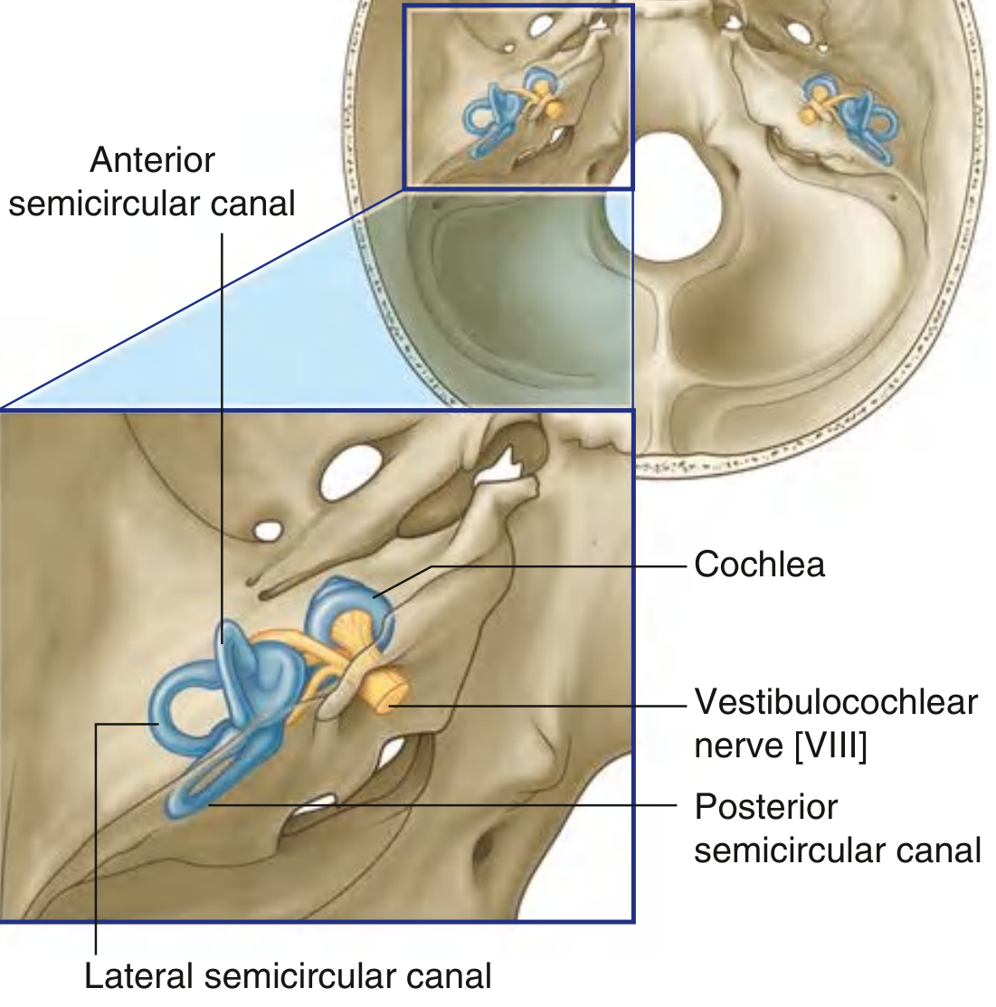
Fig. 8.130 Location of the Internal Ear in the Temporal Bone - Gray's Anatomy for Students. The anterior, posterior, and lateral semicircular canals are shown alongside the cochlea and vestibulocochlear nerve [VIII].
1. Vestibule
- Central part of the bony labyrinth
- Located medial to the middle ear, anterior to the semicircular canals, and posterior to the cochlea
- Lateral wall contains the oval window (fenestra vestibuli) - sealed by the stapes footplate
- The round window (fenestra cochleae) is on the lower part of the labyrinthine wall, sealed by the secondary tympanic membrane - this allows pressure equalization during ossicular motion
- Contains the utricle and saccule of the membranous labyrinth
- A narrow canal - the vestibular aqueduct - connects the vestibule to the posterior cranial fossa
2. Semicircular Canals
- Three bony canals: anterior (superior), posterior, and lateral (horizontal)
- Each forms about two-thirds of a circle, with one end expanded as an ampulla
- Oriented at right angles to each other so they detect rotation in all three planes
- Each canal opens into the vestibule at both ends (5 openings total - the anterior and posterior share a common crus)
3. Cochlea
- Coiled bony structure projecting anteriorly from the vestibule
- Winds 2.5 to 2.75 turns around a central bony pillar - the modiolus
- The modiolus contains the cochlear nerve fibers
- A thin bony shelf - the osseous spiral lamina - projects from the modiolus and supports the basilar membrane
- The cochlea has three fluid-filled compartments (scalae):
- Scala vestibuli (perilymph) - continuous with the vestibule; superior
- Scala media / cochlear duct (endolymph) - contains the organ of Corti; middle
- Scala tympani (perilymph) - ends at the round window; inferior
- Scala vestibuli and scala tympani communicate at the apex via the helicotrema
Membranous Labyrinth
The membranous labyrinth is the functional, endolymph-filled portion. It consists of:
Vestibular apparatus (balance):
- Utricle - detects horizontal linear acceleration and head tilts; connected to the semicircular ducts
- Saccule - detects vertical linear acceleration; connected to the cochlear duct
- Both have a macula containing hair cells embedded in an otolithic membrane with calcium carbonate crystals (otoliths/otoconia)
Semicircular ducts (within the bony canals):
- Each has a crista ampullaris in the ampulla - a ridge of hair cells embedded in a gel-like cupula
- Rotation of the head moves endolymph, deflecting the cupula, depolarizing hair cells
- The three ducts (oriented 90° to each other) signal angular acceleration in all planes
Cochlear duct (scala media):
- The organ of Corti sits on the basilar membrane within the cochlear duct
- Contains inner hair cells (1 row, ~3,500 cells - heavily innervated, primary sensory transducers) and outer hair cells (3-5 rows, ~12,000 cells - amplify and tune mechanical responses)
- Hair cell stereocilia are embedded in the overlying tectorial membrane
- Sound-induced basilar membrane vibration deflects stereocilia -> opens mechanically-gated ion channels -> depolarization -> signals transmitted via CN VIII to the brain
- Tonotopy: high frequencies are encoded at the base of the cochlea; low frequencies at the apex
Fluids of the Inner Ear
| Fluid | Location | Ionic Composition | Produced by |
|---|---|---|---|
| Perilymph | Bony labyrinth (scala vestibuli, scala tympani) | High Na⁺, low K⁺ (like ECF) | Filtrate from blood/CSF |
| Endolymph | Membranous labyrinth (cochlear duct, utricle, saccule, semicircular ducts) | High K⁺, low Na⁺ (like ICF) | Stria vascularis of cochlea |
The endocochlear potential (+80 mV in the cochlear duct) drives K⁺ into hair cells upon stereocilia deflection, powering the transduction current.
Innervation of the Inner Ear
The vestibulocochlear nerve (CN VIII) divides into:
- Cochlear nerve - from the spiral ganglion within the modiolus; carries auditory signals
- Vestibular nerve - from the vestibular ganglion (Scarpa's ganglion); carries balance signals from utricle, saccule, and semicircular ducts
Both divisions enter the internal acoustic meatus and proceed to the brainstem.
Sound Transmission Pathway (Summary)
Sound waves → External acoustic meatus → Tympanic membrane (vibrates) → Malleus → Incus → Stapes footplate → Oval window → Perilymph of scala vestibuli → Basilar membrane deflection → Organ of Corti hair cells → CN VIII (cochlear nerve) → Cochlear nuclei (brainstem) → Auditory cortex
The round window moves in the opposite direction to the oval window, acting as a pressure relief valve for the incompressible perilymph.
Sources: Gray's Anatomy for Students (9780323934237), Junqueira's Basic Histology 17e (9781264930395), TeachMeAnatomy, Medscape Ear Anatomy.
This is a shared conversation. Sign in to Orris to start your own chat.