
Callosities
Reading File
Searching sources...
Finding Sources
Finding Sources
Reading File
Reading File
Reading File
Reading File
Loading Image
Here is a comprehensive overview of Callosities from authoritative dermatology and surgery textbooks.
Callosities (Callus / Tyloma / Intractable Plantar Keratosis)
Synonyms
- Callosity, tyloma, intractable plantar keratosis
- Compare with corn (clavus / heloma): heloma durum (hard corn), heloma molle (soft corn)
Definition
A callosity is a raised, thickened patch of hyperkeratosis occurring in areas subject to excessive wear, tear, and repeated minor trauma. It is a nonpenetrating, circumscribed hyperkeratosis produced by pressure, occurring most commonly on the palms and soles - especially over bony prominences.
- S Das Manual of Clinical Surgery, 13th ed.
- Andrews' Diseases of the Skin, p. 52
Pathogenesis
Bony protuberances (condyles of metatarsal and phalangeal bones) create outward pressure on the skin. Footwear and physical activity (walking, running) create counterpressure at these same sites. The repeated friction and pressure leads to hyperkeratosis, which further increases local pressure - creating a self-perpetuating cycle of friction, pressure, and thickening until a callus develops.
Histologically: increased thickening of the epidermis, particularly the stratum corneum and the granular layer.
- Dermatology 2-Volume Set 5e, p. 1904
- S Das Manual of Clinical Surgery
Callus vs. Corn - Key Distinction
| Feature | Callus | Corn (Clavus) |
|---|---|---|
| Shape | Broad-based, diffuse thickening | Circumscribed, cone-shaped (apex inward) |
| Central core | Absent | Present (translucent, whitish-yellow) |
| Dermatoglyphics | Accentuated (preserved) | Interrupted |
| Pain | Usually mild | More painful (core presses on nerves) |
| Location | Soles, palms, pressure points | Dorsum of toes (hard); web spaces (soft) |
| Spontaneous regression | Yes, when pressure removed | Yes, when causative factor removed |
- Andrews' Diseases of the Skin, p. 52
- Dermatology 5e
Clinical Features
Feet:
Broad keratotic plaques, often under the metatarsal heads. The most common sites are the sole and heel.
Hands:
Form from repetitive occupational or recreational trauma - usually asymptomatic.
Occupation/Activity-specific callosities are important clinical stigmata:
- Surfer's nodules
- Boxer's knuckle pads
- Jogger's toe / jogger's nipple
- Rower's rump
- PlayStation thumb
- Milker's callus
- Tennis toe (tip of 2nd/3rd toe with transverse ridging and splinter hemorrhages in the nail - due to repeated shoe-tip collision)
- Prayer callus / the yoga sign
- Neck callosities of violinists
- Pillar knocker's knuckles
- Bowler's hand
- Russell sign - calluses/abrasions on the dorsum of the hand over the MCP and IP joints; a clue to bulimia nervosa
- Andrews' Diseases of the Skin, p. 52
Tennis Toe (Illustration)
This is an example of a sport-specific callus on the second toe:
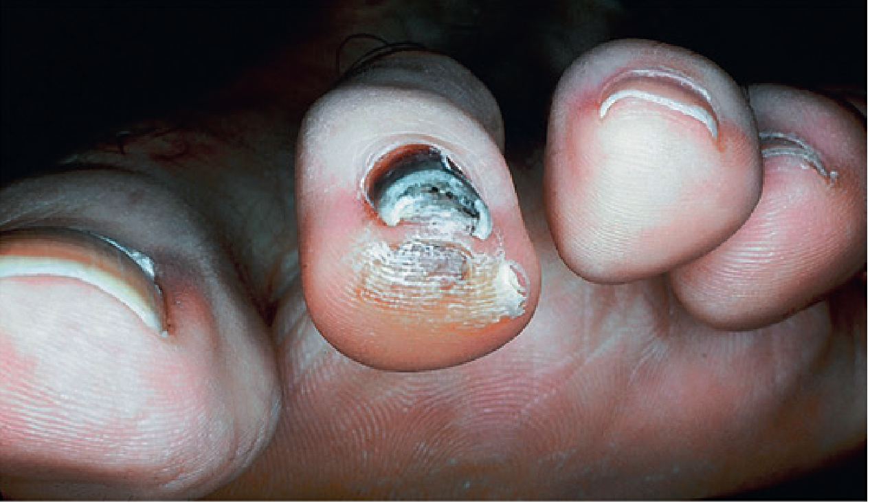
Fig. 3.33 Tennis toe - Andrews' Diseases of the Skin
Differential Diagnosis
The main differential is plantar wart (verruca plantaris):
| Feature | Callus | Plantar Wart |
|---|---|---|
| Dermatoglyphics | Accentuated | Interrupted |
| On paring | Layers of yellowish keratin | Multiple punctate intracorneal hemorrhages (bleeding points) |
| Pain | With perpendicular pressure | With lateral squeeze |
| Capillary loops | Absent | Visible as black dots |
- Dermatology 5e, p. 1904
Forensic Significance
Callosities have forensic value in occupational identification of unknown bodies or injury assessments: cuts, scars, callosities, and hyperkeratosis of the hands indicate manual labourers; needle puncture marks on the left index finger are seen in tailors; coal miners show characteristic "blue scars."
- Essentials of Forensic Medicine and Toxicology, 36th ed.
Treatment
Treatment is directed at symptomatic relief AND correction of the biomechanical cause:
- Paring of the callosity - provides prompt relief; paring a corn also removes its central core
- Filing after warm water soaking - can be self-performed periodically
- Keratolytics:
- 40% salicylic acid plasters
- 12% ammonium lactate lotion
- Urea-containing creams
- Padding - soft cushions (silicone sheet, sheepskin) to reduce friction
- Properly fitted footwear and orthotics
- If conservative measures fail: radiograph to look for exostoses (bony spurs) + orthopedic surgery referral for exostosis removal
- Andrews' Diseases of the Skin, p. 52
- Dermatology 5e
- S Das Manual of Clinical Surgery
Key Points Summary
- Callosity = diffuse hyperkeratosis from repeated friction/pressure, no central core
- Corn = circumscribed, cone-shaped with an inward-pointing core that causes pain
- Both disappear when the causative factor is removed
- Distinguish from plantar warts by dermatoglyphics and paring findings
- Occupation-specific callosities serve as clinical and forensic stigmata
- Russell sign (knuckle calluses) is a clue to bulimia nervosa
- Treatment: paring + keratolytics + correct footwear/biomechanics
Ibd
Searching sources...
Finding Sources
Finding Sources
Reading File
Reading File
Finding Sources
Finding Sources
Reading File
Reading File
Reading File
Finding Sources
Reading File
Reading File
Finding Sources
Finding Sources
Reading File
Finding Sources
Reading File
Finding Sources
Reading File
Loading Image
Finding Sources
Here is a comprehensive overview of Inflammatory Bowel Disease (IBD) compiled from multiple authoritative medical textbooks.
Inflammatory Bowel Disease (IBD)
Definition
IBD refers to two chronic idiopathic inflammatory disorders: Ulcerative Colitis (UC) and Crohn Disease (CD). No single clinical, endoscopic, or histologic finding is absolutely diagnostic for either alone. Approximately 10% of cases are labeled "IBD-undetermined" when Crohn colitis cannot be distinguished from UC.
- Goldman-Cecil Medicine, International Edition
Distribution Pattern - Key Diagram
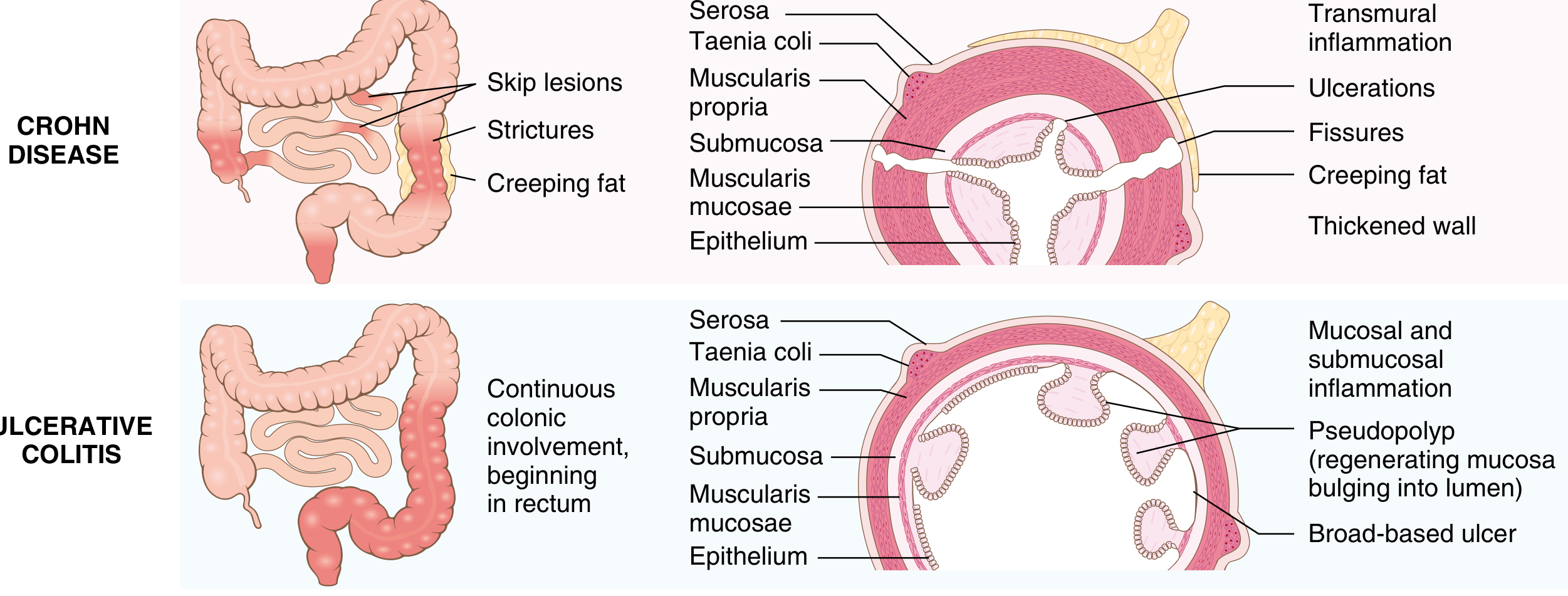
Fig. 17.35 - Distribution of lesions in IBD. Robbins & Cotran Pathologic Basis of Disease
Epidemiology
- Highest incidence: North America, UK, northern Europe
- Incidence is rising globally, including Africa, South America, and Asia
- Peak onset: 2nd to 4th decade (second peak in the 7th decade)
- No gender preference for either disease
- Highest risk group: White and Ashkenazi Jewish people of Eastern European descent
- UC incidence: ~19/100,000 person-years (North America); ~24/100,000 (Europe)
- CD incidence: ~20/100,000 person-years (North America); ~13/100,000 (Europe)
Environmental risk factors:
- Smoking: worsens Crohn disease but is protective in UC
- NSAIDs: associated with new-onset IBD and flares
- Appendectomy (for inflammatory indication): protective against UC development
- Hygiene hypothesis: improved sanitation and altered gut microbiome reduce development of regulatory immune responses
- Goldman-Cecil Medicine; Robbins & Cotran Pathology; Maingot's Abdominal Operations
Pathogenesis
IBD results from the interaction of three major pathways in genetically susceptible individuals:
-
Genetic predisposition - Over 200 IBD-associated risk alleles identified; however, they account for <20% of disease risk
- NOD2 (encodes intracellular sensor of bacterial muramyl dipeptide): most strongly associated with Crohn disease in European populations
- IL-10 / IL-10R mutations: cause monogenic "very early onset IBD" (infantile colitis)
- ~30% of IBD loci overlap with type 1 diabetes, ankylosing spondylitis, and psoriasis
-
Immune dysregulation - Dysregulated innate and adaptive immune responses involving dendritic cells, macrophages, CD4+ T cells, neutrophils, and pro-inflammatory cytokines (TNF-α, IL-6, IL-12, IL-23)
-
Host-microbiome interaction - Altered gut microbiota (dysbiosis) initiates and perpetuates mucosal inflammatory responses; antibiotic use that disrupts the microbiome increases IBD risk
- Robbins, Cotran & Kumar Pathologic Basis of Disease; Goldman-Cecil Medicine
Crohn Disease vs. Ulcerative Colitis - Comparative Table
| Feature | Crohn Disease | Ulcerative Colitis |
|---|---|---|
| Bowel region | Any GI tract (ileum + colon most common) | Colon only |
| Distribution | Skip lesions (discontinuous) | Diffuse, continuous from rectum |
| Rectal involvement | Spared in ~50% | Always involved (hallmark) |
| Wall appearance | Thick, "hose-pipe" | Normal thickness |
| Depth of inflammation | Transmural | Limited to mucosa/submucosa |
| Ulcers | Deep, knife-like, fissuring | Superficial, broad-based |
| Pseudopolyps | Moderate | Marked |
| Granulomas | Yes (~35%) | No |
| Fistulae/sinuses | Yes | No |
| Fibrosis/strictures | Marked | Rare |
| Serositis | Marked | Mild to none |
| Creeping fat | Yes | No |
| Perianal fistula | Yes (with colonic disease) | No |
| Fat/vitamin malabsorption | Yes (terminal ileum) | No |
| Toxic megacolon | No | Yes |
| Recurrence after surgery | Common | No (curative with colectomy) |
| Malignant potential | With colonic involvement | Yes (significant) |
- Robbins & Cotran Pathologic Basis of Disease, Table 17.8
Clinical Features
Ulcerative Colitis
- Relapsing-remitting course of bloody diarrhea with stringy mucoid material
- Lower abdominal cramping, temporarily relieved by defecation
-
50% of patients have clinically mild disease, but almost all experience at least one relapse in 10 years
- Historically ~30% required colectomy within 3 years; rate has fallen sharply with biologics
- Severe attack = toxic megacolon: colonic dilation >6 cm + systemic toxicity (fever, tachycardia, leukocytosis)
- Triggered by anticholinergics, antimotility agents, narcotics, antidepressants
Crohn Disease
- More variable presentation: chronic nonbloody diarrhea, abdominal pain, weight loss
- Perianal disease: fistulas, abscesses, skin tags
- Complications: obstruction (strictures), fistulas (enteroenteric, enterovesical, enterovaginal, enterocutaneous), abscesses
- Terminal ileal involvement leads to: B12 deficiency, fat malabsorption, bile salt malabsorption
Extraintestinal Manifestations (EIM)
About one-third of IBD patients develop EIMs. Having one EIM increases the risk of developing another. Many parallel intestinal activity and respond to IBD treatment.
| Organ System | Manifestations |
|---|---|
| Skin | Erythema nodosum, pyoderma gangrenosum |
| Eyes | Episcleritis, scleritis, uveitis |
| Joints | Peripheral arthritis, sacroiliitis |
| Spine | Ankylosing spondylitis |
| Liver/biliary | Primary sclerosing cholangitis (PSC - more with UC) |
| Bone | Osteoporosis |
| Vascular | DVT/PE (60% increased risk), cerebral sinus thrombosis, mesenteric ischemia |
| Neurologic | Peripheral neuropathy (immune-mediated, metronidazole toxicity, or vitamin deficiency) |
- Rosen's Emergency Medicine; Goldman-Cecil Medicine
Colorectal Cancer Risk in IBD
- UC: Cancer risk starts at 7-8 years of disease; rises ~10% per decade; can reach 34% at 30 years with universal colitis
- Risk correlates with: extent of disease (pancolitis > left-sided > proctitis) and duration
- Crohn disease with colonic involvement: 4-20x increased CRC risk
- Precursor lesion: dysplasia (low-grade → high-grade → invasive carcinoma)
- Dysplasia features: crypt architectural abnormalities, nuclear enlargement, hyperchromasia, pseudostratification ("picket fence" appearance), back-to-back glands
- Sleisenger & Fordtran's Gastrointestinal and Liver Disease
Treatment Overview
Step-Up Approach
Mild-to-moderate disease:
- 5-ASA (aminosalicylates): Mesalamine - first-line for mild-moderate UC (oral + rectal formulations); less effective in CD
- Corticosteroids: For induction of remission in moderate-severe flares; not used for maintenance
Moderate-to-severe disease:
- Immunomodulators: Azathioprine (AZA), 6-mercaptopurine (6-MP), methotrexate - for maintenance of remission
Biologic therapies:
- Anti-TNF agents: Infliximab, adalimumab, certolizumab (CD), golimumab (UC)
- Anti-integrin: Vedolizumab (gut-selective; blocks lymphocyte trafficking)
- Anti-IL-12/23: Ustekinumab
- JAK inhibitors: Tofacitinib (UC), upadacitinib
Antibiotics: Metronidazole, ciprofloxacin - for perianal Crohn disease or infectious complications
Surgery
- UC: ~40% of patients ultimately require surgery; proctocolectomy with ileal pouch-anal anastomosis (IPAA/J-pouch) is standard - colectomy is curative for intestinal disease
- CD: Surgery is not curative (recurrence is common); reserved for complications (obstruction, fistula, abscess, perforation, failure of medical therapy)
Differential Diagnosis
Key conditions to exclude:
- Infectious colitis (Salmonella, Campylobacter, C. difficile, Entamoeba)
- Irritable bowel syndrome (IBS)
- Ischemic colitis
- Radiation colitis
- Diverticular disease
- Colorectal cancer
- Appendicitis
Serologic markers for differentiation:
- p-ANCA (perinuclear antineutrophil cytoplasmic antibody): present in ~70% of UC
- ASCA (anti-Saccharomyces cerevisiae antibody): associated with Crohn disease
- Maingot's Abdominal Operations; Rosen's Emergency Medicine
Key Points
- IBD = UC + Crohn disease; 10% indeterminate
- UC: continuous, mucosal, rectum always involved; Crohn: skip, transmural, anywhere in GI tract
- NOD2 is the strongest single genetic risk factor for CD
- Smoking worsens CD but is protective in UC
- EIMs affect ~1/3 of patients; PSC is most closely linked to UC
- Dysplasia surveillance colonoscopy is mandatory in long-standing IBD due to CRC risk
- Biologics (anti-TNF, vedolizumab, ustekinumab) have dramatically reduced colectomy rates in UC
This is a shared conversation. Sign in to Orris to start your own chat.