I now have comprehensive textbook content and excellent surgical images from Kanski's. I have enough to write the full, detailed guide. Let me update the todos and compile:---
GLAUCOMA - Comprehensive Guide
1. DEFINITION
Glaucoma is a group of optic neuropathies characterised by:
- Progressive loss of retinal ganglion cells (RGCs) and their axons
- Characteristic optic disc cupping (loss of neuroretinal rim)
- Visual field defects corresponding to nerve fibre loss
- Usually associated with elevated intraocular pressure (IOP >21 mmHg), though not always
"Glaucoma is characterised by progressive loss of retinal nerve fiber layer tissue with corresponding visual field loss. The optic nerve acquires a characteristic loss of the neuroretinal rim, frequently referred to as 'cupping'." - Goodman & Gilman's, p.1480
Normal IOP = 10-21 mmHg. The damage is irreversible - once lost, ganglion cells cannot regenerate.
2. ETIOLOGICAL CLASSIFICATION
A. Primary Glaucoma (no identifiable underlying cause)
- Primary Open-Angle Glaucoma (POAG) - most common type worldwide
- Primary Angle-Closure Glaucoma (PACG) - common in Asians
- Normal-Tension Glaucoma (NTG) - IOP consistently <22 mmHg
- Ocular Hypertension (OHT) - elevated IOP without disc or field damage
B. Secondary Glaucoma (identifiable cause)
| Type | Mechanism |
|---|
| Pseudoexfoliative glaucoma | Exfoliative material clogs trabecular meshwork - commonest cause of secondary OAG |
| Pigmentary glaucoma | Iris pigment granules block TM |
| Neovascular glaucoma | New vessels from diabetes/CRVO occlude angle |
| Steroid-induced | Corticosteroids increase TM resistance |
| Uveitic glaucoma | Inflammatory cells + PAS block angle |
| Traumatic glaucoma | Angle recession, ghost cells |
| Lens-induced | Phacomorphic, phacolytic |
C. Developmental / Congenital Glaucoma
- Primary congenital (trabeculodysgenesis) - presents at birth
- Associated with Sturge-Weber syndrome, aniridia, Axenfeld-Rieger syndrome
3. CLINICAL CLASSIFICATION (Based on Angle Status)
The anterior chamber angle (gonioscopic appearance) is the cornerstone of clinical classification:
| Feature | Open-Angle | Angle-Closure |
|---|
| Gonioscopy | Trabecular meshwork visible | Angle obscured by iris |
| IOP rise mechanism | Increased TM resistance | Mechanical blockage of TM |
| Onset | Insidious, chronic | Can be acute, subacute, or chronic |
| Symptoms | Asymptomatic until late | Can be symptomatic (acute) |
| Pupil | Normal | Mid-dilated, fixed (acute) |
Shaffer Grading of Angle (Gonioscopy):
| Grade | Angle Width | Risk |
|---|
| 4 | 35-45° (wide open) | No closure risk |
| 3 | 25-35° | No closure risk |
| 2 | 20° | Possible closure |
| 1 | 10° | Probable closure |
| 0 | Closed | Closed angle |
Van Herick slit-lamp grading (without gonioscopy) is also used - peripheral anterior chamber depth compared to corneal thickness, from Kanski's Table 11.4.
4. PRIMARY OPEN-ANGLE GLAUCOMA (POAG)
Definition
A chronic, progressive optic neuropathy with open anterior chamber angles, raised IOP (>21 mmHg), characteristic disc cupping, and corresponding visual field loss - in the absence of any secondary cause.
Epidemiology & Risk Factors
- Most common type (accounts for ~74% of all glaucoma)
- Affects 1-2% of people over 40; prevalence rises with age
- Risk factors:
- IOP >21 mmHg (most modifiable factor)
- Age >40 years
- Family history (first-degree relative)
- African ancestry (higher prevalence and severity)
- Myopia (>5D)
- Diabetes mellitus
- Hypertension
- Thin central corneal thickness (<555 µm)
Pathophysiology
- Increased resistance to aqueous outflow through the trabecular meshwork (TM)
- Results in raised IOP → mechanical compression + ischaemia of optic nerve axons at lamina cribrosa
- Selective loss of large ganglion cells (type M) first
- Raised IOP causes axonal compression at the lamina cribrosa - the sieve-like plate at the optic nerve head
- Progressive retinal nerve fibre layer (RNFL) thinning → optic disc cupping → visual field loss
Clinical Features
Symptoms:
- Asymptomatic in early and moderate disease - "the silent thief of sight"
- Peripheral vision lost first (patient unaware - bilateral loss)
- Central vision preserved until late (Snellen acuity normal until advanced)
- No pain, no redness (distinguishes from acute angle closure)
- Terminal stage: tunnel vision → total blindness
Signs - Optic Disc Changes:
- Increased cup:disc ratio (CDR) >0.6 (normal CDR <0.5 in most)
- Asymmetric CDR between the two eyes (>0.2 difference is significant)
- Loss of neuroretinal rim (thinning, especially at inferior and superior poles - "ISNT rule" reversal)
- Disc haemorrhages (Drance haemorrhages) - splinter haemorrhages at disc margin
- Notching of the neuroretinal rim
- Nasal displacement of blood vessels
- RNFL defects visible on OCT or red-free photography
Signs - Intraocular Pressure:
- IOP >21 mmHg (but 30-40% of POAG have IOP in normal range - Normal Tension Glaucoma)
- IOP fluctuation is important
- Measured by Goldmann applanation tonometry
Signs - Visual Field:
See progressive sequence:
- Paracentral scotoma (early - near fixation, within 10-20° from center)
- Nasal step (Rönne's nasal step) - at horizontal meridian, nasal field
- Seidel's scotoma - elongation of blind spot
- Arcuate (Bjerrum) scotoma - arc-shaped scotoma following RNFL bundles from blind spot to nasal step
- Ring (double arcuate) scotoma - upper and lower arcuate join
- Tunnel vision - only central and temporal islands remain
- Total blindness
"Most important defects in glaucoma occur centrally - within a 30° radius from the fixation point." - Kanski's Clinical Ophthalmology
Signs - Gonioscopy:
- Open angle throughout
- May show increased TM pigmentation
Complications:
- Irreversible blindness (leading cause of irreversible blindness globally)
- Loss of quality of life, falls, driving incapacity
- Central vision loss (late)
5. PRIMARY ANGLE-CLOSURE GLAUCOMA (PACG)
Definition
Glaucoma with obstruction of aqueous outflow by apposition or adhesion of the peripheral iris to the trabecular meshwork (iridotrabecular contact - ITC in ≥3 quadrants), associated with glaucomatous optic neuropathy.
Epidemiology & Risk Factors
- More common in East Asians, Inuit, South Asians
- Predominantly affects women (4:1 ratio)
- Typically over 50 years
- Risk factors:
- Hyperopia (shallow anterior chamber, small crowded eye)
- Short axial length
- Large crystalline lens
- Narrow anterior chamber angle
- Large lens vault
- Plateau iris configuration
- Family history
Mechanism - Relative Pupillary Block (Most Common)
From Kanski's: "Failure of physiological aqueous flow through the pupil leads to a pressure differential between the anterior and posterior chambers, with resultant anterior bowing of the iris."
- Aqueous produced by ciliary body
- Cannot pass through narrow pupil (pupillary block)
- Pressure builds in posterior chamber > anterior chamber
- Peripheral iris bows forward (iris bombé)
- Peripheral iris apposes trabecular meshwork → blocks outflow
- IOP rises acutely
Other mechanisms: Plateau iris (thick peripheral iris), phacomorphic (large lens), lens subluxation.
Clinical Subtypes
| Subtype | Features |
|---|
| Primary angle-closure suspect (PACS) | Narrow angle on gonioscopy, no PAS, no disc/field damage |
| Primary angle closure (PAC) | Narrow angle + elevated IOP or PAS, but no glaucomatous damage |
| PACG | Narrow angle + glaucomatous optic neuropathy/field damage |
| Acute angle closure (AAC) | Sudden complete closure - ophthalmic emergency |
ACUTE ANGLE CLOSURE - Clinical Features (Emergency)
Symptoms:
- Sudden, severe eye pain (often excruciating)
- Blurred vision with coloured halos around lights (corneal oedema)
- Headache, nausea, vomiting (may mimic GI emergency or migraine)
- Photophobia
Signs:
- Ciliary flush (circumcorneal redness) + conjunctival injection
- Corneal oedema - hazy, steamy cornea
- Shallow anterior chamber (anterior chamber angle closed)
- Mid-dilated, oval, fixed pupil (ischaemia of iris sphincter)
- Elevated IOP - often 40-80 mmHg
- Disc hyperaemia (early) → cupping (if prolonged)
- Fellow eye typically has a shallow chamber (risk factor)
Complications of acute AAC:
- Glaukomflecken - grey-white anterior subcapsular lens opacities (pathognomonic of previous acute attack)
- Permanent optic nerve damage
- Peripheral anterior synechiae (PAS)
- Iris atrophy
- Chronic angle-closure
CHRONIC ANGLE CLOSURE - Clinical Features
- Gradual, insidious progression (similar to POAG but with narrow angle)
- Extensive PAS formation over time
- May present as advanced glaucoma without acute attack history
- More common in Asia
6. INVESTIGATIONS / INVESTIGATIVE PROCEDURES
1. Tonometry - IOP Measurement
Goldmann Applanation Tonometry (GAT) - Gold Standard
- Measures the force required to flatten (applanate) a 3.14 mm area of cornea
- Accounts for corneal curvature
- IOP values influenced by central corneal thickness (CCT) - thin corneas underestimate IOP
- Normal: 10-21 mmHg
Other methods: Non-contact tonometry (air-puff), Icare rebound tonometry, Perkins (portable), Schiotz (indentation - now rarely used)
2. Gonioscopy (Assessment of Anterior Chamber Angle)
The single most important investigation to classify glaucoma.
- Uses a gonioscopic contact lens + slit lamp
- Directly visualises the anterior chamber angle structures:
- Schwalbe's line (anterior border of TM)
- Trabecular meshwork (non-pigmented and pigmented)
- Scleral spur
- Ciliary body band
- Iris
- Van Herick grade (screening without contact lens) assesses peripheral AC depth
- Identifies open vs. closed angle, PAS, neovascularisation, pigment, recession
3. Optic Disc Assessment
Direct ophthalmoscopy / Slit-lamp biomicroscopy with 78D or 90D lens:
- CDR measurement (vertical > horizontal is significant)
- Neuroretinal rim assessment (ISNT rule: Inferior > Superior > Nasal > Temporal thickness - normally)
- Disc haemorrhages
- Disc margin: sharp or blurred
Optic Disc Photography (stereo disc photos):
- Baseline documentation
- Serial comparison to detect progression
OCT (Optical Coherence Tomography):
- Measures RNFL thickness (retinal nerve fibre layer) - most sensitive early test
- Detects RNFL loss before visual field changes (structural before functional)
- Ganglion Cell Complex (GCC) analysis
- Optic disc cube - CDR, rim area, disc area
- HRT (Heidelberg Retinal Tomograph) - alternative for disc topography
4. Visual Field Testing (Perimetry)
Automated Static Perimetry (Humphrey Visual Field Analyser) - Gold Standard:
- Most commonly used: 24-2 pattern (tests 24° temporal, 30° nasal)
- 30-2 pattern: broader field testing
- 10-2 pattern: central 10° - for advanced glaucoma monitoring
Key indices:
- Mean Deviation (MD): overall field loss compared to age-matched normals
- Pattern Standard Deviation (PSD): localised defect
- Visual Field Index (VFI): % of normal visual function
- Glaucoma Hemifield Test (GHT): compares superior and inferior hemifields
Progression analysis: GPA (Guided Progression Analysis) identifies rate of field loss.
"24-2 is a routinely used glaucoma-orientated pattern. The 10-2 pattern facilitates more detailed monitoring of the extent of damage, especially in advanced glaucoma." - Kanski's
Goldmann kinetic perimetry - used for advanced/peripheral field assessment.
5. Central Corneal Thickness (Pachymetry)
- Thin CCT (<555 µm) → underestimates IOP, independent risk factor for POAG progression
- Thick CCT → overestimates IOP
6. Ultrasound Biomicroscopy (UBM) & Anterior Segment OCT (AS-OCT)
- High-resolution imaging of anterior segment
- Identifies angle configuration, iris plateau, lens position
- Particularly useful in PACG assessment
7. Diurnal IOP Curve
- IOP measured at multiple time points in a single day
- IOP typically peaks in early morning
- Peak-to-trough variation >8 mmHg is significant
7. MEDICAL MANAGEMENT OF GLAUCOMA
Goal: Reduce IOP to a "target IOP" that prevents further optic nerve damage.
- Target IOP = usually 30-50% reduction from baseline IOP
- "Reduction of IOP by at least 30% appears to have the best chance of preventing further optic nerve damage." - Wills Eye Manual
Stepwise approach: Start with monotherapy, add agents if inadequate control.
A. PROSTAGLANDIN ANALOGUES (First-Line)
Drugs: Latanoprost 0.005%, Bimatoprost 0.01%/0.03%, Travoprost 0.004%, Tafluprost 0.0015%
Mechanism:
- Bind to FP (prostaglandin F₂α) receptors → activate G-protein-linked PLC-IP₃-Ca²⁺ pathway
- Increase uveoscleral (non-conventional) outflow by remodelling extracellular matrix of ciliary muscle (matrix metalloproteinase release)
- Latanoprostene bunod also donates nitric oxide → relaxes TM cytoskeleton → also increases conventional outflow
IOP reduction: 25-35% (most potent class)
Dosing: Once daily at bedtime (q.h.s.)
Side effects:
- Increased iris pigmentation (irreversible in hazel/mixed-colour eyes)
- Hypertrichosis - lengthening, thickening, darkening of lashes
- Periorbital hyperpigmentation and fat atrophy (deepening of upper lid sulcus)
- Conjunctival hyperaemia (bimatoprost > latanoprost)
- Anterior uveitis, cystoid macular oedema (use caution in at-risk eyes)
- Contraindicated in pregnancy
"Due to their once-daily dosing, low incidence of systemic side effects, and potent IOP-lowering effect, PG analogues have largely replaced β-adrenergic receptor antagonists as first-line medical therapy for glaucoma." - Goodman & Gilman's
B. TOPICAL BETA-BLOCKERS
Drugs:
| Drug | Selectivity | Dosing |
|---|
| Timolol 0.25%, 0.5% | Non-selective (β₁ + β₂) | Once or twice daily |
| Levobunolol 0.5% | Non-selective | Once or twice daily |
| Carteolol 1%, 2% | Non-selective (with ISA) | Twice daily |
| Betaxolol 0.5% | β₁-selective | Twice daily (less effective) |
Mechanism:
- Block β₂ receptors on ciliary body epithelium → reduce intracellular cAMP → decrease aqueous humour production (by ~40-50%)
- Also may decrease ocular blood flow → reduced ultrafiltration
IOP reduction: 20-25%
Side effects (local):
- Ocular hyperaemia, dry eye, punctate keratopathy
Side effects (systemic) - most important:
- Bronchoconstriction (contraindicated in asthma, COPD) - β₂ blockade of lungs
- Bradycardia, heart block, hypotension - β₁ blockade of heart
- Worsening of congestive heart failure
- Masking of hypoglycaemia in diabetics
- CNS: depression, fatigue, impotence
- Betaxolol (β₁-selective) has less pulmonary risk but also less IOP reduction
Contraindications: Asthma, COPD, 2nd/3rd degree heart block, sinus bradycardia, decompensated heart failure, myasthenia gravis.
C. CARBONIC ANHYDRASE INHIBITORS (CAIs)
Drugs:
Topical: Dorzolamide 2%, Brinzolamide 1%
Systemic (oral): Acetazolamide 250 mg (tabs) / 500 mg (SR capsules); Methazolamide 25-50 mg
Mechanism:
- Inhibit carbonic anhydrase (CA-II and CA-IV) in ciliary body epithelium
- Reduces production of HCO₃⁻ and H⁺ ions needed for aqueous humour secretion
- Decreases aqueous production by 40-60%
IOP reduction:
- Topical: 15-20%
- Oral acetazolamide: 25-30% (more potent; used for acute angle closure)
Topical side effects:
- Local stinging, burning
- Bitter/metallic taste (brinzolamide < dorzolamide)
- Superficial punctate keratopathy
Systemic (acetazolamide) side effects:
- Metabolic acidosis (bicarbonate depletion)
- Hypokalaemia
- Paraesthesia (tingling of fingers/toes) - very common
- Renal calculi (uric acid/calcium stones)
- Anorexia, nausea, malaise
- Aplastic anaemia (rare but serious)
- Contraindicated in sulfa allergy, sickle cell disease, hepatic or renal failure
Use in acute angle closure: Oral/IV acetazolamide is used urgently to reduce IOP rapidly.
D. ADRENERGIC DRUGS (Alpha-2 Agonists)
Drugs: Brimonidine 0.1%, 0.15%, 0.2%; Apraclonidine 0.5%, 1%
Mechanism:
- Selective α₂-adrenergic receptor agonists
- Dual mechanism:
- Decrease aqueous production (pre-synaptic α₂ reduces NE release, activates postsynaptic α₂ on ciliary body)
- Increase uveoscleral outflow (possibly via local PG production)
- Brimonidine is lipophilic → good corneal penetration; crosses blood-brain barrier → neuroprotective effects under investigation
- Apraclonidine is hydrophilic → does not cross BBB, used for short-term only
IOP reduction: 18-25%
Dosing: 2-3 times daily
Side effects:
- Local allergic/follicular conjunctivitis (especially apraclonidine - up to 50% with long-term use; brimonidine ~10-15%)
- Ocular hyperaemia, eyelid retraction
- Systemic (brimonidine crossing BBB): Dry mouth, fatigue, drowsiness, headache
- Infants: Brimonidine causes severe CNS depression/apnoea - absolutely contraindicated in children <2 years (neonates/infants)
- Contraindicated with MAO inhibitors
E. MIOTICS - PILOCARPINE
Drug: Pilocarpine 1%, 2%, 4% (eye drops); also available as 4% gel
Mechanism:
- Muscarinic M3 receptor agonist (parasympathomimetic/cholinergic)
- Stimulates the ciliary muscle → contracts → pulls scleral spur → opens trabecular meshwork pores → increases conventional (trabecular) aqueous outflow
- Miosis (pupil constriction) via sphincter pupillae contraction → pulls peripheral iris away from angle → useful in angle closure
- In angle closure: pilocarpine contracts sphincter → breaks pupillary block → narrows pupil → pulls iris from angle
IOP reduction: 20-25%
Dosing: 3-4 times daily (short duration of action)
Side effects:
- Miosis → dim vision (especially in dark), difficulty driving at night
- Induced myopia - ciliary muscle spasm causes accommodation
- Browache/headache - ciliary muscle spasm (especially in young patients)
- Retinal detachment risk - pilocarpine-induced miosis pulls on vitreous (caution in high myopes and peripheral retinal degeneration)
- Increased salivation, sweating (systemic if absorbed)
- Rarely: posterior synechiae with prolonged use in inflamed eyes
Current use: Mainly second/third line. Historically first-line but replaced by PG analogues and beta-blockers due to side effects. Still important in acute angle closure (pilocarpine 2-4% to break attack once IOP is partially reduced and pupil responds).
Contraindications: Uveitis (risk of posterior synechiae), high myopia, previous retinal detachment.
Drug Class Summary Table
| Drug Class | Mechanism | IOP Reduction | Key S/E | Dosing |
|---|
| Prostaglandin analogues | ↑ uveoscleral outflow | 25-35% | Iris pigment, lash growth | Once daily (PM) |
| Beta-blockers | ↓ aqueous production | 20-25% | Bronchoconstriction, bradycardia | BD |
| CAI (topical) | ↓ aqueous production | 15-20% | Stinging, metallic taste | TDS |
| CAI (oral) | ↓ aqueous production | 25-30% | Metabolic acidosis, paraesthesia | BD-QDS |
| Alpha-2 agonists | ↓ production + ↑ uveoscleral | 18-25% | Allergic conjunctivitis, CNS in infants | BD-TDS |
| Pilocarpine | ↑ trabecular outflow + miosis | 20-25% | Browache, dim vision, myopia | QDS |
Fixed combinations (to improve compliance): Timolol + dorzolamide (Cosopt), Timolol + brimonidine (Combigan), Timolol + latanoprost (Xalacom).
8. SURGICAL MANAGEMENT
A. PERIPHERAL IRIDECTOMY (PI) / LASER PERIPHERAL IRIDOTOMY (LPI)
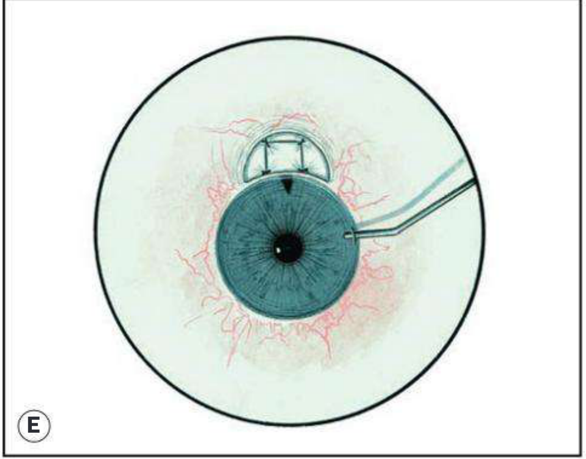
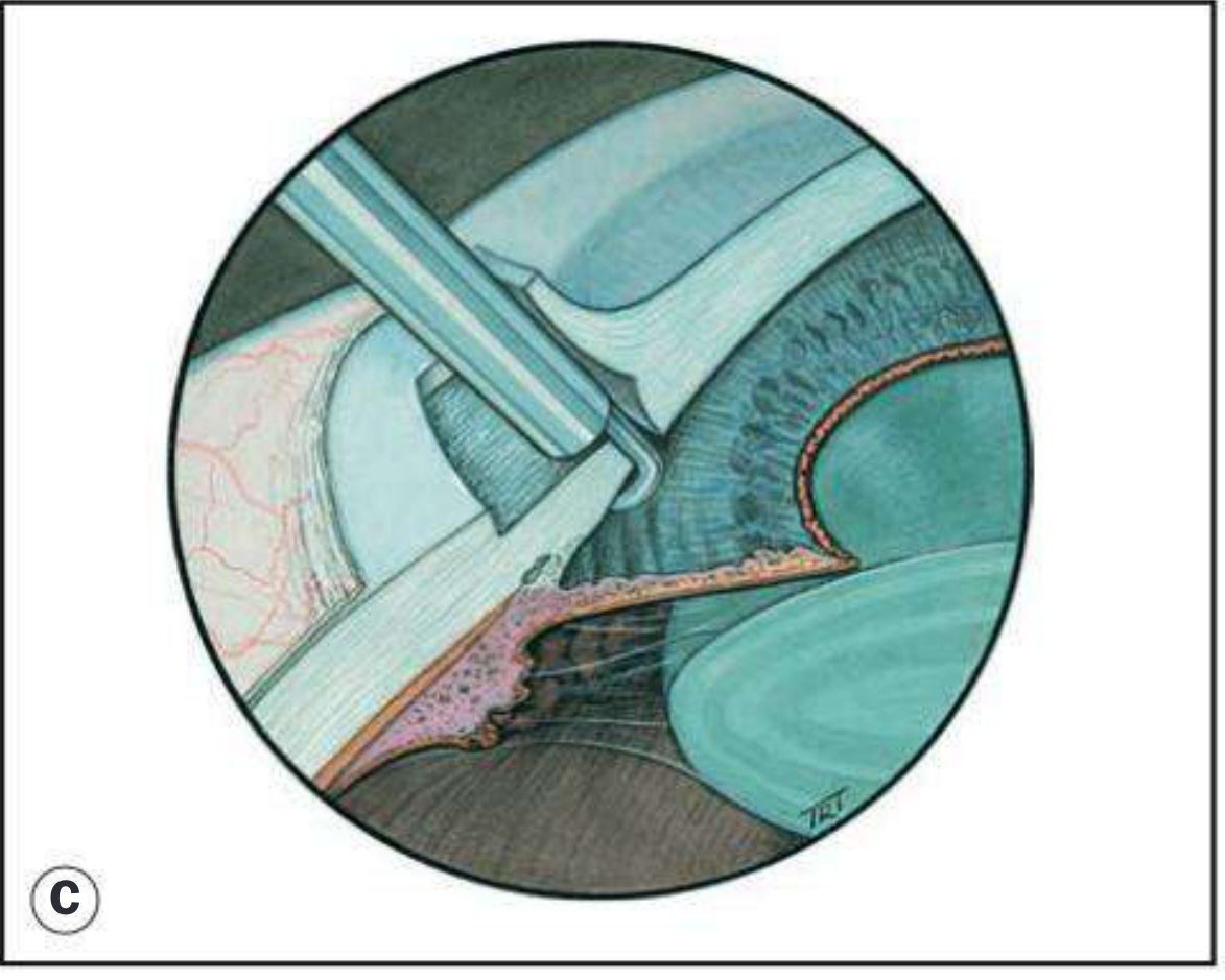
Fig. 11.72(D) - Peripheral iridectomy during trabeculectomy (Kanski's)
Intraoperative view of trabeculectomy with peripheral iridectomy
Indication:
- Primary treatment for angle-closure glaucoma with pupillary block
- Prophylaxis in fellow eye after acute angle closure
- Acute angle-closure glaucoma (after IOP is medically controlled)
- Chronic PACG, narrow angle (PACS/PAC)
Mechanism:
- Creates a full-thickness hole in the peripheral iris → establishes direct communication between posterior and anterior chambers → equalises pressure differential → iris falls back → angle opens
Laser PI (Nd:YAG LPI) - Preferred Method:
- Anaesthetic drops instilled
- Pre-treatment with pilocarpine 2% (to thin the iris) and apraclonidine 0.5-1% (to prevent IOP spike)
- Via a Abraham or Wise iridotomy lens, Nd:YAG pulses delivered to thin/crypt area of iris (usually superior, at 10-2 o'clock position, covered by upper lid)
- Full-thickness hole created through iris stroma and pigment epithelium
- Confirmed by visualising flow of pigment into anterior chamber
- Post-laser: apraclonidine, topical steroids for 1 week, review at 1 hour and 1 week
Surgical (Incisional) Peripheral Iridectomy:
- Performed under LA via limbal incision
- Iris delivered, excised with scissors, reposited
- Now rarely performed alone (usually incorporated into trabeculectomy or cataract surgery)
Outcome:
- "Usually anatomically relieved by peripheral iridotomy, which equalizes anterior and posterior chamber pressure." - Kanski's
- Highly effective for pupillary block mechanism
- Less effective if substantial PAS already formed (trabecular damage persists)
- Fellow eye should be treated prophylactically
B. TRABECULECTOMY (Filtration Surgery)
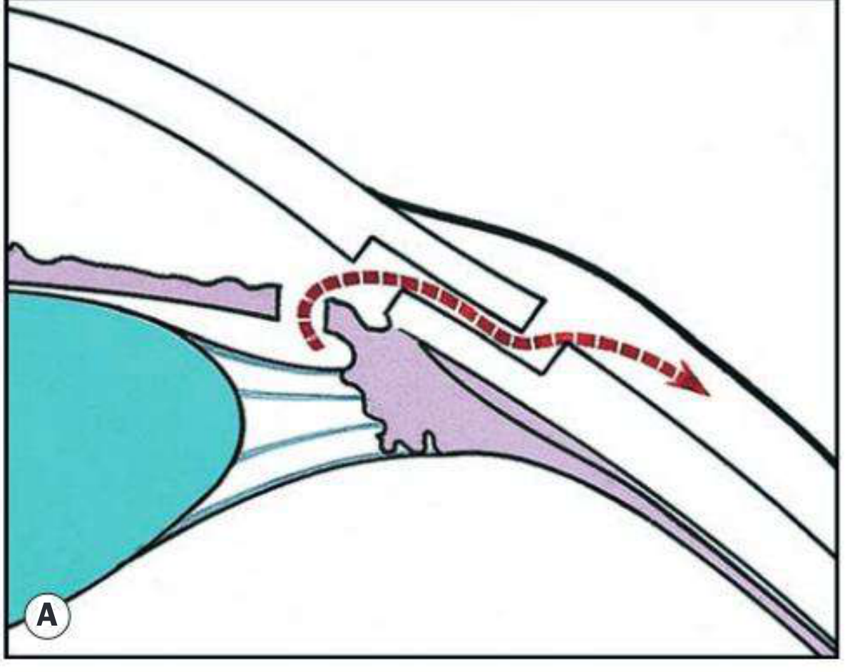
Fig. 11.71A - Pathway of aqueous egress following trabeculectomy (Kanski's)
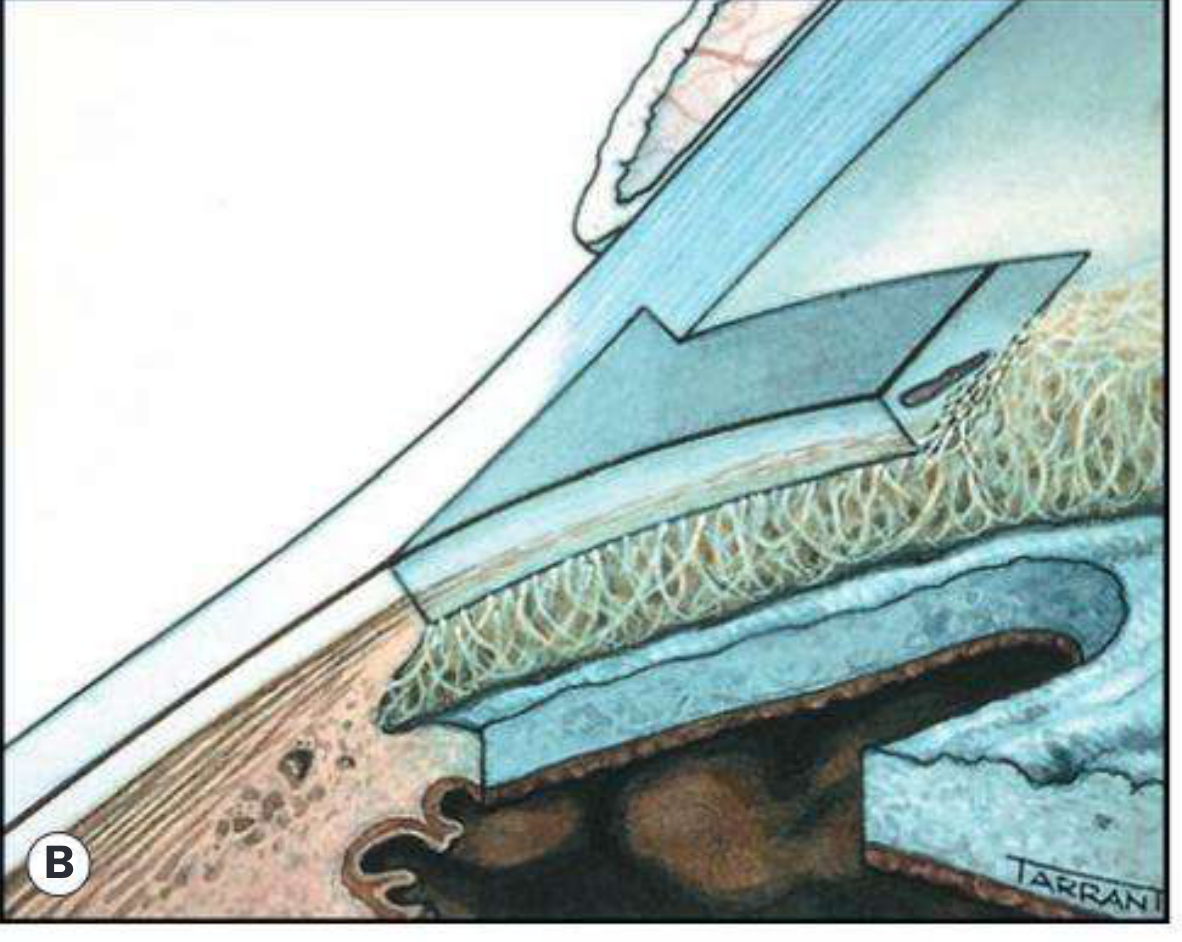
Fig. 11.71B - Schematic cross-section showing scleral flap and sclerostomy (Kanski's)
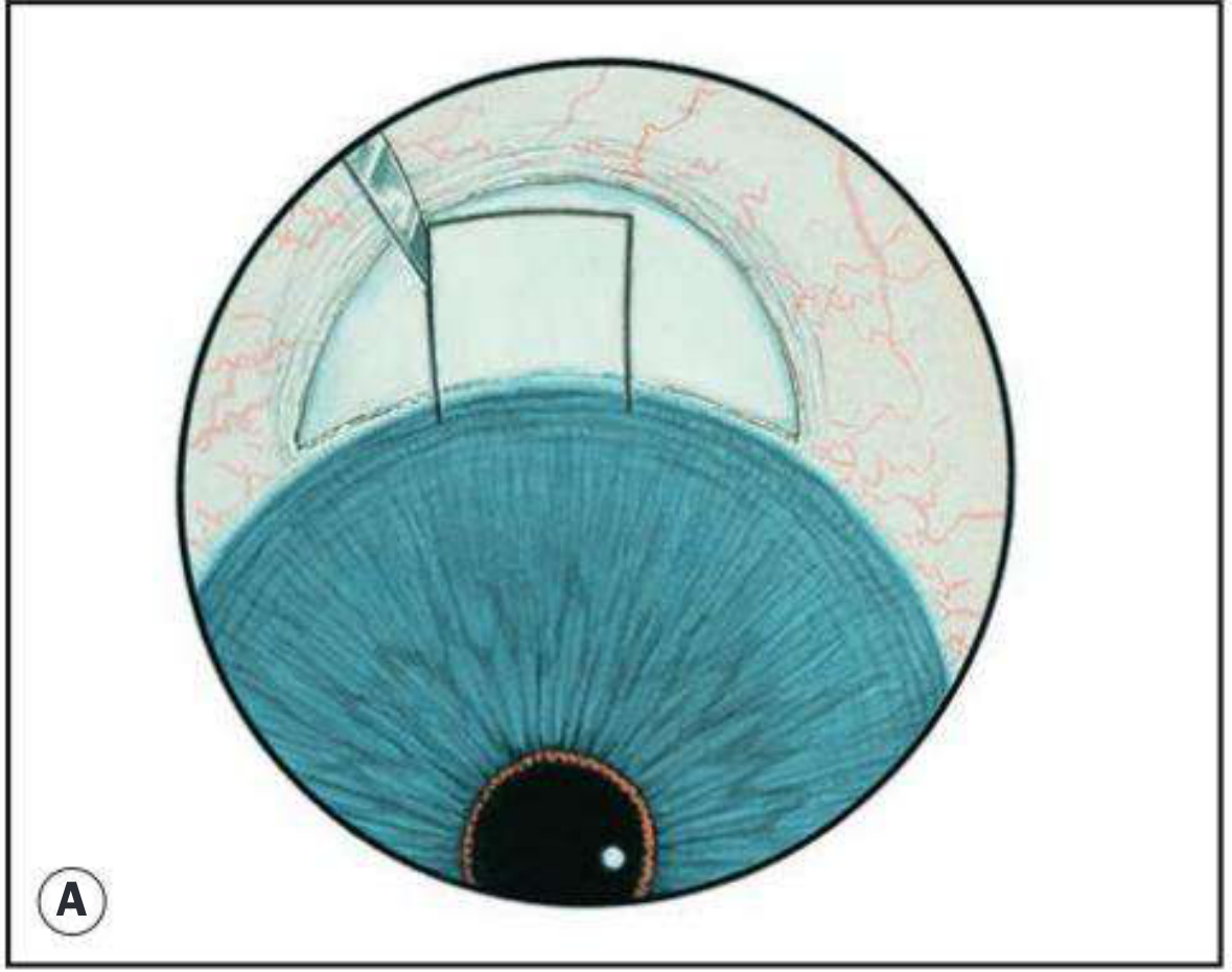
Fig. 11.72C - Peripheral iridectomy step during trabeculectomy (Kanski's)
Indication:
- POAG or PACG with inadequate IOP control on maximum medical therapy
- Advanced glaucoma with rapid progression
- Poor compliance with drops
- May be first-line for very advanced disease at presentation
Principle:
Creates a new drainage pathway bypassing the blocked trabecular meshwork - aqueous flows from the anterior chamber through a surgical opening, under a scleral flap, and into the subconjunctival space (bleb), where it is absorbed.
Technique:
- Conjunctival flap created - either limbus-based (incision at limbus) or fornix-based (incision at fornix)
- Partial-thickness superficial scleral flap dissected (3-4 mm x 3 mm, one-half to one-third scleral thickness) toward the limbus
- Antimetabolites applied (see below): Mitomycin C (MMC) or 5-Fluorouracil (5-FU) on sponge under conjunctiva to prevent fibrosis
- Deep sclerectomy punch (Kelley punch or Maquet punch) removes a block of deep sclera, TM, and inner wall of Schlemm's canal beneath the flap - creates the sclerostomy
- Peripheral iridectomy performed (to prevent iris incarceration into sclerostomy)
- Balanced salt solution injected to confirm flow
- Scleral flap is sutured (releasable sutures allow post-op bleb titration)
- Conjunctiva closed watertight
Post-operative management:
- Topical steroids (4x daily) and antibiotics
- Avoid strenuous activity
- Bleb massage - to improve filtration
- Laser suture lysis or suture removal to adjust IOP
- Atropine 1% drop (if no iridectomy, pilocarpine 2% used)
Antimetabolites (Adjuncts):
- Mitomycin C (MMC) - DNA cross-linker, potent antiproliferative; applied 0.2-0.5 mg/mL for 1-5 minutes under the scleral flap; preferred for higher-risk eyes (secondary glaucoma, young patients, repeat surgery)
- 5-Fluorouracil (5-FU) - antimetabolite, less potent than MMC; used as subconjunctival injections post-op or intra-operatively
Risk factors for surgical failure:
- Previous failed trabeculectomy or MIGS
- Previous conjunctival or cataract surgery
- Secondary glaucoma (inflammatory, neovascular, post-traumatic)
- Young patients (more fibrosis)
- Afro-Caribbean descent (higher failure rate)
Complications:
| Early | Late |
|---|
| Hypotony (over-drainage) | Bleb failure/fibrosis |
| Choroidal detachment | Blebitis/endophthalmitis |
| Shallow anterior chamber | Late hypotony |
| Hyphaema | Cataract progression |
| Wound leak (positive Seidel test) | Bleb dysaesthesia |
| Malignant glaucoma (ciliolenticular block) | Symptomatic bleb |
Malignant Glaucoma (Aqueous Misdirection):
"A rare but serious complication of trabeculectomy in patients with primary angle closure... caused by anterior rotation of the ciliary processes and iris root (ciliolenticular block) leading to posterior misdirection of aqueous." - Kanski's
- Signs: Shallow AC, high IOP, absent bleb, negative Seidel test
- Treatment: Atropine 1% + phenylephrine 10% → IV mannitol → Nd:YAG disruption of anterior hyaloid → pars plana vitrectomy
C. CYCLODESTRUCTIVE PROCEDURES
Principle: Destroy a portion of the ciliary body (ciliary epithelium) to reduce aqueous humour production - a destructive, non-filtration approach to IOP reduction.
Indications:
- Refractory/end-stage glaucoma where other methods have failed
- Blind or painful eye where IOP control is needed for comfort
- Eyes where filtration surgery has repeatedly failed
- Neovascular glaucoma
- Aphakic/pseudophakic glaucoma
1. Trans-Scleral Cyclophotocoagulation (TS-CPC) / Cyclodiode Laser
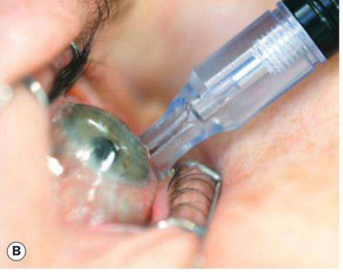
Fig. 11.68B - Cyclodiode probe during laser application (Kanski's)
- Most commonly used cyclodestructive procedure
- Uses an 810 nm semiconductor diode laser delivered trans-sclerally via a G-probe (optic fibre probe)
- Energy selectively absorbed by melanin in ciliary pigment epithelium → coagulation necrosis of ciliary processes → reduced aqueous production
- Probe placed 1.2-1.5 mm posterior to limbus (overlying the ciliary body)
- Technique: Under LA or GA, 15-25 applications of 1500-2000 mW for 1.5-2 seconds, spaced around the globe (avoiding the 3 and 9 o'clock positions where long ciliary nerves/vessels run)
- Treat about 180-270° of the circumference to avoid excessive hypotony
Complications:
- Hypotony and phthisis bulbi (over-treatment) - most serious
- Chronic uveitis
- Sympathetic ophthalmitis (rare)
- Visual acuity reduction (often limited by underlying disease anyway)
- Suprachoroidal haemorrhage
- Cystoid macular oedema
2. Endoscopic Cyclophotocoagulation (ECP)
- Laser applied directly under endoscopic vision to ciliary processes through a limbal or pars plana incision
- More precise, less collateral damage than TS-CPC
- Fibrinous uveitis, hyphaema, and CME in ~5%
- Often combined with cataract surgery (phaco-ECP)
3. Cyclocryotherapy (Cyclocryocoagulation)
- A nitrous oxide or liquid nitrogen cryoprobe (-80°C) applied trans-sclerally to freeze ciliary body
- Destroys ciliary epithelium via freeze-thaw cycles
- Largely replaced by cyclodiode due to higher complication rates
- More collateral damage, greater risk of hypotony and phthisis
- Still used when laser unavailable
9. MANAGEMENT OVERVIEW
POAG Management Algorithm
Confirmed POAG
↓
Set Target IOP (30-50% reduction)
↓
1st Line: Topical PG analogue (latanoprost, bimatoprost) q.h.s.
↓ (if target not reached)
Add: Beta-blocker (timolol) BD OR Alpha-2 agonist (brimonidine)
↓ (if still uncontrolled)
Add: Topical CAI (dorzolamide/brinzolamide) or fixed combinations
↓ (if maximum medical therapy fails)
Laser Trabeculoplasty (SLT/ALT)
↓ (if uncontrolled)
Trabeculectomy (with MMC) ± MIGS
↓ (refractory/end-stage)
Cyclodestructive procedures (cyclodiode)
ACUTE ANGLE CLOSURE - Emergency Management
Immediate steps:
1. IV Acetazolamide 500 mg (or oral 500 mg + 250 mg bd)
2. Topical Timolol 0.5% + Apraclonidine 1%
3. Topical Pilocarpine 2% (once IOP <50 mmHg, pupil can respond)
4. IV/oral Mannitol (20%, 1 g/kg over 30 min) if IOP still high
5. Analgesia + antiemetics
↓
When IOP controlled + cornea clear:
6. Laser Peripheral Iridotomy (Nd:YAG) to both eyes
↓
Long-term: Gonioscopy, PACG treatment as indicated
10. COMPARISON TABLE: POAG vs PACG
| Feature | POAG | PACG (Acute) |
|---|
| Onset | Insidious | Sudden (acute) |
| Symptoms | None (until late) | Severe pain, halos, nausea |
| IOP | Mildly-moderately elevated | Very high (40-80 mmHg) |
| Anterior chamber | Normal depth | Shallow |
| Gonioscopy | Open angle | Closed angle |
| Cornea | Clear | Steamy/oedematous |
| Pupil | Normal | Mid-dilated, fixed |
| Pain | No | Severe |
| Epidemiology | African, Western | Asian, female, hyperope |
| Primary treatment | Topical drops → trabeculectomy | Peripheral iridotomy |
Sources: Kanski's Clinical Ophthalmology 10th Ed., Goodman & Gilman's Pharmacological Basis of Therapeutics, Wills Eye Manual 7th Ed., Katzung's Basic & Clinical Pharmacology 16th Ed., Robbins & Kumar Basic Pathology