
Mandible and bones of skull physiotherapy notes for university exams
Reading File
Now let me search the medical library for mandible and skull bones content in parallel:
Finding Sources
Finding Sources
Finding Sources
Reading File
Reading File
Reading File
Reading File
Reading File
Reading File
Reading File
Now I have comprehensive content from Gray's Anatomy for Students. Let me fetch the key skull diagrams for visual clarity:
Loading Image
Loading Image
Excellent - I have rich anatomical content and beautiful diagrams. Now let me compile comprehensive physiotherapy-focused notes:
Mandible and Bones of the Skull - Physiotherapy Notes
Source: Gray's Anatomy for Students
1. Overview of the Skull
The skull has 22 bones (excluding the ossicles of the ear). Most bones are connected by sutures (immovable fibrous joints). The only mobile bone of the skull is the mandible (via the temporomandibular joints).
The skull is divided into:
| Division | Bones |
|---|---|
| Calvaria (skullcap) | Frontal, paired parietal, temporal, parts of sphenoid and occipital |
| Base of cranium | Parts of sphenoid, temporal, and occipital |
| Facial skeleton (viscerocranium) | Paired nasal, palatine, lacrimal, zygomatic, maxillae, inferior nasal conchae + unpaired vomer |
| Lower jaw | Mandible (NOT part of the cranium or facial skeleton - it is separate) |
Key exam point: The skull has 3 pairs of synovial joints: 2x temporomandibular joints (TMJs) + 3 ossicles on each side (malleus-incus, incus-stapes joints in the middle ear).
2. Anterior View of the Skull
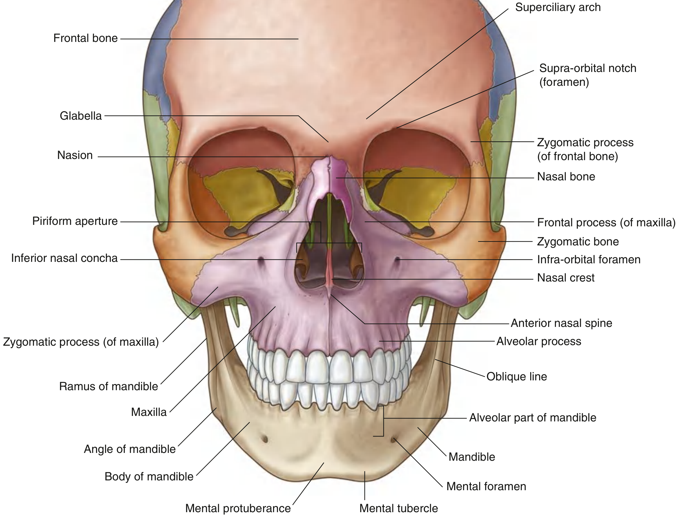
Frontal Bone
- Forms the forehead and superior rim of each orbit
- Superciliary arches - raised ridges above orbital rim (more pronounced in males)
- Glabella - small depression between the superciliary arches
- Supra-orbital foramen/notch - transmits supra-orbital nerve and vessels (visible in medial part of superior orbital rim)
- Zygomatic process of frontal bone - forms the upper lateral rim of the orbit
Nasal Bones
- Two small paired bones forming the bridge of the nose
Zygomatic Bone
- Cheekbone; forms lateral orbital rim and zygomatic arch
Maxilla (Paired)
- Forms upper jaw
- Contains the alveolar process (bears upper teeth)
- Infra-orbital foramen - transmits infra-orbital nerve and vessels, below the orbital margin
- Anterior nasal spine - midline projection at the base of the nasal aperture
- Frontal process of maxilla - forms medial orbital margin
Inferior Nasal Concha
- Scroll-like bone visible within the nasal (piriform) aperture
3. Lateral View of the Skull
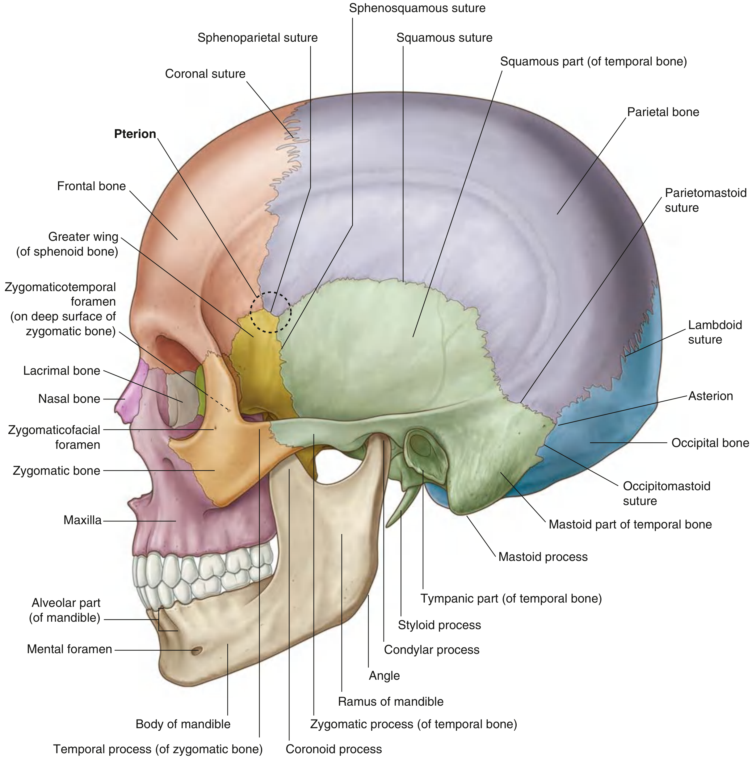
Bones of the Lateral Calvaria
- Frontal → Parietal at the coronal suture
- Parietal → Occipital at the lambdoid suture
- Frontal → Greater wing of sphenoid → Parietal at the sphenoparietal suture
- Sphenoid → Temporal at the sphenosquamous suture
- Temporal → Parietal at the squamous suture
Key Landmark - Pterion
The pterion is the H-shaped junction of four bones: frontal, parietal, sphenoid (greater wing), and temporal. It is the thinnest part of the skull and overlies the middle meningeal artery. Clinical relevance: a blow here can rupture the middle meningeal artery causing an extradural haematoma - a physiotherapy red flag.
Temporal Bone
- Has squamous, mastoid, tympanic, and petrous parts
- Mastoid process - prominent posterior projection; attachment for sternocleidomastoid and other neck muscles
- Styloid process - thin projection, attachment for stylohyoid, styloglossus, stylopharyngeus
- Zygomatic process - forms the zygomatic arch with the temporal process of the zygomatic bone
- Asterion - junction of occipital, parietal, and temporal bones (posteriorly)
Sutures - Summary Table
| Suture | Bones Joined |
|---|---|
| Coronal | Frontal + Parietal |
| Sagittal | Left + Right Parietal |
| Lambdoid | Parietal + Occipital |
| Squamous | Temporal + Parietal |
| Sphenoparietal | Sphenoid + Parietal |
| Sphenosquamous | Sphenoid + Temporal |
| Occipitomastoid | Occipital + Mastoid part of temporal |
| Parietomastoid | Parietal + Mastoid part of temporal |
4. Posterior View of the Skull
Occipital Bone
- Main bone of the posterior skull
- Articulates with parietal bones at the lambdoid suture and temporal bones at the occipitomastoid sutures
- Wormian (sutural) bones - small island bones along the lambdoid suture
- External occipital protuberance - midline bony bump; the most prominent point is called the inion
- Superior nuchal lines - curved ridges extending laterally from the protuberance; muscle attachment
- Inferior nuchal lines - ~2.5 cm below superior lines; further muscle attachment
- External occipital crest - ridge running from protuberance toward the foramen magnum
Temporal Bones (Posterior View)
- Mastoid process is the prominent feature
- Mastoid notch - on the inferomedial border of the mastoid process; attachment for posterior belly of digastric muscle
5. Superior View of the Skull (Calvaria/Calva)
Bones visible: Frontal, paired Parietal, Occipital
- Coronal suture: Frontal ↔ Parietal
- Sagittal suture: Left Parietal ↔ Right Parietal (midline)
- Lambdoid suture: Parietal ↔ Occipital
- Bregma - junction of sagittal and coronal sutures (site of anterior fontanelle in newborns)
- Lambda - junction of sagittal and lambdoid sutures (site of posterior fontanelle in newborns)
- Parietal foramina - paired small foramina lateral to sagittal suture posteriorly
Structure of Calvaria (Diploe)
The calvarial bones have a unique three-layer structure:
- External table - dense compact bone
- Diploë - spongy cancellous bone layer (contains red bone marrow)
- Internal table - dense compact bone
6. Base of Skull - Inferior View
Divided into three parts:
| Part | Key Features |
|---|---|
| Anterior | Teeth, hard palate, alveolar arch of maxillae |
| Middle | Behind hard palate to anterior foramen magnum; foramen ovale, spinosum, lacerum, carotid canal |
| Posterior | Foramen magnum, occipital condyles, mastoid processes |
7. The Mandible - Detailed
The mandible is the only mobile bone of the skull and is the bone of the lower jaw.
Parts of the Mandible
| Part | Description |
|---|---|
| Body | Horizontal part; left and right halves fused at mandibular symphysis anteriorly |
| Ramus (x2) | Quadrangular vertical plates; meet body at the angle of mandible |
| Coronoid process | Anterior superior projection of ramus; attachment for temporalis muscle |
| Condylar process | Posterior superior projection; forms the TMJ with the temporal bone |
| Mandibular notch | Between coronoid and condylar processes |
External (Lateral) Surface Features
- Base of mandible - lower border of body
- Alveolar part - upper part of body, bears the teeth; resorbed when teeth are removed
- Mental protuberance - midline swelling where the two halves meet (the "chin")
- Mental tubercles - slight bumps just lateral to mental protuberance
- Mental foramen - midway between upper alveolar border and lower base of mandible; transmits mental nerve and vessels (branch of inferior alveolar nerve/artery)
- Oblique line - ridge from the front of the ramus onto the body; attachment for muscles that depress the lower lip
Internal (Medial) Surface Features
- Mandibular symphysis - site of fusion; visible as a small vertical ridge externally
- Superior and inferior mental spines (genial spines) - two pairs of small spines posterior to symphysis; attachments for genioglossus (superior) and geniohyoid (inferior) muscles
- Mylohyoid line - raised ridge running posterosuperiorly from midline; attachment for mylohyoid muscle (floor of mouth)
- Sublingual fossa - shallow depression above anterior 1/3 of mylohyoid line; houses sublingual salivary gland
- Submandibular fossa - depression below posterior 2/3 of mylohyoid line; houses submandibular salivary gland
- Retromolar triangle - small triangular area posterior to last molar; attachment for pterygomandibular raphe
- Shallow groove for lingual nerve - between last molar tooth and mylohyoid line
Ramus Features
- Mandibular foramen - large foramen on medial surface of ramus; transmits inferior alveolar nerve and vessels into the inferior alveolar canal
- Lingula - small bony projection at the anterior edge of mandibular foramen; attachment for sphenomandibular ligament
8. Key Foramina and Their Contents (Exam Table)
| Foramen | Location | Contents |
|---|---|---|
| Supra-orbital | Frontal bone, superior orbital rim | Supra-orbital nerve (V1) + vessels |
| Infra-orbital | Maxilla, below orbital rim | Infra-orbital nerve (V2) + vessels |
| Mental | Lateral body of mandible, midway | Mental nerve (V3) + vessels |
| Mandibular | Medial surface of ramus | Inferior alveolar nerve + vessels |
Remember the "3 on a line" rule: Supra-orbital, infra-orbital, and mental foramina are roughly vertically aligned - useful clinically for nerve blocks.
9. Fontanelles (Physiotherapy and Developmental Context)
In the fetus and newborn, unossified membranous gaps allow head deformation during birth and postnatal brain growth.
| Fontanelle | Location | Closure |
|---|---|---|
| Anterior (bregmatic) | Junction of coronal + sagittal sutures (bregma) | 18-24 months |
| Posterior (lambdoid) | Junction of sagittal + lambdoid sutures (lambda) | 2-3 months |
| Anterolateral (sphenoidal) | Pterion region | ~3 months |
| Posterolateral (mastoid) | Asterion region | ~12-18 months |
A bulging anterior fontanelle indicates raised intracranial pressure; a sunken fontanelle indicates dehydration.
10. Physiotherapy Clinical Relevance
| Structure | Clinical Relevance |
|---|---|
| Pterion | Thinnest skull point; trauma here → middle meningeal artery rupture → extradural haematoma |
| Mastoid process | SCM attachment; tenderness in mastoiditis; palpated in cervical spine assessment |
| Occipital protuberance / nuchal lines | Attachment for trapezius, semispinalis capitis; key landmarks in postural assessment |
| Mental foramen | Numbness over chin/lower lip may indicate mental nerve compression or mandibular pathology |
| Mandibular condyle | Core of the TMJ; dysfunction = temporomandibular disorder (TMD) - treated by physiotherapists |
| Coronoid process | Temporalis attachment; trismus after fracture or scarring |
| Mylohyoid line | Floor of mouth anatomy; relevant in swallowing therapy |
| Calvaria structure (diploë) | Stress fracture knowledge; internal table more brittle → contre-coup fractures |
| Sutures | Cranial sacral therapy uses these; also important in detecting craniosynostosis |
11. Muscles Attaching to the Mandible (Physiotherapy Muscle Chart)
| Muscle | Attachment on Mandible | Action |
|---|---|---|
| Temporalis | Coronoid process + anterior ramus | Elevation + retraction of mandible |
| Masseter | Lateral surface of ramus + angle | Elevation of mandible |
| Medial pterygoid | Medial surface of ramus + angle | Elevation + protrusion |
| Lateral pterygoid | Condylar process / articular disc | Depression + protrusion |
| Genioglossus | Superior genial spines | Depresses/protrudes tongue |
| Geniohyoid | Inferior genial spines | Elevates hyoid, depresses mandible |
| Mylohyoid | Mylohyoid line | Elevates floor of mouth, depresses mandible |
| Digastric (anterior belly) | Digastric fossa (near symphysis) | Depresses mandible, elevates hyoid |
| Buccinator | Alveolar part (lateral) + pterygomandibular raphe | Compresses cheek; keeps food on teeth |
Quick Revision Summary
-
22 skull bones (excl. ossicles): 1 frontal, 2 parietal, 2 temporal, 1 occipital, 1 sphenoid, 1 ethmoid + facial bones
-
Only movable skull bone = mandible
-
Sutures = fibrous immovable joints; TMJ = only synovial joint involving skull + mandible
-
Pterion = thinnest skull region, H-junction of 4 bones, overlies middle meningeal artery
-
Bregma = anterior fontanelle site; Lambda = posterior fontanelle site
-
Mandible parts: body (alveolar + base), ramus, angle, coronoid process, condylar process
-
Mental foramen = on body; Mandibular foramen = on ramus (medial)
-
Mylohyoid line = important oral floor muscle attachment on mandible's medial surface
-
Gray's Anatomy for Students, pp. 979-998, 1229-1231
This is a shared conversation. Sign in to Orris to start your own chat.