
I wanna learn how to read MRI give me some images with diagmosis with commin like ligament labrum spinal cord injury fracture and other common conditions with images and how to find that easily
Reading File
Finding Sources
Finding Sources
Finding Sources
Finding Sources
Finding Sources
Reading File
Finding Sources
Finding Sources
Finding Sources
Finding Sources
Finding Sources
Searching the Web
MRI reading guide musculoskeletal ligament labrum spinal cord fracture imaging findings
I found the key sections in Grainger & Allison's Diagnostic Radiology (the main radiology textbook). Let me read the MRI sections on shoulder, labrum, knee ligaments, and spine.
Reading File
Reading File
Excellent! I'm finding rich content and images. Let me read more sections - shoulder labrum, ACL details, and spine sections.
Reading File
Reading File
Reading File
Reading File
Reading File
Reading File
Reading File
Reading File
I now have enough content to write a comprehensive MRI reading guide. Let me also fetch the key MRI images from the textbook to include visually.Here is your complete practical guide to reading musculoskeletal MRI, built from Grainger & Allison's Diagnostic Radiology (the leading radiology textbook).
How to Read Musculoskeletal MRI: A Practical Guide
First - Understand MRI Signal Basics
Before looking at pathology, you need to know what normal tissue looks like on each sequence. Two sequences dominate MSK MRI:
| Sequence | Fluid appears | Fat appears | Bone marrow | Use for |
|---|---|---|---|---|
| T1-weighted | Dark (low signal) | Bright (high signal) | Bright | Anatomy, bone marrow replacement, fracture age |
| T2-weighted / PD fat-sat | Bright (high signal) | Dark (suppressed) | Intermediate | Edema, tears, fluid, inflammation |
The golden rule: On fat-saturated T2/PD sequences, anything bright = water/fluid/edema = abnormal (usually). Normal ligaments and tendons are uniformly dark on all sequences.
1. SHOULDER - Rotator Cuff & Labrum
How to approach the shoulder MRI
- Look at sagittal oblique images for rotator cuff thickness and continuity
- Look at coronal oblique for full-thickness tears and subacromial space
- Look at axial images for the labrum (anterior and posterior), biceps tendon, and subscapularis
Rotator Cuff Tears
The supraspinatus is the most commonly torn tendon. The cuff can show:
- Tendinopathy - intermediate T2 signal within the tendon (thickened, disorganized collagen)
- Partial thickness tear - high T2 signal not spanning full thickness (articular surface tears are most common)
- Full thickness tear - fluid signal traversing the tendon from articular to bursal surface; creates an abnormal communication between the glenohumeral joint and subacromial bursa
- Massive tear - involves 2+ tendons or measures ≥5 cm; associated with tendon retraction and fatty muscle atrophy
Quick tip: On coronal images, if you see fluid in the subacromial bursa AND in the glenohumeral joint with no normal tendon tissue separating them = full-thickness tear until proven otherwise.
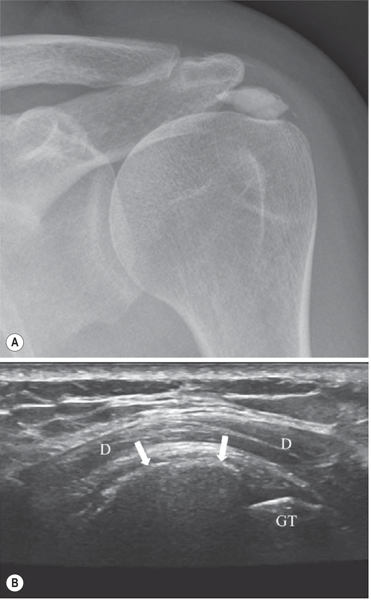
Normal shoulder: AP radiograph (left) and ultrasound (right) showing calcific tendinitis with calcium deposit in the supraspinatus tendon (white arrows). On MRI, this calcification appears as a focus of low signal.
Labral Tears - Bankart Lesion
The labrum is a fibrocartilaginous ring that deepens the glenoid socket. It should appear as a uniformly dark, triangular structure on axial T1 or T2 images.
Bankart lesion = anteroinferior labral tear caused by anterior shoulder dislocation
- MRI finding: fluid/contrast signal extending between the glenoid and the labrum (primary sign)
- The labrum may be displaced, frayed, or absent at the 3-6 o'clock position (anterior-inferior quadrant)
- Best seen on axial images, especially on MR arthrography (gadolinium injected into joint)
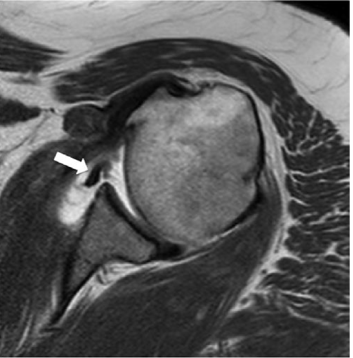
Bankart Lesion (axial T1 MR arthrogram): High signal contrast extends deep to the anteroinferior labrum (white arrow), indicating labral detachment. Compare with the normal dark posterior labrum.
Hill-Sachs defect = impaction fracture of the posterosuperior humeral head (golf ball dents when it hits the glenoid rim). Seen as a notch/depression on the posterolateral humeral head on axial images.
SLAP Tear (Superior Labrum Anterior to Posterior) = tear at the biceps anchor (12 o'clock). Seen as high-signal fluid extending into the superior labrum on coronal fat-sat images.
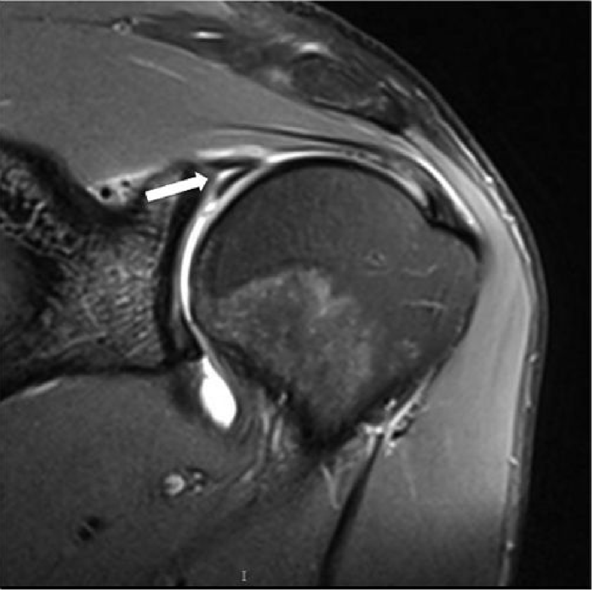
SLAP Tear (coronal FS T2 MR arthrogram): High signal contrast in the substance of the superior labrum at the biceps anchor.
2. KNEE - Ligaments & Menisci
How to approach the knee MRI
- Sagittal is your workhorse: ACL, PCL, meniscal posterior horns
- Coronal: MCL, LCL complex, meniscal bodies, tibial plateau
- Axial: patellofemoral compartment, popliteal structures
Anterior Cruciate Ligament (ACL)
Normal ACL: a tight, parallel-fibered band running from the posteromedial lateral femoral condyle to the anterior tibial intercondylar eminence - dark on all sequences, straight course on sagittal.
ACL tear signs:
- Complete tear: discontinuity of fibers + increased signal within the torn stump + laxity or horizontal orientation of the remaining fibers
- Partial tear: signal change and laxity with some fibers still intact
Secondary signs (very useful - don't miss these):
- "Kissing contusions" - bone marrow edema (bright T2) at the posterolateral tibial plateau + lateral femoral condyle = classic pivot shift injury pattern
- Anterior tibial translation ≥7 mm relative to the femur
- PCL buckling
- Segond fracture - tiny avulsion off lateral tibial rim (highly specific for ACL tear; subtle on MRI, better on X-ray)
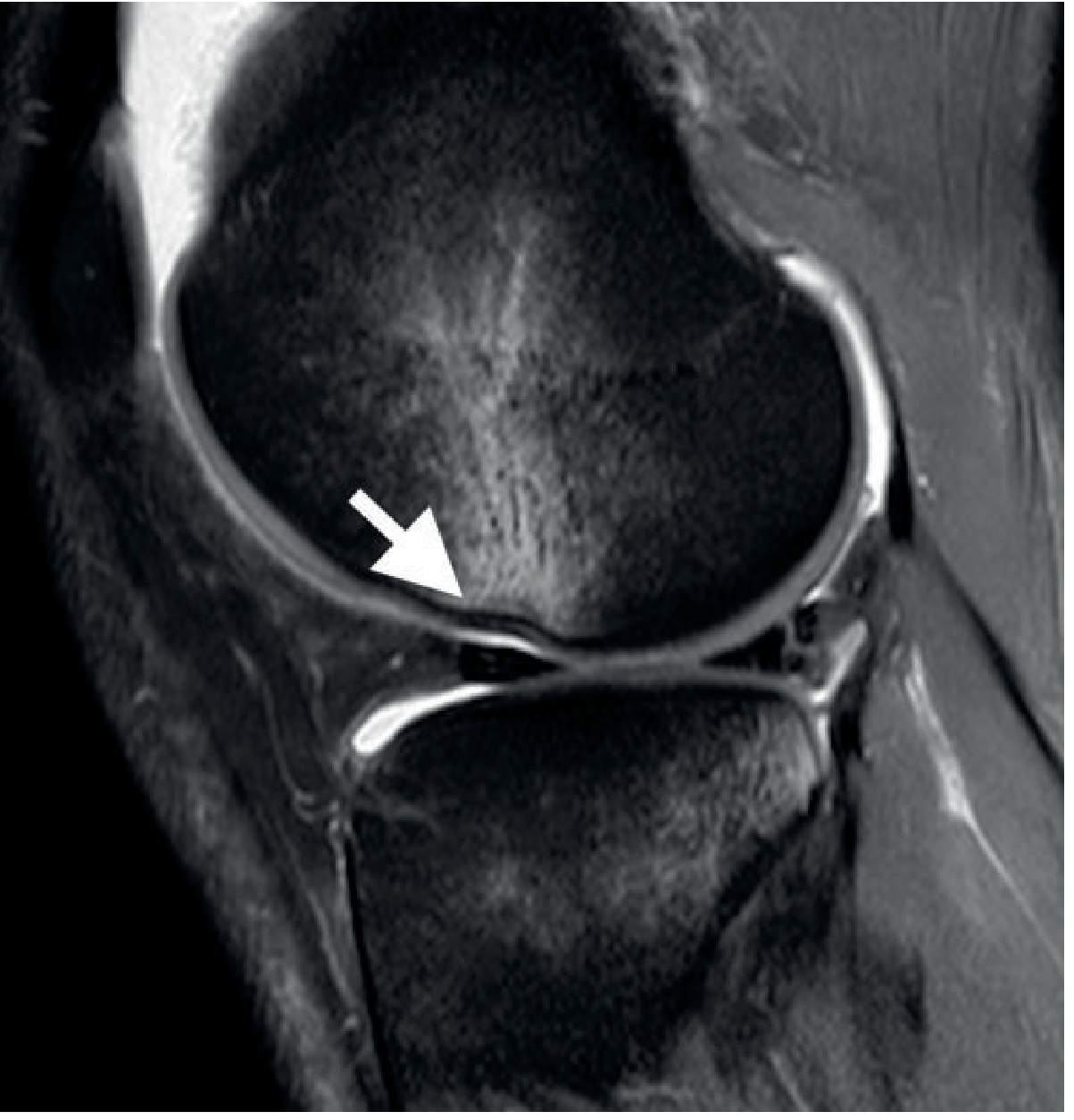
Complete ACL Tear (sagittal PD fat-sat): Retracted distal ACL fibers with no intact proximal fibers (arrow). The normal "straight taut band" appearance is completely lost.
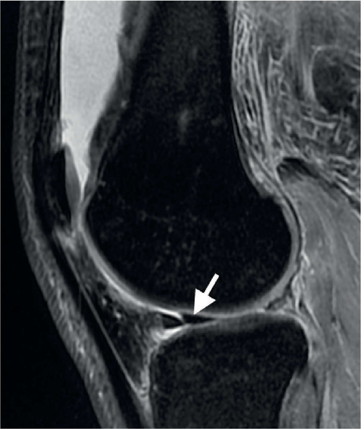
Pivot Shift Injury with ACL Tear (sagittal PD fat-sat): Bright bone marrow edema (contusions) at the lateral femoral condyle and posterolateral tibial plateau. Small subcortical impaction fracture of the lateral femoral condyle (arrow).
Posterior Cruciate Ligament (PCL)
Normal PCL: a thick, low-signal curved band - easy to find on sagittal images. More robust than ACL and less commonly torn.
- Torn by "dashboard injury" (posterior force on bent knee)
- Partial middle-third tears are more common than complete tears
- Increased T2 signal + laxity = sprain; complete disruption = complete tear
Double PCL sign: on sagittal images, you see the PCL and a second parallel dark structure just anterior to it = displaced bucket handle meniscal tear has flipped into the intercondylar notch.
Medial Collateral Ligament (MCL)
Normal MCL: a dark band on the medial joint line on coronal images.
- Tears show fluid signal within or around the ligament
- Grade 1: edema around ligament (periligamentous fluid), ligament intact
- Grade 2: partial tear - signal within the substance
- Grade 3: complete tear - disrupted fibers with fluid gap
O'Donoghue's unhappy triad: ACL + MCL + medial meniscus - caused by valgus stress. One of the most common combined knee injuries.
Menisci
Normal menisci: uniformly dark on all sequences. Any intrinsic signal = abnormal if it reaches an articular surface.
Grading meniscal signal:
- Grade 1-2: intrasubstance signal that does NOT touch the articular surface = degeneration (not a true tear)
- Grade 3: high signal extending to an articular surface = TEAR
Common tear patterns:
| Tear type | MRI appearance |
|---|---|
| Horizontal tear | Linear high signal parallel to tibial plateau |
| Longitudinal/vertical tear | Vertical cleft in the peripheral zone |
| Radial / parrot beak | Linear signal from the free edge inward |
| Bucket handle | Displaced fragment flips into intercondylar notch; "double PCL sign" |
| Ghost meniscus | Absent/empty meniscus slice on radial tear |
| Flipped meniscus | Posterior horn fragment lying adjacent to anterior horn |
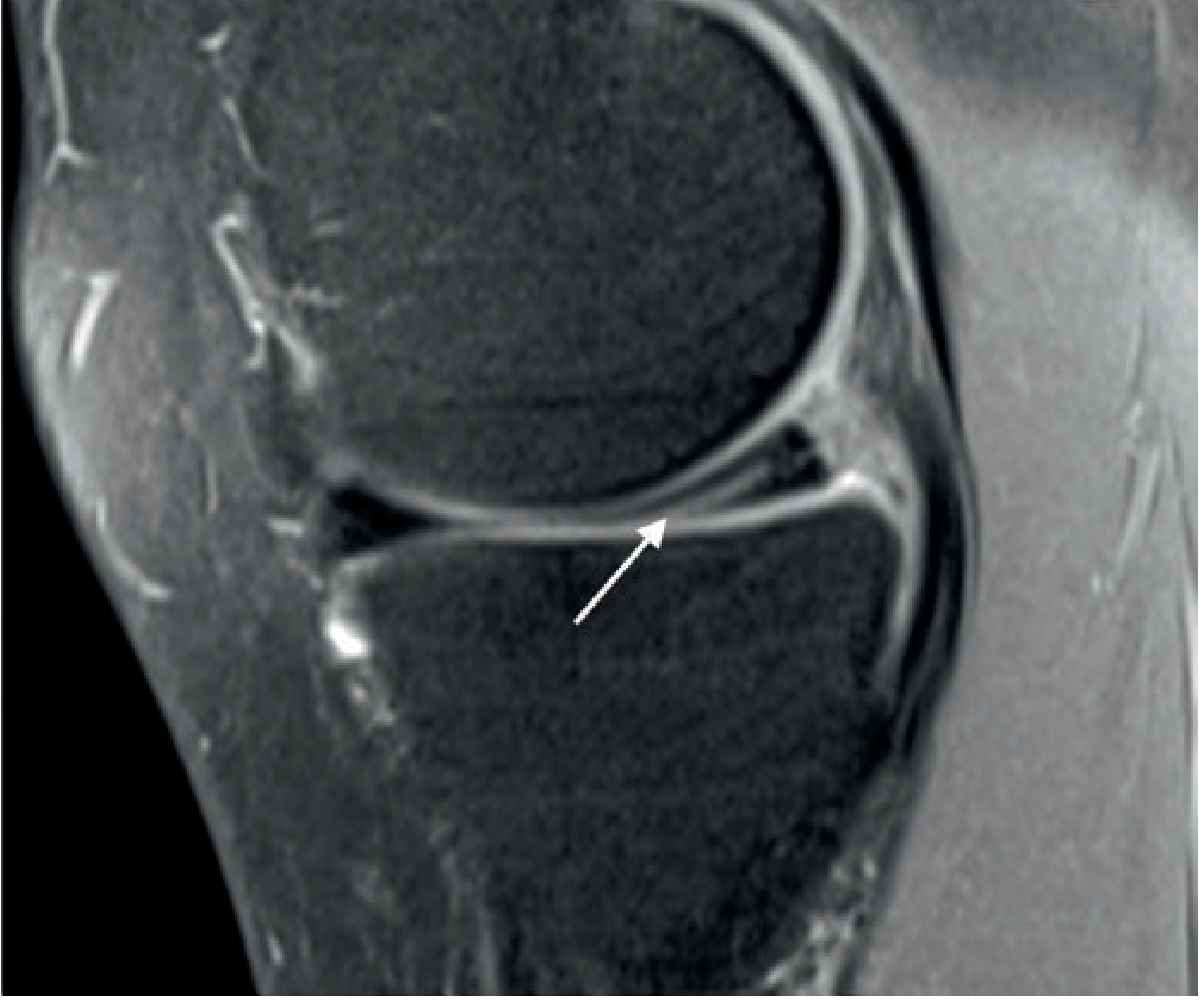
Horizontal Tear of Medial Meniscus (sagittal PD fat-sat): Linear high signal undersurface tear of the posterior horn (arrow). Horizontal tears can form parameniscal cysts.
3. SPINE - Cord, Disc, and Fractures
How to approach the spine MRI
- Sagittal T1: see anatomy, vertebral body height, marrow signal
- Sagittal T2: disc hydration (bright = healthy), cord signal, nerve root compression
- Axial T2/GRE: neural foramina, disc herniation level, cord cross-section
Spinal Cord Injury
MRI is the only modality that directly images the spinal cord. Key cord findings:
| Finding | Signal | Meaning |
|---|---|---|
| Normal cord | Intermediate T1, low-intermediate T2 | No injury |
| Cord edema | Normal T1, increased T2 | Contusion - better prognosis |
| Cord hemorrhage | Decreased T2 (blood products) | Hemorrhagic contusion - worse prognosis |
| Cord transection | Complete signal discontinuity | Catastrophic injury |
| Chronic myelomalacia | Low T1, high T2 (cystic) | Old cord injury with cavitation |
Tip: Hemorrhage within the cord on MRI significantly worsens prognosis compared to edema alone. This distinction is one of MRI's most important roles in spinal trauma.
Disc Herniation and Nerve Root Compression
Healthy disc: bright white on T2 (high water content). Degenerated disc: dark/desiccated on T2.
Types of disc pathology:
- Disc bulge: broad, diffuse extension beyond vertebral endplates
- Disc protrusion: focal herniation, base wider than the dome
- Disc extrusion: herniated material extends beyond disc space
- Sequestered fragment (free fragment): disc material separated from parent disc - can migrate up/down canal
Canal and foraminal stenosis: Measure the AP diameter of the spinal canal and look for obliteration of the CSF (white T2 fluid) around the cord/cauda equina. Loss of epidural fat on T1 and obliteration of the T2 bright perineural sleeve = foraminal stenosis.
Osteoporotic/Insufficiency Vertebral Fractures
MRI is excellent for:
- Dating fractures - acute fractures show diffuse low T1 + high T2 marrow edema; chronic fractures have normal marrow signal
- Differentiating benign vs. malignant collapse - benign osteoporotic fractures preserve posterior element signal and show a fluid sign; malignant (metastatic) fractures replace all marrow, involve posterior elements, and show an epidural mass
Wedge/endplate/crush fracture types:
- Wedge: anterior height loss, common at thoracolumbar junction (T12-L1)
- Crush: >40% height loss (grade 3 = severe)
- Fractures above T7 = suspect metastasis, not osteoporosis
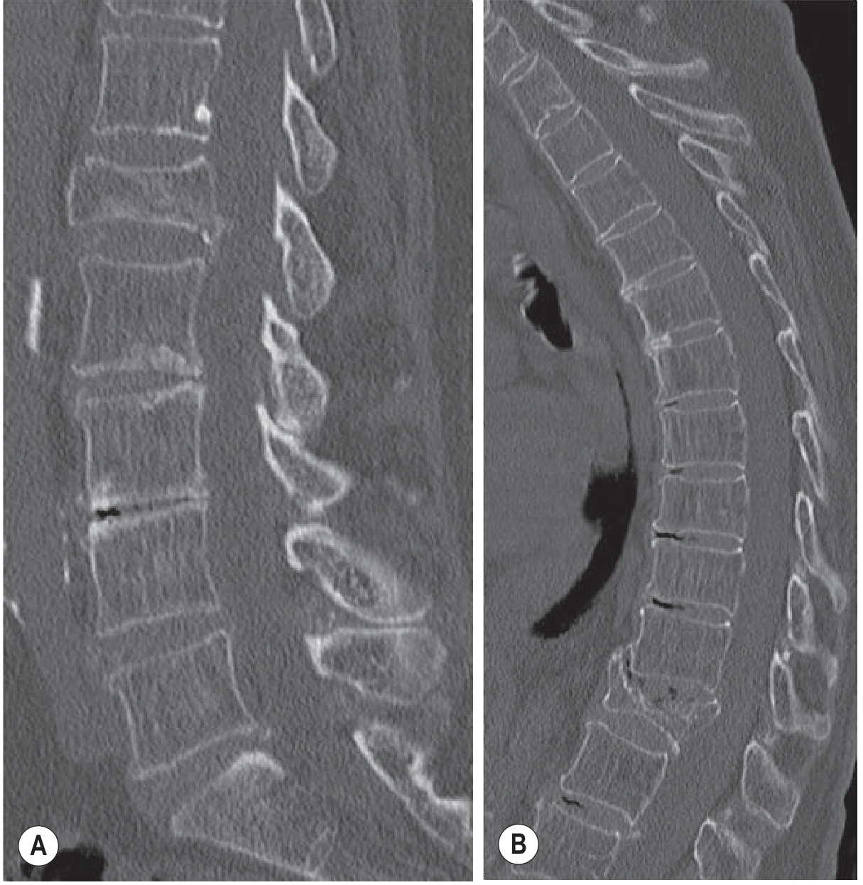
Osteoporotic Vertebral Fractures (sagittal CT): Severe compression fractures at L1 and T12 with loss of vertebral height. MRI would show marrow edema (low T1, high T2) in acute phase.
4. ANKLE - Ligament Tears
Lateral ankle ligaments (most commonly injured)
The ATFL (anterior talofibular ligament) is torn first in ankle inversion, followed by the CFL (calcaneofibular ligament). On MRI:
- Normal ligament: dark band
- Acute tear: wavy, high-signal, thickened, or absent ligament
- Chronic tear: thickened, irregular, heterogeneous
- Bone marrow edema adjacent to the tear site (look for bright T2 in lateral talus/fibula)
Osteochondral Lesion of the Talus (OLT)
A common missed diagnosis after ankle sprain. Look for:
- Subchondral bone marrow edema (bright T2) in the talar dome
- A dark fracture line beneath the articular surface
- Fluid "undercutting" the fragment = unstable lesion requiring surgery
5. Quick Reference - "Red Flags" on MSK MRI
| Sign | Look for | Diagnosis |
|---|---|---|
| Fluid deep to labrum | Axial shoulder | Bankart/labral tear |
| Missing ACL or horizontal ACL | Sagittal knee | ACL tear |
| Kissing contusions LFC + posterolateral tibia | Sagittal knee | Pivot shift / ACL tear |
| Double PCL sign | Sagittal knee | Bucket handle meniscal tear |
| Grade 3 meniscal signal | Any knee plane | Meniscal tear |
| Cord T2 hyperintensity | Sagittal spine | Cord contusion/myelopathy |
| Marrow low T1 + high T2 in vertebral body | Sagittal spine | Acute fracture or malignancy |
| Absent ATFL + lateral talar edema | Axial ankle | Lateral ligament tear |
| Fluid in subacromial bursa + GHJ | Coronal shoulder | Full-thickness cuff tear |
Key Practical Tips for MRI Reading
- Always use multiple planes - a structure visible in only one plane may be artifact
- Compare signal to known references - disc fluid (T2 bright), cortical bone (T2 dark), fat (T1 bright before fat-sat)
- Follow the structure - trace a ligament or tendon through all slices in its best plane
- Correlate with clinical history - mechanism of injury predicts which structures to scrutinize first
- MR arthrography (gadolinium injected into joint) dramatically improves sensitivity for partial labral tears and partial cuff tears - particularly useful for shoulder and hip
- Fat saturation is your friend - it eliminates the bright fat signal and makes edema/fluid POP
*Sources: Grainger & Allison's Diagnostic Radiology, 7th ed., Chapters 39 (Musculoskeletal MRI) and 43 (Metabolic Bone Disease); supplemented by PMC review on spinal MRI [PMC4957861]*That's your comprehensive MRI reading guide, sourced directly from Grainger & Allison's Diagnostic Radiology (the gold-standard radiology reference). Here's a quick summary of what was covered:
What you got:
-
MRI signal basics - T1 vs T2/fat-sat and what each looks like for fluid, fat, bone, and soft tissue
-
Shoulder - Rotator cuff tears (partial vs full-thickness), Bankart labral lesions with actual MRI arthrogram image, SLAP tears, Hill-Sachs defect
-
Knee ligaments & menisci - Complete ACL tear with image, pivot-shift contusion pattern, PCL tears, MCL grading, all meniscal tear types (horizontal, bucket handle, ghost meniscus, flipped meniscus) with images
-
Spine - Cord edema vs hemorrhage (prognostic difference), disc herniation types, osteoporotic vs malignant vertebral collapse on MRI
-
Ankle - ATFL/CFL lateral ligament tears, osteochondral lesions of the talus
-
Red flags cheat sheet - 9 key signs to always check and what they mean
Most important habit: Always review all 3 planes before calling something normal - many tears are only visible in one specific plane.
This is a shared conversation. Sign in to Orris to start your own chat.