
Explain to me this chapter of breast in... Stenberg by Gregory R. Bean and Kimberly H. Ellison.
Sternberg's Diagnostic Surgical Pathology breast chapter Gregory Bean Kimberly Ellison
https://dokumen.pub/mills-and-sternbergs-diagnostic-surgical…
breast pathology histology surgical pathology diagnostic
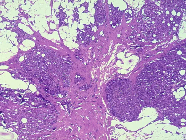
Breast tissue histology obtained by surgical biopsy demonstrating a radial scar (complex sclerosing lesion) with associated ductal hyperplasia. The lesion features a central densely fibrotic core with radiating strands of oriented, compressed ducts embedded in fibrous and myxoid stroma. Ductal epithelium shows hyperplastic changes consistent with mild ductal hyperplasia; there is no overt cytologic atypia or invasion observed. The overall architecture resembles a starburst lesion on gross imaging but is evaluated here on hematoxylin-eosin stained sections at multiple magnifications (4x–40x). The fibrous stroma is variably hyalinized, with entrapped glands that may be distorted in a stellate configuration. The differential diagnosis includes tubular carcinoma and other sclerosing lesions; however, the absence of infiltrative growth and high-grade cytology supports a benign radial scar, though associations with occult or nearby carcinoma have been reported. Clinically, radial scars larger than 1 cm carry an increased risk of carcinoma development, particularly tubular carcinoma, and surgical excision or close radiologic-pathologic correlation is advised for larger lesions or those with atypia. This image is relevant for pathology education, radiologic-pathologic correlation, differential diagnosis training, and risk-stratified breast lesion assessment in a diagnostic pathology workflow. Correlate with imaging features and patient age to guide management decisions in clinical practice.
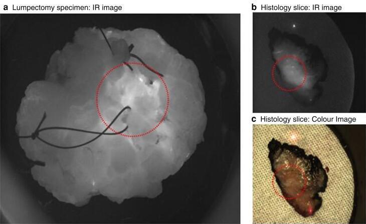
A clinical diagnostic image panel showing ex-vivo breast lumpectomy specimens during surgical pathology processing. Panel (a) presents a whole lumpectomy specimen viewed via infrared (IR) imaging; the tissue appears as a multi-lobulated, hyper-reflective mass against a dark background, with surgical orientation sutures visible and a red dotted circle demarcating the tumor location found 1 mm deep to the surface. Panels (b) and (c) compare an transverse histology slice of the tissue. Image (b) shows the slice in IR, highlighting differences in tissue reflectivity, while image (c) is a standard color photograph showing the specimen's heterogeneous texture and dark peripheral margins on a textured background. In both (b) and (c), a red dotted circle identifies the tumor focus within the medial-to-lateral orientation of the slice. The images illustrate the application of infrared imaging in surgical oncology and pathology for tumor localization and margin assessment.
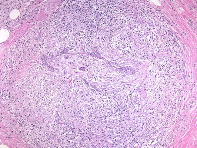
Breast tissue histology image obtained from hematoxylin and eosin stained section viewed under light microscopy of a granulomatous mastitis specimen. The specimen is from breast tissue showcasing destruction of native architecture with a cuff of inflammatory infiltrate surrounding residual distorted ducts, consistent with periductal granulomatous inflammation. The granulomas are typified by aggregates of epithelioid histiocytes, occasional Langhans-type giant cells, and a surrounding lymphoplasmacytic infiltrate. Ductal structures appear distorted or obliterated within a dense inflammatory milieu; there is little or no evident caseous necrosis in this field. This pattern can arise from various etiologies, including infectious agents such as Mycobacterium tuberculosis, non-tuberculous mycobacteria, fungi, brucella, or parasitic organisms, as well as idiopathic granulomatous mastitis or a granulomatous response adjacent to mammary carcinoma. Clinically, granulomatous mastitis may mimic breast carcinoma due to focal mass effect and architectural distortion, underscoring the importance of correlating morphology with microbiological studies and imaging. The diagnostic significance lies in distinguishing inflammatory/granulomatous breast disease from neoplasia, guiding appropriate testing (culture, PCR, fungal stains) and therapy (antimicrobials, corticosteroids, or surgical management). This image is relevant for educational purposes in pathology, surgical oncology, radiology, and breast disease research and serves as a reference for differential diagnosis in granulomatous breast lesions.
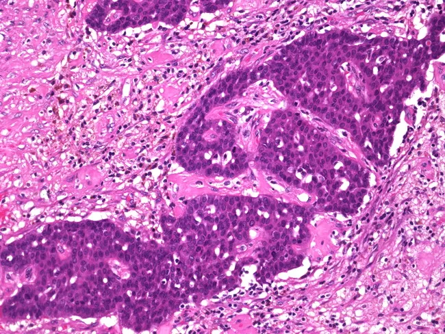
Imaging modality: bright-field light microscopy of hematoxylin and eosin–stained breast tissue section; high-quality histology slide prepared from a surgical pathology specimen. Anatomical focus: invasive papillary carcinoma of the breast with tumor cells forming arborizing papillary fronds and invading adjacent desmoplastic stroma. Precise localization within mammary gland ducts and lobules, showing extension beyond the tumor borders and evidence of stromal desmoplasia, inflammation, and fibrotic change. Cellular morphology includes malignant epithelial cells with vesicular to hyperchromatic nuclei, conspicuous nucleoli, and mitotic activity distributed throughout papillary cores and surrounding nests. Fibrovascular cores within papillary structures are embedded in a collagen-rich, reactive stroma; hemorrhage and scar-like fibrosis are variably present in areas of postbiopsy change. Notable features indicating invasion include disruption of basement membrane continuity and penetration into adjacent breast parenchyma. Diagnostic significance lies in differentiating in situ from invasive disease and in informing staging (T stage) and prognosis; the desmoplastic reaction supports invasive behavior. Differential considerations include papillary in situ carcinoma with invasion-like features, invasive ductal carcinoma with papillary morphology, and benign papillary lesions with reactive stroma. Clinically relevant applications encompass surgical planning (wide excision vs mastectomy), adjuvant therapy decisions, and correlation with radiologic findings for comprehensive cancer management. Educational value for teaching.
Chapter 9: Breast - Mills & Sternberg's Diagnostic Surgical Pathology (Bean & Allison)
1. Normal Breast Anatomy & Histology
- Lobular unit (TDLU - Terminal Duct Lobular Unit): The functional unit of the breast, consisting of acini (small glands) drained by an intralobular duct, which drains into the extralobular terminal duct. This is where most breast pathology originates.
- Ductal system: Terminal ducts coalesce into segmental ducts and finally into the major lactiferous sinuses opening at the nipple.
- Two cell layers: All normal ducts and lobules have an inner luminal epithelial layer and an outer myoepithelial layer (important in distinguishing in situ from invasive carcinoma - invasion means loss of the myoepithelial layer).
- Hormonal changes: The breast is exquisitely hormone-responsive. Cyclical changes, pregnancy, and lactation all cause recognizable histologic patterns.
2. Non-Neoplastic Breast Conditions
Fibrocystic Changes
- Cysts (simple and apocrine): Fluid-filled, lined by flat or apocrine epithelium. No increased cancer risk.
- Adenosis: Increased number of acini per lobule. When sclerosing (sclerosing adenosis), it can mimic invasive carcinoma but myoepithelial cells are preserved.
- Apocrine metaplasia: Cells with abundant eosinophilic granular cytoplasm and small nucleoli - benign.
Columnar Cell Lesions (CCLs)
- Columnar cell change / columnar cell hyperplasia: Lobules dilated with secretions, lined by columnar cells with apical snouts. Low/no risk.
- Flat epithelial atypia (FEA): Columnar cells with low-grade cytologic atypia. Clonal proliferation; associated with low-grade DCIS and tubular carcinoma. Warrants surgical excision when found on core biopsy.
Usual Ductal Hyperplasia (UDH)
- Benign proliferation with heterogeneous, "streaming" architecture, irregular slit-like peripheral spaces.
- Polyclonal; slight (~1.5-2x) risk increase for breast cancer.
- Distinguished from atypical ductal hyperplasia (ADH) by its cytologic and architectural heterogeneity.
Atypical Ductal Hyperplasia (ADH)
- Shares architectural and cytologic features with low-grade DCIS but is limited in extent (< 2 mm or involves < 2 duct spaces).
- ~4-5x increased relative risk of breast cancer (both breasts).
- Management: Surgical excision if found on core biopsy due to upgrade rate to DCIS or invasive carcinoma.
Atypical Lobular Hyperplasia (ALH) & Lobular Carcinoma In Situ (LCIS)
- Both are lobular neoplasia - a neoplastic proliferation of small, discohesive cells filling and expanding lobular acini.
- Cells lose E-cadherin expression (important IHC marker).
- ALH: Involves < 50% of acini in a lobular unit. ~4-5x increased risk.
- Classic LCIS: > 50% of acini involved. ~8-10x increased risk. Bilateral risk marker. Management controversial - some advocate excision, others surveillance.
- Pleomorphic LCIS: Large cells with high-grade nuclei and sometimes necrosis. Behaves more like DCIS; typically excised.
Radial Scar / Complex Sclerosing Lesion
- Star-shaped (stellate) lesion with a central fibrous core from which ducts radiate outward - mimics invasive carcinoma both grossly and radiologically.
- Myoepithelial cells are preserved throughout - key to recognizing this as benign.
- Associated with ADH and ALH; upgraded carcinoma found in ~5-8% at excision.
Inflammatory / Reactive Conditions
- Acute mastitis / abscess: Usually puerperal, Staph aureus.
- Periductal mastitis / duct ectasia: Dilated ducts filled with secretions, periductal fibrosis and chronic inflammation - linked to smoking.
- Granulomatous mastitis: Idiopathic (most common in reproductive-age women) vs. infectious (TB, fungal). Must exclude carcinoma.
3. Benign Neoplasms
Fibroadenoma
- Most common benign breast tumor, typically in women 15-35.
- Biphasic: epithelial glands (two cell layers) embedded in cellular stroma.
- Two growth patterns: intracanalicular (stroma compresses ducts into slit-like spaces) and pericanalicular (stroma grows around open, rounded ducts).
- Cellular fibroadenoma: if stroma is more cellular, must be distinguished from phyllodes tumor by the absence of stromal overgrowth.
- Juvenile fibroadenoma: Large, rapidly growing variant in adolescents.
Phyllodes Tumor
- Fibroepithelial tumor with leaf-like stromal fronds projecting into epithelium-lined spaces (the classic "phyllo" = leaf architecture).
- Graded as benign, borderline, or malignant based on:
- Stromal cellularity
- Stromal atypia
- Mitotic count (key: > 10/10 HPF = malignant)
- Stromal overgrowth (stroma without epithelium in a 4x field)
- Tumor margins (infiltrative vs. pushing)
- Malignant phyllodes has distant metastatic potential (typically sarcomatous elements go to lungs); axillary node mets are rare.
Intraductal Papilloma
- Fibrovascular fronds lined by both epithelial and myoepithelial cells within a duct.
- Central papilloma: Single, in large ducts near nipple; presents with bloody nipple discharge.
- Peripheral papilloma: Multiple, in TDLU region; higher risk of carcinoma.
- Atypical papilloma: has ADH/ALH within it - upgrade rate on excision is significant.
4. Carcinoma In Situ
Ductal Carcinoma In Situ (DCIS)
- Low grade: Small, uniform cells with round nuclei, inconspicuous nucleoli, no necrosis. Grows slowly.
- Intermediate grade: Between the two.
- High grade: Large pleomorphic cells, prominent nucleoli, frequent mitoses, often central comedo-type necrosis with calcification (visible on mammography as linear/branching calcifications).
Lobular Carcinoma In Situ (LCIS) - see above
5. Invasive Breast Carcinoma
General Principles
- Staging: Uses TNM. Size (pT), nodes (pN - number of involved nodes, micrometastasis vs. macrometastasis, sentinel node), and metastasis (pM).
- Core biomarkers (IHC panel): Every invasive carcinoma must be tested for:
- ER (Estrogen Receptor) - positive if ≥ 1% of nuclei stain
- PR (Progesterone Receptor)
- HER2 (Human Epidermal Growth Factor Receptor 2) - IHC 0/1+/2+/3+; 2+ requires FISH/ISH reflex testing
- Ki-67 (proliferation index)
| Subtype | ER | PR | HER2 | Features |
|---|---|---|---|---|
| Luminal A | + | + | - | Low Ki-67; best prognosis |
| Luminal B | + | +/- | - or + | High Ki-67 or HER2+ |
| HER2-enriched | - | - | + | Aggressive; targeted therapy |
| Triple-negative | - | - | - | BRCA1-like; aggressive; chemotherapy |
Invasive Ductal Carcinoma (NST - No Special Type)
- Most common type (~70-80%).
- Diagnosis of exclusion when the tumor does not fit a special type.
- Hard gritty texture grossly (desmoplastic stroma).
- Graded by Nottingham (Elston-Ellis) grade:
- Tubule formation (1-3 points)
- Nuclear pleomorphism (1-3 points)
- Mitotic count (1-3 points)
- Total 3-4 = Grade 1 (well); 5-6 = Grade 2 (moderate); 7-9 = Grade 3 (poorly differentiated)
Special Histologic Types (with distinct biologic behavior)
- Tubular carcinoma: Well-formed angulated tubules, single epithelial layer (no myoepithelium), desmoplastic stroma. Always Grade 1, ER+. Excellent prognosis.
- Mucinous (colloid) carcinoma: Clusters of tumor cells floating in abundant extracellular mucin. ER+. Low-grade.
- Cribriform carcinoma: Invasive cribriform architecture. Excellent prognosis.
- Invasive lobular carcinoma (ILC): Discohesive single-file ("Indian file") cell infiltration; loss of E-cadherin. Difficult to detect on imaging. Bilateral risk. More likely to metastasize to peritoneum, GI tract, meninges, ovary.
- Invasive papillary carcinoma
- Solid papillary carcinoma
- Micropapillary carcinoma: Clusters of cells inside clear spaces (reverse polarity). Highly lymphovascular invasive; high node metastasis rate.
- Metaplastic carcinoma: Triple-negative; heterologous differentiation (squamous, spindle cell, osseous, chondroid). Poor prognosis, chemotherapy resistance.
Inflammatory Carcinoma
- Clinical diagnosis (red, warm, edematous breast resembling mastitis); pathologic confirmation by dermal lymphatic emboli.
- Rapidly fatal without neoadjuvant chemotherapy; T4d by definition.
6. Prognostic and Predictive Factors
- Tumor size: Single strongest prognostic factor after nodes.
- Lymph node status: Number of positive nodes, extranodal extension.
- Histologic grade (Nottingham): Grade 3 has 3-4x worse prognosis than Grade 1.
- Lymphovascular invasion (LVI): Presence of tumor emboli in vessels near the invasive edge.
- Margins: Key for lumpectomy specimens. Current consensus is "no ink on tumor" for invasive carcinoma (SSO-ASTRO).
- Genomic tests: Oncotype DX (Recurrence Score) for ER+/HER2- patients - determines chemotherapy benefit. MammaPrint, Prosigna (PAM50) also discussed.
7. Pathologic Handling & Reporting
- Core needle biopsy: How to assess adequacy, how to triage for ancillary studies.
- Lumpectomy/wide local excision: Margin inking, serial sectioning, radiograph correlation for calcifications.
- Mastectomy: Specimen orientation, skin and nipple sampling, axillary tail.
- Sentinel lymph node (SLN): Sectioning protocol, cytokeratin IHC when H&E negative.
- Post-neoadjuvant chemotherapy specimens: Assessment of residual disease using Miller-Payne grade or Residual Cancer Burden (RCB). Pathologic complete response (pCR) = no residual invasive carcinoma in breast or nodes.
- Synoptic (CAP) reporting: Mandatory elements vs. conditional elements per College of American Pathologists protocols.
8. Special Topics
- Male breast: Gynecomastia (florid vs. fibrous phase); male breast carcinoma (almost always ER+, tends to be higher stage at diagnosis).
- Nipple/Paget disease: Malignant cells in nipple epidermis; virtually always associated with underlying DCIS or invasive carcinoma.
- BRCA1/BRCA2-associated carcinomas: BRCA1 tumors tend to be high-grade, triple-negative, with pushing margins, prominent lymphocytic infiltrate, and syncytial pattern (medullary-like). BRCA2 tumors are more heterogeneous.
- Metastases to the breast: From contralateral breast, melanoma, lymphoma, lung, ovary. Key clue: absence of in-situ component.
- Neoadjuvant therapy changes: Tumor bed fibrosis, foamy macrophages, hemosiderin, ghost outlines of tumor cells - how to map and measure residual disease.
Key Immunohistochemistry Summary
| Marker | Use |
|---|---|
| E-cadherin | Ductal (+) vs. Lobular (-) |
| p63, calponin, SMMHC, CK5/6 | Myoepithelial cells (absent = invasion) |
| ER, PR, HER2 | Biomarker profiling |
| Ki-67 | Proliferation index |
| CK7, GCDFP-15, mammaglobin | Confirm breast origin in metastasis |
| SOX10 | Myoepithelial and metaplastic carcinoma |
Make notes of this chapter.
ls -la /tmp/workspace/ 2>/dev/null || mkdir -p /tmp/workspace && echo "workspace ready"
mkdir -p /tmp/workspace/breast-pathology-notes && python3 << 'PYEOF' from docx import Document from docx.shared import Pt, Inches, RGBColor from docx.enum.text import WD_ALIGN_PARAGRAPH from docx.oxml.ns import qn from docx.oxml import OxmlElement doc = Document() # ── Page margins ── from docx.shared import Inches section = doc.sections[0] section.top_margin = Inches(1) section.bottom_margin = Inches(1) section.left_margin = Inches(1.1) section.right_margin = Inches(1.1) # ── Default Normal style ── normal = doc.styles["Normal"] normal.font.name = "Calibri" normal.font.size = Pt(11) # ── Heading styles ── h1_style = doc.styles["Heading 1"] h1_style.font.name = "Calibri" h1_style.font.size = Pt(16) h1_style.font.bold = True h1_style.font.color.rgb = RGBColor(0x1F, 0x49, 0x7D) h2_style = doc.styles["Heading 2"] h2_style.font.name = "Calibri" h2_style.font.size = Pt(13) h2_style.font.bold = True h2_style.font.color.rgb = RGBColor(0x2E, 0x74, 0xB5) h3_style = doc.styles["Heading 3"] h3_style.font.name = "Calibri" h3_style.font.size = Pt(12) h3_style.font.bold = True h3_style.font.color.rgb = RGBColor(0x1F, 0x49, 0x7D) # ── Helper functions ── def add_h1(text): doc.add_heading(text, level=1) def add_h2(text): doc.add_heading(text, level=2) def add_h3(text): doc.add_heading(text, level=3) def add_para(text, bold=False, italic=False, indent=0): p = doc.add_paragraph() if indent: p.paragraph_format.left_indent = Inches(indent * 0.25) run = p.add_run(text) run.bold = bold run.italic = italic return p def add_bullet(text, level=0, bold_prefix=None): style_name = "List Bullet" if level == 0 else "List Bullet 2" p = doc.add_paragraph(style=style_name) p.paragraph_format.left_indent = Inches(0.25 + level * 0.2) if bold_prefix: run_bold = p.add_run(bold_prefix) run_bold.bold = True p.add_run(text) else: p.add_run(text) return p def add_space(): p = doc.add_paragraph() p.paragraph_format.space_after = Pt(2) def add_rule(): p = doc.add_paragraph() pPr = p._p.get_or_add_pPr() pBdr = OxmlElement('w:pBdr') bottom = OxmlElement('w:bottom') bottom.set(qn('w:val'), 'single') bottom.set(qn('w:sz'), '6') bottom.set(qn('w:space'), '1') bottom.set(qn('w:color'), '2E74B5') pBdr.append(bottom) pPr.append(pBdr) def add_table_2col(headers, rows, col_widths=(2.5, 4.0)): table = doc.add_table(rows=1+len(rows), cols=2) table.style = "Table Grid" hdr_cells = table.rows[0].cells for i, h in enumerate(headers): hdr_cells[i].text = h for run in hdr_cells[i].paragraphs[0].runs: run.bold = True hdr_cells[i].paragraphs[0].alignment = WD_ALIGN_PARAGRAPH.CENTER tc = hdr_cells[i]._tc tcPr = tc.get_or_add_tcPr() shd = OxmlElement('w:shd') shd.set(qn('w:val'), 'clear') shd.set(qn('w:color'), 'auto') shd.set(qn('w:fill'), 'BDD7EE') tcPr.append(shd) for r_idx, row_data in enumerate(rows): row_cells = table.rows[r_idx+1].cells for c_idx, cell_text in enumerate(row_data): row_cells[c_idx].text = cell_text # set column widths for row in table.rows: row.cells[0].width = Inches(col_widths[0]) row.cells[1].width = Inches(col_widths[1]) doc.add_paragraph() # ════════════════════════════════════════════════════════════ # TITLE PAGE # ════════════════════════════════════════════════════════════ title_p = doc.add_paragraph() title_p.alignment = WD_ALIGN_PARAGRAPH.CENTER title_run = title_p.add_run("CHAPTER 9: BREAST") title_run.bold = True title_run.font.size = Pt(20) title_run.font.color.rgb = RGBColor(0x1F, 0x49, 0x7D) title_run.font.name = "Calibri" sub_p = doc.add_paragraph() sub_p.alignment = WD_ALIGN_PARAGRAPH.CENTER sub_run = sub_p.add_run("Mills & Sternberg's Diagnostic Surgical Pathology, 7th Edition") sub_run.italic = True sub_run.font.size = Pt(13) sub_run.font.name = "Calibri" sub_run.font.color.rgb = RGBColor(0x2E, 0x74, 0xB5) auth_p = doc.add_paragraph() auth_p.alignment = WD_ALIGN_PARAGRAPH.CENTER auth_run = auth_p.add_run("Authors: Gregory R. Bean & Kimberly H. Allison (Stanford University)") auth_run.font.size = Pt(11) auth_run.font.name = "Calibri" auth_run.font.color.rgb = RGBColor(0x40, 0x40, 0x40) doc.add_paragraph() add_rule() doc.add_paragraph() # ════════════════════════════════════════════════════════════ # SECTION 1 – NORMAL ANATOMY & HISTOLOGY # ════════════════════════════════════════════════════════════ add_h1("1. Normal Breast Anatomy & Histology") add_bullet("Functional unit: Terminal Duct Lobular Unit (TDLU) — acini + intralobular duct", bold_prefix="TDLU: ") add_bullet("Ductal hierarchy: Acini → intralobular duct → extralobular terminal duct → segmental duct → lactiferous sinus → nipple") add_bullet("Two-cell layer rule: ALL normal ducts and lobules have inner LUMINAL epithelial cells + outer MYOEPITHELIAL cells", bold_prefix="Two-cell layer: ") add_bullet("Loss of myoepithelium = invasion — key diagnostic principle") add_bullet("Stroma: specialized loose intralobular stroma + denser interlobular fibrous stroma") add_bullet("Hormone-responsive: cyclical changes, pregnancy (acinar hyperplasia), lactation (secretory change)") add_space() # ════════════════════════════════════════════════════════════ # SECTION 2 – NON-NEOPLASTIC CONDITIONS # ════════════════════════════════════════════════════════════ add_h1("2. Non-Neoplastic Breast Conditions") add_h2("2.1 Fibrocystic Changes") add_bullet("Most common breast finding in premenopausal women (20–50 yrs)") add_bullet("Cysts: fluid-filled, lined by flat or apocrine epithelium; simple cysts = NO increased cancer risk") add_bullet("Apocrine metaplasia: abundant eosinophilic granular cytoplasm, small round nucleoli → BENIGN") add_bullet("Adenosis: increased number of acini per lobule") add_bullet(" Sclerosing adenosis: distorted acini in fibrous stroma — mimics invasive carcinoma BUT myoepithelium preserved", level=1) add_space() add_h2("2.2 Columnar Cell Lesions (CCL)") add_bullet("Spectrum of TDLU lesions — extremely common on core needle biopsy") add_table_2col( ["Lesion", "Key Features / Risk"], [ ("Columnar Cell Change (CCC)", "1–2 cell layers of columnar cells with apical snouts; dilated acini with secretions; NO atypia; NO increased risk"), ("Columnar Cell Hyperplasia (CCH)", "> 2 cell layers; same cytology; NO atypia; slight risk"), ("Flat Epithelial Atypia (FEA)", "Columnar cells with LOW-GRADE nuclear atypia; clonal; associated with low-grade DCIS & tubular carcinoma; EXCISE on core biopsy"), ], col_widths=(2.2, 4.3) ) add_h2("2.3 Ductal Hyperplasia Spectrum") add_h3("Usual Ductal Hyperplasia (UDH)") add_bullet("Benign polyclonal proliferation") add_bullet("Architecture: irregular peripheral slit-like spaces; heterogeneous cell population ('streaming')") add_bullet("Risk: ~1.5–2x relative risk for breast cancer (both breasts)") add_space() add_h3("Atypical Ductal Hyperplasia (ADH)") add_bullet("Shares cytologic and architectural features with LOW-GRADE DCIS") add_bullet("Distinction from low-grade DCIS: ADH is LIMITED — < 2 mm OR involves < 2 duct spaces") add_bullet("Risk: ~4–5x increased relative risk (bilateral)") add_bullet("Management: SURGICAL EXCISION when found on core biopsy (upgrade rate to DCIS/invasive ~15–30%)") add_space() add_h2("2.4 Lobular Neoplasia (ALH & LCIS)") add_para("Both share the same neoplastic process — differ only in extent.", italic=True) add_bullet("Cells: small, uniform, discohesive, round nuclei, inconspicuous nucleoli") add_bullet("KEY IHC: E-cadherin NEGATIVE (loss of cell-to-cell adhesion molecule)") add_bullet("CK8/18: cytoplasmic staining; distends and fills acini ('clover-leaf' pattern)") add_table_2col( ["Feature", "ALH vs. LCIS"], [ ("Definition", "ALH: < 50% of acini in a lobule involved; LCIS: ≥ 50% or distended acini"), ("Cancer risk", "ALH: ~4–5x; Classic LCIS: ~8–10x relative risk"), ("Laterality", "BILATERAL risk marker — indicates systemic risk, not local"), ("Classic LCIS management", "Controversial: some centers observe; others excise (upgrade ~3%)"), ("Pleomorphic LCIS", "Large cells, high-grade nuclei ± necrosis; behaves like DCIS → EXCISE"), ], col_widths=(2.2, 4.3) ) add_h2("2.5 Radial Scar / Complex Sclerosing Lesion") add_bullet("Gross/mammographic: stellate (star-shaped) lesion — MIMICS invasive carcinoma") add_bullet("Histology: central fibrous/elastotic core + radiating distorted ducts and lobules") add_bullet("Myoepithelium PRESERVED throughout — benign") add_bullet("May harbor ADH, ALH, or carcinoma within it → upgrade rate ~5–8% on excision") add_bullet("Management: surgical excision recommended") add_space() add_h2("2.6 Inflammatory / Reactive Conditions") add_bullet("Acute mastitis/abscess: puerperal; Staphylococcus aureus; treat with antibiotics ± drainage") add_bullet("Periductal mastitis / duct ectasia: dilated ducts, periductal fibrosis, chronic inflammation; associated with smoking") add_bullet("Granulomatous mastitis: idiopathic (reproductive-age women, post-partum) vs. infectious (TB, fungal, brucella); mimics carcinoma clinically") add_bullet("Fat necrosis: post-trauma or post-surgery; oil cysts, foamy macrophages, foreign-body giant cells; can appear as spiculated mass on imaging") add_space() # ════════════════════════════════════════════════════════════ # SECTION 3 – BENIGN NEOPLASMS # ════════════════════════════════════════════════════════════ add_h1("3. Benign Neoplasms") add_h2("3.1 Fibroadenoma") add_bullet("Most common benign breast tumor; peak 15–35 yrs") add_bullet("Biphasic: epithelial glands (two cell layers) + cellular stroma") add_bullet("Architectural patterns:") add_bullet("Intracanalicular: stroma compresses ducts into slit-like spaces", level=1) add_bullet("Pericanalicular: stroma grows around open, rounded ducts", level=1) add_bullet("Cellular fibroadenoma: more cellular stroma → must exclude phyllodes tumor") add_bullet("Juvenile fibroadenoma: large, rapidly growing; adolescents; highly cellular") add_space() add_h2("3.2 Phyllodes Tumor") add_bullet("Fibroepithelial tumor; leaf-like (phyllo = leaf) stromal fronds projecting into epithelium-lined cleft spaces") add_bullet("Graded: Benign / Borderline / Malignant based on 5 parameters:") add_table_2col( ["Parameter", "Benign → Malignant"], [ ("Stromal cellularity", "Mild → Marked"), ("Stromal atypia", "None/mild → Moderate/severe"), ("Mitotic count", "< 5 → > 10 per 10 HPF (malignant ≥ 10)"), ("Stromal overgrowth", "Absent → Present (stroma fills 4x field without epithelium)"), ("Tumor margins", "Pushing/circumscribed → Infiltrative"), ], col_widths=(2.5, 4.0) ) add_bullet("Malignant phyllodes: distant mets (lungs) via hematogenous spread; axillary nodes rarely involved") add_bullet("Treatment: wide local excision with clear margins; mastectomy if large") add_space() add_h2("3.3 Intraductal Papilloma") add_bullet("Fibrovascular fronds lined by BOTH epithelial AND myoepithelial cells (benign)") add_table_2col( ["Type", "Features"], [ ("Central (solitary)", "Large ducts near nipple; unilateral bloody/serous nipple discharge; lower cancer risk"), ("Peripheral (multiple)", "Located in TDLU region; bilateral; higher risk of associated carcinoma"), ("Atypical papilloma", "ADH/ALH within papilloma; significant upgrade rate → surgical excision mandatory"), ], col_widths=(2.0, 4.5) ) # ════════════════════════════════════════════════════════════ # SECTION 4 – CARCINOMA IN SITU # ════════════════════════════════════════════════════════════ add_h1("4. Carcinoma In Situ") add_h2("4.1 Ductal Carcinoma In Situ (DCIS)") add_bullet("Malignant epithelial cells confined to ducts/lobules — basement membrane INTACT") add_bullet("Nuclear grading is the cornerstone of management:") add_table_2col( ["Grade", "Features"], [ ("Low grade", "Small, uniform cells; round nuclei; inconspicuous nucleoli; NO necrosis; slow growth; ER+"), ("Intermediate grade", "Between low and high"), ("High grade", "Large pleomorphic cells; prominent nucleoli; frequent mitoses; comedo-type CENTRAL NECROSIS + calcification (linear/branching on mammography); often ER−"), ], col_widths=(1.8, 4.7) ) add_h3("Architectural Patterns of DCIS") add_bullet("Comedo: solid growth with central necrosis and calcification") add_bullet("Cribriform: punched-out spaces with rigid 'cookie-cutter' appearance") add_bullet("Micropapillary: tufts/club-shaped projections without fibrovascular cores") add_bullet("Solid: sheets of cells filling duct spaces") add_bullet("Clinging: 1–2 cell layers along duct wall") add_space() add_h3("Key Practical Points") add_bullet("Extent of DCIS determines surgery (lumpectomy + RT vs. mastectomy)") add_bullet("Microinvasion: foci of invasion ≤ 1 mm beyond basement membrane within a DCIS background — staged pT1mi") add_bullet("Margin for DCIS (lumpectomy): ≥ 2 mm (SSO-ASTRO-ASCO 2016 guideline)") add_space() add_h2("4.2 Lobular Carcinoma In Situ (LCIS) — see Section 2.4") add_bullet("Covered under Lobular Neoplasia above") add_bullet("Pleomorphic LCIS — treat like DCIS (excise with clear margins)") add_space() # ════════════════════════════════════════════════════════════ # SECTION 5 – INVASIVE CARCINOMA # ════════════════════════════════════════════════════════════ add_h1("5. Invasive Breast Carcinoma") add_h2("5.1 Core Biomarkers — Mandatory on ALL Invasive Carcinomas") add_table_2col( ["Marker", "Details"], [ ("ER (Estrogen Receptor)", "POSITIVE if ≥ 1% of tumor nuclei stain (Allred or H-score); guide for endocrine therapy (tamoxifen, aromatase inhibitors)"), ("PR (Progesterone Receptor)", "Additional predictor of endocrine sensitivity; low PR with high ER → possible ESR1 mutation"), ("HER2", "IHC: 0 (negative), 1+ (negative), 2+ (equivocal → FISH/ISH), 3+ (positive = strong complete membrane staining > 10% of cells); targeted therapy (trastuzumab, pertuzumab)"), ("Ki-67", "Proliferation index; > 20% = high proliferation; used for luminal subtyping and chemotherapy decisions"), ], col_widths=(2.0, 4.5) ) add_h2("5.2 Molecular Subtypes") add_table_2col( ["Subtype", "ER | PR | HER2 | Ki-67 | Prognosis / Treatment"], [ ("Luminal A", "ER+ | PR+ | HER2− | Ki67 low | Best prognosis; endocrine therapy alone often sufficient"), ("Luminal B (HER2−)", "ER+ | PR+/− | HER2− | Ki67 HIGH | Intermediate; may need chemotherapy + endocrine Rx"), ("Luminal B (HER2+)", "ER+ | PR+/− | HER2+ | Variable | Anti-HER2 + endocrine therapy"), ("HER2-enriched", "ER− | PR− | HER2+ | High | Aggressive; anti-HER2 + chemotherapy"), ("Triple-Negative (TNBC)", "ER− | PR− | HER2− | High | Aggressive; chemotherapy; BRCA1-like; PARP inhibitors if BRCA mutated"), ], col_widths=(1.8, 4.7) ) add_h2("5.3 Nottingham (Elston-Ellis) Histologic Grading") add_para("Applied to ALL invasive breast carcinomas. Three parameters, each scored 1–3:", bold=True) add_table_2col( ["Parameter", "Score 1 | Score 2 | Score 3"], [ ("Tubule formation", "> 75% tubules | 10–75% | < 10%"), ("Nuclear pleomorphism", "Small, uniform | Moderate variation | Marked variation"), ("Mitotic count (10 HPF)", "Dependent on microscope field area — refer to published tables"), ], col_widths=(2.5, 4.0) ) add_bullet("Total 3–4 = Grade 1 (well differentiated) — best prognosis") add_bullet("Total 5–6 = Grade 2 (moderately differentiated)") add_bullet("Total 7–9 = Grade 3 (poorly differentiated) — worst prognosis") add_space() add_h2("5.4 Invasive Ductal Carcinoma, No Special Type (NST)") add_bullet("Most common; ~70–80% of all invasive breast cancers") add_bullet("Diagnosis of EXCLUSION (tumor lacks features of a special type)") add_bullet("Gross: hard, gritty, irregular mass with stellate margins; fibrous ('desmoplastic') stroma") add_bullet("Histology: irregular infiltrating glands, cords, nests, or sheets; varying grade") add_space() add_h2("5.5 Special Histologic Types") add_para("Require > 90% of the tumor to show the special type pattern to qualify (otherwise NST).", italic=True) add_space() add_h3("A. Favorable Prognosis Types") add_table_2col( ["Type", "Pathology / Features"], [ ("Tubular carcinoma", "Well-formed angulated tubules; single epithelial layer (NO myoepithelium); desmoplastic stroma; always Grade 1; ER+; axillary mets rare; excellent prognosis"), ("Mucinous (colloid)", "Clusters of low-grade cells FLOATING in abundant extracellular mucin pools; ER+; older women; favorable prognosis"), ("Cribriform", "Invasive cribriform architecture; Grade 1; ER+; excellent prognosis; often coexists with tubular carcinoma"), ], col_widths=(2.0, 4.5) ) add_h3("B. Intermediate / Mixed Types") add_table_2col( ["Type", "Key Features"], [ ("Invasive Lobular Carcinoma (ILC)", "Discohesive single-file 'Indian file' infiltration; targetoid pattern around ducts; E-cadherin NEGATIVE; difficult to detect on mammography; bilateral risk; atypical mets: peritoneum, GI, meninges, ovary"), ("Solid papillary carcinoma", "Expansile nodules of cells; fibrovascular cores; often ER+; borderline in situ vs. invasive"), ("Invasive papillary carcinoma", "Papillary fronds invading stroma; lack myoepithelium within fronds"), ], col_widths=(2.0, 4.5) ) add_h3("C. Aggressive Types") add_table_2col( ["Type", "Key Features"], [ ("Micropapillary carcinoma", "Small clusters of cells in clear lacunar spaces (reverse polarity: apical surface faces stroma); HIGH rate lymphovascular invasion; early nodal mets; poor prognosis"), ("Metaplastic carcinoma", "Triple-negative; heterologous differentiation — squamous, spindle cell, chondroid, osseous elements; chemotherapy-resistant; poor prognosis; high Ki-67; p63+ in squamous type"), ("Inflammatory carcinoma", "CLINICAL diagnosis: red, warm, edematous breast (peau d'orange); PATHOLOGIC confirmation: dermal lymphatic tumor emboli; pT4d by definition; requires neoadjuvant chemotherapy before surgery"), ], col_widths=(2.0, 4.5) ) add_space() # ════════════════════════════════════════════════════════════ # SECTION 6 – PROGNOSTIC & PREDICTIVE FACTORS # ════════════════════════════════════════════════════════════ add_h1("6. Prognostic & Predictive Factors") add_table_2col( ["Factor", "Clinical Significance"], [ ("Tumor size (pT)", "Single strongest prognostic factor after nodal status; pT1 ≤ 2 cm; pT2 2–5 cm; pT3 > 5 cm"), ("Lymph node status (pN)", "Most important prognostic factor; pN0 (node-negative); report: total nodes examined, number positive, extra-nodal extension (ENE); isolated tumor cells < 0.2 mm = pN0(i+)"), ("Histologic grade (Nottingham)", "Grade 3 has 3–4x worse prognosis than Grade 1 independent of size/stage"), ("Lymphovascular invasion (LVI)", "Tumor emboli in lymphatics/vessels near invasive front; adverse factor; predicts nodal involvement"), ("Margins (lumpectomy)", "SSO-ASTRO 2014: 'no ink on tumor' = adequate margin for invasive carcinoma; ≥ 2 mm preferred for DCIS"), ("Oncotype DX (Recurrence Score)", "21-gene assay; ER+/HER2−/node-negative or 1–3 positive nodes; RS 0–25 = low risk (no chemo benefit); RS ≥ 26 = high risk (chemo benefit); TAILORx/RxPONDER trials"), ("MammaPrint (70-gene)", "Binary: low vs. high genomic risk; MINDACT trial; used for ER+ node-negative cases"), ("PAM50 / Prosigna", "Molecular subtype classification + ROR (Risk of Recurrence) score"), ], col_widths=(2.3, 4.2) ) # ════════════════════════════════════════════════════════════ # SECTION 7 – SPECIMEN HANDLING & PATHOLOGIC REPORTING # ════════════════════════════════════════════════════════════ add_h1("7. Specimen Handling & Pathologic Reporting") add_h2("7.1 Core Needle Biopsy (CNB)") add_bullet("Minimum 2–3 cores for adequacy") add_bullet("Correlate histology with imaging (radiologic-pathologic concordance)") add_bullet("If CNB shows benign result but imaging is suspicious → DISCORDANT → repeat biopsy") add_bullet("Triage tissue for IHC on first core if carcinoma likely") add_space() add_h2("7.2 Lumpectomy / Wide Local Excision") add_bullet("Ink all 6 surfaces with different colors (or per institutional protocol)") add_bullet("Specimen radiograph to confirm calcifications are included") add_bullet("Serial bread-loaf sectioning at 3–5 mm intervals; section perpendicular to closest margin") add_bullet("Map tumor location (clock position, distance from nipple)") add_space() add_h2("7.3 Mastectomy") add_bullet("Orient specimen with sutures/clips from surgeon") add_bullet("Sample: nipple-areola complex, skin overlying tumor, all quadrants, deep margin") add_bullet("If prophylactic: extensive sectioning (higher yield for occult lesions)") add_space() add_h2("7.4 Sentinel Lymph Node (SLN)") add_bullet("Intraoperative: frozen section or touch prep cytology") add_bullet("Permanent: 3 H&E levels ± cytokeratin IHC (AE1/AE3 or CAM5.2) if H&E negative") add_bullet("Classification:") add_bullet("pN0(i−): no mets", level=1) add_bullet("pN0(i+): isolated tumor cells ≤ 0.2 mm", level=1) add_bullet("pN1mi: micrometastasis > 0.2 mm but ≤ 2 mm", level=1) add_bullet("pN1a: macrometastasis > 2 mm", level=1) add_space() add_h2("7.5 Post-Neoadjuvant Therapy Specimens") add_bullet("Map tumor bed: identify fibrosis, foamy macrophages, hemosiderin, ghost cells") add_bullet("Assess for residual disease:") add_table_2col( ["Response System", "Method"], [ ("Pathologic Complete Response (pCR)", "No residual invasive carcinoma in breast OR nodes; strongest predictor of favorable outcome for TNBC and HER2+"), ("Miller-Payne Grade (1–5)", "Grade 5 = no residual invasive cells (pCR); Grade 1 = no reduction in cellularity"), ("Residual Cancer Burden (RCB)", "Composite score of: tumor bed size, % in situ, % invasive, lymph node status; RCB-0 = pCR; RCB-III = extensive residual disease"), ], col_widths=(2.3, 4.2) ) add_h2("7.6 Synoptic (CAP Protocol) Reporting — Mandatory Elements") add_bullet("Procedure type and laterality") add_bullet("Tumor site, size (largest dimension), focality (unifocal vs. multifocal)") add_bullet("Histologic type and Nottingham grade") add_bullet("In-situ component: type, grade, % of tumor") add_bullet("Lymphovascular invasion") add_bullet("Margins: status and distance to closest margin") add_bullet("Pathologic TNM staging (pT, pN)") add_bullet("Biomarkers: ER, PR, HER2 (IHC + ISH), Ki-67") add_space() # ════════════════════════════════════════════════════════════ # SECTION 8 – SPECIAL TOPICS # ════════════════════════════════════════════════════════════ add_h1("8. Special Topics") add_h2("8.1 Male Breast") add_bullet("Gynecomastia: benign enlargement; florid phase (ductal hyperplasia, edematous stroma) vs. fibrous phase (dense stroma, minimal ducts); associated with estrogen excess, drugs, liver disease") add_bullet("Male breast carcinoma: rare (~1% of all breast cancers); almost always ER+; tends to present at higher stage; treated similarly to female breast cancer") add_space() add_h2("8.2 Paget Disease of the Nipple") add_bullet("Large malignant cells (Paget cells) within the nipple epidermis") add_bullet("Paget cells: abundant pale cytoplasm, prominent nucleoli, CK7+, HER2+, mucin+") add_bullet("Virtually ALWAYS associated with underlying DCIS (most common) or invasive carcinoma") add_bullet("May appear as eczematoid nipple change clinically — biopsy is diagnostic") add_space() add_h2("8.3 BRCA1/BRCA2-Associated Carcinomas") add_table_2col( ["Gene", "Typical Pathology"], [ ("BRCA1", "High grade; triple-negative (TNBC); pushing margins; medullary-like (syncytial growth, prominent lymphocytic infiltrate); high mitotic rate; 'BRCAness' signature"), ("BRCA2", "More heterogeneous; often ER+; higher-grade than sporadic ER+ tumors; lobular carcinoma also seen"), ], col_widths=(1.8, 4.7) ) add_space() add_h2("8.4 Metastases TO the Breast") add_bullet("Sources: contralateral breast, melanoma, lymphoma/leukemia, lung, ovary, GI") add_bullet("Key clue: ABSENT in situ component (no DCIS/LCIS) in surrounding tissue") add_bullet("IHC panel helps: CK7/CK20, GCDFP-15, mammaglobin, ER, HER2, S100, Melan-A") add_space() add_h2("8.5 Neoadjuvant Therapy Response — Pathologic Changes") add_bullet("Tumor bed: fibrous scar + foamy macrophages + hemosiderin + ghost tumor outlines") add_bullet("Residual cells may show: enlarged nuclei, cytoplasmic vacuolation, multinucleation") add_bullet("MAP entire tumor bed to detect scattered residual foci") add_space() # ════════════════════════════════════════════════════════════ # SECTION 9 – KEY IHC SUMMARY # ════════════════════════════════════════════════════════════ add_h1("9. Key Immunohistochemistry Summary") add_table_2col( ["Marker", "Purpose / Interpretation"], [ ("E-cadherin", "POSITIVE: ductal lesions (IHC membranous); NEGATIVE: lobular lesions (ILC, LCIS, ALH)"), ("p63", "Myoepithelial (nuclear); absent in invasive carcinoma; positive in squamous metaplastic CA"), ("Calponin / SMMHC / CK5/6", "Myoepithelial markers; confirm myoepithelial layer in benign lesions"), ("CK5/6 (high-mol-weight CK)", "Also positive in UDH (mosaic staining); negative in ADH/DCIS (uniform)"), ("ER / PR", "Biomarker + confirm breast origin in metastases"), ("HER2 (Her2/neu)", "Biomarker; also positive in Paget disease"), ("Ki-67", "Proliferation index; low < 14%; high > 20%"), ("CK7 / GCDFP-15 / Mammaglobin", "Confirm primary breast origin in metastasis workup"), ("SOX10", "Myoepithelial cells; metaplastic (especially spindle) carcinoma"), ("P53", "Mutant (diffuse strong or complete absence) in high-grade DCIS and TNBC"), ], col_widths=(2.2, 4.3) ) # ════════════════════════════════════════════════════════════ # SECTION 10 – QUICK MNEMONICS & HIGH-YIELD FACTS # ════════════════════════════════════════════════════════════ add_h1("10. High-Yield Facts & Exam Mnemonics") add_h2("Risk Stratification of Benign Lesions (Dupont & Page)") add_table_2col( ["Category", "Lesion | Relative Risk"], [ ("No increased risk (1x)", "Simple cysts, adenosis, apocrine metaplasia, mild hyperplasia, fibroadenoma (simple)"), ("Slightly increased (~1.5–2x)", "Moderate/florid UDH, sclerosing adenosis, papilloma, fibroadenoma with complex features"), ("Moderately increased (~4–5x)", "ADH, ALH"), ("Markedly increased (~8–10x)", "LCIS (classic)"), ], col_widths=(2.2, 4.3) ) add_h2("Mnemonic: 'GRADE' for Nottingham Grade Components") add_bullet("G — Glands (tubule formation)") add_bullet("R — Round nuclei? (nuclear pleomorphism)") add_bullet("A — Active divisions (mitotic count)") add_bullet("D — Determine score (add up: 3–4 = G1, 5–6 = G2, 7–9 = G3)") add_bullet("E — Enter grade in synoptic report") add_space() add_h2("Mnemonic: 'PLACE' for Phyllodes Grading Parameters") add_bullet("P — Pushing vs. infiltrative Margins") add_bullet("L — Leaf-like architecture present in all grades") add_bullet("A — Atypia (stromal)") add_bullet("C — Cellularity (stromal)") add_bullet("E — Eight (8) = rough cutoff; > 10 mitoses/10HPF = malignant") add_space() add_h2("Classic Metastatic Patterns of Invasive Lobular Carcinoma") add_bullet("'GPMB' — GI tract, Peritoneum, Meninges (leptomeningeal carcinomatosis), Bone marrow, Ovary/uterus") add_bullet("Diffuse signet-ring cells in gastric wall → can mimic linitis plastica (must exclude metastatic lobular CA)") add_space() add_h2("Key Numbers to Remember") add_table_2col( ["Fact", "Value"], [ ("Microinvasion definition", "≤ 1 mm beyond basement membrane (pT1mi)"), ("ADH vs. low-grade DCIS cutoff", "< 2 mm or < 2 duct spaces = ADH"), ("DCIS margin (lumpectomy)", "≥ 2 mm (SSO-ASTRO-ASCO 2016)"), ("Invasive CA margin (lumpectomy)", "'No ink on tumor' (SSO-ASTRO 2014)"), ("SLN macrometastasis", "> 2 mm"), ("SLN micrometastasis", "> 0.2 mm and ≤ 2 mm"), ("Isolated tumor cells (ITC)", "≤ 0.2 mm = pN0(i+)"), ("Malignant phyllodes mitotic count", "≥ 10/10 HPF"), ("HER2 3+ (IHC) positive", "> 10% of cells with strong complete membrane staining"), ("Ki-67 high proliferation", "> 20% (used for Luminal A vs. B distinction)"), ], col_widths=(2.8, 3.7) ) # FOOTER doc.add_paragraph() add_rule() footer_p = doc.add_paragraph() footer_p.alignment = WD_ALIGN_PARAGRAPH.CENTER footer_run = footer_p.add_run("Notes prepared from: Mills & Sternberg's Diagnostic Surgical Pathology, 7th Ed. | Chapter 9: Breast | Bean GR & Allison KH") footer_run.italic = True footer_run.font.size = Pt(9) footer_run.font.color.rgb = RGBColor(0x70, 0x70, 0x70) # SAVE out_path = "/tmp/workspace/breast-pathology-notes/Chapter9_Breast_Bean_Allison_Notes.docx" doc.save(out_path) print(f"Saved: {out_path}") PYEOF
python3 -c " from docx import Document doc = Document('/tmp/workspace/breast-pathology-notes/Chapter9_Breast_Bean_Allison_Notes.docx') headings = [p.text for p in doc.paragraphs if p.style.name.startswith('Heading')] print('Headings found:', len(headings)) for h in headings: print(' -', h) print('Total paragraphs:', len(doc.paragraphs)) tables = doc.tables print('Tables:', len(tables)) "
Chapter9 Breast Bean Allison Notes
Word Document · DOCX
| Section | Content |
|---|---|
| 1 | Normal anatomy & histology — TDLU, two-cell layer rule |
| 2 | Non-neoplastic conditions — fibrocystic changes, columnar cell lesions, UDH, ADH, lobular neoplasia (ALH/LCIS), radial scar, inflammatory conditions |
| 3 | Benign neoplasms — fibroadenoma, phyllodes tumor (with grading table), intraductal papilloma |
| 4 | Carcinoma in situ — DCIS nuclear grading, architectural patterns, microinvasion, margins |
| 5 | Invasive carcinoma — biomarkers, molecular subtypes, Nottingham grading, NST, all special types (tubular, mucinous, ILC, micropapillary, metaplastic, inflammatory) |
| 6 | Prognostic factors — TNM, grade, LVI, margins, Oncotype DX, MammaPrint |
| 7 | Specimen handling — CNB, lumpectomy, mastectomy, SLN classification, post-NAC assessment (RCB/Miller-Payne), CAP synoptic reporting |
| 8 | Special topics — male breast, Paget disease, BRCA1/BRCA2 patterns, metastases to breast |
| 9 | IHC summary table — E-cadherin, myoepithelial markers, ER/PR/HER2, Ki-67, lineage markers |
| 10 | High-yield facts — Dupont & Page risk stratification, mnemonics (GRADE, PLACE), key numbers table |
Notes of chapter Thyroid, Pathology of Thyroid from Mills and Sternberg's Diagnostic Surgical Pathology.
Mills Sternberg Diagnostic Surgical Pathology chapter 13 thyroid parathyroid Baloch Livolsi contents
ls -la /tmp/workspace/
mkdir -p /tmp/workspace/thyroid-pathology-notes && python3 << 'PYEOF' from docx import Document from docx.shared import Pt, Inches, RGBColor from docx.enum.text import WD_ALIGN_PARAGRAPH from docx.oxml.ns import qn from docx.oxml import OxmlElement doc = Document() # ── Page margins ── section = doc.sections[0] section.top_margin = Inches(1) section.bottom_margin = Inches(1) section.left_margin = Inches(1.1) section.right_margin = Inches(1.1) # ── Default Normal style ── normal = doc.styles["Normal"] normal.font.name = "Calibri" normal.font.size = Pt(11) # ── Heading styles ── h1s = doc.styles["Heading 1"] h1s.font.name = "Calibri"; h1s.font.size = Pt(16); h1s.font.bold = True h1s.font.color.rgb = RGBColor(0x17, 0x37, 0x5E) h2s = doc.styles["Heading 2"] h2s.font.name = "Calibri"; h2s.font.size = Pt(13); h2s.font.bold = True h2s.font.color.rgb = RGBColor(0x2F, 0x75, 0xB6) h3s = doc.styles["Heading 3"] h3s.font.name = "Calibri"; h3s.font.size = Pt(12); h3s.font.bold = True h3s.font.color.rgb = RGBColor(0x1F, 0x49, 0x7D) # ── Helpers ── def h1(t): doc.add_heading(t, level=1) def h2(t): doc.add_heading(t, level=2) def h3(t): doc.add_heading(t, level=3) def para(text, bold=False, italic=False): p = doc.add_paragraph() r = p.add_run(text) r.bold = bold; r.italic = italic return p def bullet(text, level=0, bp=None): sty = "List Bullet" if level == 0 else "List Bullet 2" p = doc.add_paragraph(style=sty) p.paragraph_format.left_indent = Inches(0.25 + level*0.2) if bp: rb = p.add_run(bp); rb.bold = True p.add_run(text) else: p.add_run(text) def sp(): doc.add_paragraph() def rule(): p = doc.add_paragraph() pPr = p._p.get_or_add_pPr() pBdr = OxmlElement('w:pBdr') bot = OxmlElement('w:bottom') bot.set(qn('w:val'), 'single'); bot.set(qn('w:sz'), '6') bot.set(qn('w:space'), '1'); bot.set(qn('w:color'), '2F75B6') pBdr.append(bot); pPr.append(pBdr) def tbl(headers, rows, cw=None): if cw is None: cw = [2.2, 4.3] t = doc.add_table(rows=1+len(rows), cols=len(headers)) t.style = "Table Grid" hcells = t.rows[0].cells for i, h in enumerate(headers): hcells[i].text = h for run in hcells[i].paragraphs[0].runs: run.bold = True hcells[i].paragraphs[0].alignment = WD_ALIGN_PARAGRAPH.CENTER tc = hcells[i]._tc tcPr = tc.get_or_add_tcPr() shd = OxmlElement('w:shd') shd.set(qn('w:val'), 'clear'); shd.set(qn('w:color'), 'auto') shd.set(qn('w:fill'), 'BDD7EE'); tcPr.append(shd) for ri, row in enumerate(rows): rc = t.rows[ri+1].cells for ci, val in enumerate(row): rc[ci].text = val for row in t.rows: for ci, w in enumerate(cw): if ci < len(row.cells): row.cells[ci].width = Inches(w) doc.add_paragraph() # ═══════════════════════════════════════════ # TITLE # ═══════════════════════════════════════════ tp = doc.add_paragraph(); tp.alignment = WD_ALIGN_PARAGRAPH.CENTER tr = tp.add_run("CHAPTER 13: PATHOLOGY OF THYROID AND PARATHYROID") tr.bold = True; tr.font.size = Pt(18); tr.font.name = "Calibri" tr.font.color.rgb = RGBColor(0x17, 0x37, 0x5E) sp2 = doc.add_paragraph(); sp2.alignment = WD_ALIGN_PARAGRAPH.CENTER s2r = sp2.add_run("Mills & Sternberg's Diagnostic Surgical Pathology, 7th Edition") s2r.italic = True; s2r.font.size = Pt(12); s2r.font.name = "Calibri" s2r.font.color.rgb = RGBColor(0x2F, 0x75, 0xB6) ap = doc.add_paragraph(); ap.alignment = WD_ALIGN_PARAGRAPH.CENTER ar = ap.add_run("Authors: Zubair W. Baloch & Virginia A. LiVolsi") ar.font.size = Pt(11); ar.font.name = "Calibri" ar.font.color.rgb = RGBColor(0x40, 0x40, 0x40) sp(); rule(); sp() # ═══════════════════════════════════════════ # 1. NORMAL THYROID # ═══════════════════════════════════════════ h1("1. Normal Thyroid — Anatomy & Histology") bullet("Weight: 20–30 g in adults; two lobes + isthmus; pyramidal lobe in ~50% of people") bullet("Blood supply: superior thyroid artery (from external carotid) + inferior thyroid artery (from thyrocervical trunk)") bullet("Recurrent laryngeal nerve runs in tracheo-esophageal groove — at risk during thyroidectomy") bullet("Histology: follicles lined by follicular cells (principal cells) surrounding colloid (thyroglobulin)") bullet("Follicular cell height varies with activity: flat (inactive) → columnar (active)") bullet("C cells (parafollicular): scattered within follicles or inter-follicular stroma; produce CALCITONIN; derived from neural crest") bullet("Normal colloid: dense, homogeneous, eosinophilic with peripheral scalloping ('resorption vacuoles') in active glands") bullet("Ultimobranchial body remnants: small nests of squamoid cells; may resemble microcarcinoma — do NOT over-diagnose") sp() # ═══════════════════════════════════════════ # 2. NON-NEOPLASTIC THYROID DISEASE # ═══════════════════════════════════════════ h1("2. Non-Neoplastic Thyroid Conditions") h2("2.1 Goiter (Non-Toxic / Simple / Multinodular)") bullet("Definition: diffuse or nodular thyroid enlargement WITHOUT primary inflammatory, neoplastic, or hyperfunctional cause") bullet("Endemic goiter: iodine deficiency (most common cause worldwide)") bullet("Sporadic goiter: dietary goitrogens, biosynthetic defects") h3("Morphologic Stages") tbl( ["Stage", "Gross / Histologic Features"], [ ("Diffuse hyperplastic", "Uniform follicular hyperplasia; tall columnar cells; reduced/absent colloid; no nodularity"), ("Early colloid (involution)", "Follicles enlarge; colloid accumulates; flattened follicular cells"), ("Multinodular goiter (MNG)", "Asymmetric nodularity; variable follicle sizes; hemorrhage, fibrosis, cystic degeneration, calcification; adenomatoid nodules"), ], cw=[2.0, 4.5] ) bullet("Adenomatoid nodule: clonal or non-clonal; LACKS true capsule (pseudo-capsule only); most common 'nodule' in MNG") bullet("Key concern: ~5% of MNG harbor an occult papillary microcarcinoma → sample all lesions") sp() h2("2.2 Graves Disease (Diffuse Toxic Goiter)") bullet("Autoimmune: TSH-receptor stimulating antibodies (TRAb/TSHR-Ab) → thyroid stimulation") bullet("Triad: hyperthyroidism + diffuse goiter + ophthalmopathy (exophthalmos)") bullet("Histology:") bullet("Diffuse follicular hyperplasia: columnar cells with papillary infoldings ('Sanderson polsters')", level=1) bullet("Scalloped colloid with marked resorption vacuoles", level=1) bullet("Lymphocytic infiltrate with germinal centers in stroma", level=1) bullet("Pre-operative iodine treatment: follicles involute → larger, colloid-filled; hyperplasia diminishes (may confuse pathologist)") bullet("Graves + Hashimoto overlap ('Hashitoxicosis'): lymphocytic infiltrate prominent; oxyphilic change") sp() h2("2.3 Hashimoto Thyroiditis (Chronic Lymphocytic / Autoimmune Thyroiditis)") bullet("Most common cause of hypothyroidism in iodine-sufficient regions") bullet("Autoantibodies: anti-TPO (microsomal Ab) and anti-thyroglobulin Ab") bullet("Epidemiology: female > male; peak 30–50 yrs; HLA-DR3, HLA-DR5") h3("Classic Histologic Features") bullet("Dense lymphocytic infiltrate with GERMINAL CENTER formation (hallmark)", bp="Germinal centers: ") bullet("Follicular atrophy: small follicles, reduced/absent colloid") bullet("Hurthle cell (oxyphilic/oncocytic) change: follicular cells with abundant granular eosinophilic cytoplasm (mitochondrial hyperplasia)") bullet("Fibrosis: variable; marked in fibrous variant") h3("Variants") tbl( ["Variant", "Features"], [ ("Classic (lymphocytic)", "Germinal centers + Hurthle cells; hypothyroidism over time"), ("Fibrous variant", "Extensive fibrosis; no germinal centers; must distinguish from Riedel thyroiditis"), ("Fibrous atrophy", "Severe fibrosis + atrophy; end-stage; small gland"), ("Juvenile (lymphocytic)", "Young patients; minimal Hurthle change; may recover function"), ("IgG4-related thyroiditis", "Dense IgG4+ plasma cell infiltrate; storiform fibrosis; overlap with Riedel"), ], cw=[2.2, 4.3] ) bullet("RISK: 60–80x increased risk of thyroid LYMPHOMA (almost always diffuse large B-cell or MALT lymphoma)") bullet("Slightly increased risk of papillary thyroid carcinoma") sp() h2("2.4 Subacute (De Quervain / Granulomatous) Thyroiditis") bullet("Post-viral (mumps, coxsackie, adenovirus); painful thyroid; self-limited") bullet("Histology: granulomatous inflammation with giant cells engulfing colloid ('follicle-consuming granulomas'); central colloid fragment surrounded by epithelioid histiocytes and multinucleated giant cells") bullet("Stages: acute granulomatous → fibrosis → recovery") bullet("Lab: elevated ESR; transient hyperthyroidism → hypothyroidism → euthyroid") sp() h2("2.5 Riedel Thyroiditis (Invasive Fibrous Thyroiditis)") bullet("Rare; dense fibrosis replacing thyroid parenchyma and INVADING adjacent soft tissue (distinguishes from Hashimoto fibrous variant)") bullet("Associated with IgG4-related disease; multifocal fibrosclerosis (retroperitoneal fibrosis, orbital fibrosis)") bullet("Histology: paucicellular dense keloid-like fibrosis; obliterative phlebitis; IgG4+ plasma cells") bullet("Clinical: rock-hard fixed thyroid mass; may compress trachea/esophagus; euthyroid or hypothyroid") sp() h2("2.6 Acute (Suppurative) Thyroiditis") bullet("Rare; bacterial (Staph, Strep, Pseudomonas) or fungal; often in immunocompromised") bullet("Histology: neutrophilic infiltrate; abscess formation; normal follicles destroyed") sp() h2("2.7 Dyshormonogenetic Goiter") bullet("Inherited biosynthetic defect (TPO, DUOX2, thyroglobulin gene mutations) → compensatory TSH elevation → goiter") bullet("Histology: microfollicular/solid pattern; nuclear atypia; can mimic carcinoma (hypercellular, atypical nuclei)") bullet("Key: despite alarming histology, behavior is BENIGN if no capsular/vascular invasion") sp() h2("2.8 Thyroid in Systemic Disease") bullet("Amyloid: Congo red+; associated with medullary thyroid carcinoma (amyloid in stroma = calcitonin-derived)") bullet("Sarcoidosis: non-caseating granulomas; rare thyroid involvement") bullet("Hemochromatosis: iron deposits in follicular cells") sp() # ═══════════════════════════════════════════ # 3. BENIGN THYROID TUMORS # ═══════════════════════════════════════════ h1("3. Benign Thyroid Tumors — Follicular Adenoma") bullet("Well-encapsulated follicular neoplasm; NO capsular or vascular invasion") bullet("Complete fibrous capsule — KEY feature distinguishing from adenomatoid nodule (no capsule) and follicular carcinoma (invasion)") h3("Architectural Variants (all benign if encapsulated + no invasion)") tbl( ["Variant", "Histology"], [ ("Normofollicular (simple)", "Follicles similar to normal thyroid"), ("Macrofollicular (colloid)", "Large colloid-filled follicles"), ("Microfollicular (fetal)", "Small follicles with scant colloid; resembles fetal thyroid"), ("Trabecular / solid (embryonal)", "Cords and trabeculae; minimal follicle formation"), ("Hurthle cell (oncocytic) adenoma", "Cells with abundant eosinophilic granular cytoplasm (oxyphilic); >75% Hurthle cells; distinct behavior"), ("Adenoma with papillary hyperplasia", "Papillary projections WITHOUT nuclear features of PTC; reactive papillae (no PTC grooves/pseudoinclusions)"), ("Lipoadenoma (thyrolipoma)", "Entrapped mature adipose tissue in follicular adenoma"), ("Signet-ring cell adenoma", "Cytoplasmic vacuoles containing thyroglobulin; benign"), ("Black (pigmented) adenoma", "Neuromelanin pigment; associated with minocycline therapy"), ], cw=[2.3, 4.2] ) bullet("Hurthle cell adenoma: if >75% Hurthle cells + encapsulated + no invasion = BENIGN; high nuclear grade alone is NOT sufficient for malignancy") sp() # ═══════════════════════════════════════════ # 4. MALIGNANT THYROID TUMORS — OVERVIEW # ═══════════════════════════════════════════ h1("4. Malignant Thyroid Tumors — Overview") tbl( ["Type", "% of Thyroid CA", "Cell of Origin", "Key Molecular"], [ ("Papillary thyroid carcinoma (PTC)", "~80–85%", "Follicular cell", "BRAF V600E (~60%), RET/PTC rearrangements"), ("Follicular thyroid carcinoma (FTC)", "~10–15%", "Follicular cell", "RAS mutations, PAX8-PPARγ rearrangement"), ("Hurthle cell (oncocytic) carcinoma", "~3–5%", "Follicular cell (oxyphilic)", "Mitochondrial DNA mutations, TERT, TP53"), ("Medullary thyroid carcinoma (MTC)", "~5%", "C cell (parafollicular)", "RET proto-oncogene mutation (sporadic + MEN2)"), ("Anaplastic (undifferentiated) carcinoma", "<2%", "Follicular cell (dedifferentiated)", "TP53, BRAF, TERT, RAS, CTNNB1"), ("Primary thyroid lymphoma", "<2%", "B lymphocyte", "Background Hashimoto; BCL2, BCL6"), ], cw=[2.2, 0.8, 1.5, 2.0] ) # ═══════════════════════════════════════════ # 5. PAPILLARY THYROID CARCINOMA (PTC) # ═══════════════════════════════════════════ h1("5. Papillary Thyroid Carcinoma (PTC)") bullet("Most common thyroid malignancy; excellent prognosis (10-yr survival >95% for low-risk tumors)") bullet("Originates from follicular cells") h2("5.1 Diagnostic Nuclear Features — The 'Orphan Annie Eye' Triad") para("NUCLEAR FEATURES are the SINE QUA NON of PTC diagnosis — present even in cells without papillary architecture:", bold=True) tbl( ["Nuclear Feature", "Description"], [ ("Nuclear clearing ('Orphan Annie eyes')", "Optically clear, ground-glass nuclei (chromatin margination artifact); most characteristic"), ("Nuclear grooves", "Linear infolding of nuclear membrane; parallel to long axis of nucleus"), ("Intranuclear pseudoinclusions", "Cytoplasmic invagination into nucleus; sharply defined round inclusion; eosinophilic"), ("Overlapping nuclei", "Nuclear crowding; loss of polarity"), ("Nuclear enlargement", "Nuclei larger than normal follicular cells"), ], cw=[2.5, 4.0] ) bullet("Mnemonic: '3 Gs' — Ground-glass, Grooves, (pseudo)inclusions") sp() h2("5.2 Architecture") bullet("Papillary fronds with TRUE fibrovascular cores (distinguish from reactive papillae which lack this)") bullet("Psammoma bodies: concentric laminated calcifications = almost PATHOGNOMONIC for PTC (appear in ~50% of cases)") bullet("Often infiltrative, non-encapsulated; lymphatic invasion common → lymph node mets frequent") sp() h2("5.3 Variants of PTC") tbl( ["Variant", "Key Features & Clinical Significance"], [ ("Classical PTC", "Standard; papillary + nuclear features; BRAF V600E ~60%; excellent prognosis"), ("Follicular variant (FVPTC)", "Follicular architecture + PTC nuclei; two subtypes:\n - Encapsulated (non-invasive): now called NIFTP (see below)\n - Infiltrative: behaves like classic PTC"), ("NIFTP (Non-invasive follicular thyroid neoplasm with papillary-like nuclear features)", "Encapsulated follicular neoplasm; PTC nuclei; NO papillae > 1%; NO invasion; BENIGN behavior; avoid over-treatment"), ("Tall cell variant", ">50% cells with height 3x width; abundant eosinophilic cytoplasm; BRAF V600E ~95%; aggressive; more extranodal extension + distant mets"), ("Columnar cell variant", "Pseudostratified columnar cells; nuclear stratification; aggressive behavior"), ("Hobnail variant", ">30% cells with apical 'hobnail' projection; loss of cohesion; aggressive; BRAF+"), ("Diffuse sclerosing variant", "Diffuse bilateral involvement; dense fibrosis; numerous psammoma bodies; squamous metaplasia; lymphocytic infiltrate; young patients; high rate lymph node mets; BRAF−, usually RET/PTC+"), ("Cribriform-morular variant", "Cribriform + solid + morular (squamoid) growth; associated with FAP (APC mutation); CTNNB1 mutations"), ("Warthin-like variant", "Oncocytic cells on papillary cores with lymphocytic infiltrate (resembles Warthin tumor of salivary gland); background Hashimoto; excellent prognosis"), ("Solid/trabecular variant", "Solid growth; seen post-Chernobyl in children; RET/PTC3 rearrangement; intermediate prognosis"), ("Microcarcinoma", "PTC ≤ 1 cm; often incidental; most indolent; active surveillance acceptable for low-risk tumors"), ], cw=[2.2, 4.3] ) sp() h2("5.4 NIFTP — Concept (Important!)") bullet("Introduced 2016 (Nikiforov et al.) — reclassification of encapsulated FVPTC") bullet("Criteria: encapsulated OR well-circumscribed + PTC nuclear features (score ≥ 2) + < 1% papillae + NO invasion + NO psammoma bodies + NO high-grade features") bullet("Clinical impact: no need for completion thyroidectomy or RAI in most cases") bullet("RAS mutations (not BRAF) predominate → different molecular profile from classic PTC") sp() h2("5.5 Molecular Alterations in PTC") tbl( ["Alteration", "Frequency / Notes"], [ ("BRAF V600E", "~60% classic PTC; tall cell variant ~95%; predicts aggressive behavior, RAI resistance"), ("RET/PTC rearrangements (RET/PTC1, 3)", "~20%; RET/PTC1 = classic PTC; RET/PTC3 = solid variant, post-radiation"), ("RAS mutations (NRAS, HRAS)", "~10–15% PTC; enriched in FVPTC/NIFTP; lower-risk"), ("TERT promoter mutations", "~10–15%; associated with aggressive behavior, especially combined with BRAF"), ("ALK rearrangements", "Rare; pediatric PTC"), ], cw=[2.2, 4.3] ) sp() h2("5.6 Prognosis & Staging (AJCC 8th ed.)") bullet("Age < 55 yrs: ANY T, ANY N, NO distant mets = Stage I; with distant mets = Stage II only") bullet("Age ≥ 55 yrs: conventional TNM staging applies") bullet("Favorable factors: age < 45, classic type, low grade, ≤ 4 cm, intrathyroidal, no vascular invasion") bullet("Adverse factors: tall cell/columnar/hobnail, extrathyroidal extension, vascular invasion, distant mets, BRAF+TERT co-mutation") sp() # ═══════════════════════════════════════════ # 6. FOLLICULAR THYROID CARCINOMA (FTC) # ═══════════════════════════════════════════ h1("6. Follicular Thyroid Carcinoma (FTC)") bullet("Second most common thyroid carcinoma; follicular cell origin") bullet("CANNOT be diagnosed on FNA — capsular/vascular invasion only seen on histology of resected specimen") bullet("Spread: HEMATOGENOUS (bone, lung, brain) — NOT lymphatic (unlike PTC)") h2("6.1 Diagnosis — Invasion is Everything") tbl( ["Feature", "Criteria"], [ ("Capsular invasion", "Full-thickness capsule penetration by tumor cells (mushroom/plug-like extension through capsule); NOT merely distortion or indentation"), ("Vascular invasion", "Tumor thrombus within endothelium-lined vessel INSIDE or BEYOND capsule; must be attached to vessel wall or have fibrin thrombus"), ], cw=[2.0, 4.5] ) bullet("Minimally invasive FTC: limited capsular invasion ± limited vascular invasion (<4 vessels); excellent prognosis (>97% 10-yr survival)") bullet("Widely invasive FTC: extensive capsular breach + multiple vascular spaces involved; poor prognosis") sp() h2("6.2 Classification by Invasion") tbl( ["Category", "Definition & Prognosis"], [ ("Minimally invasive — capsule only", "Capsular invasion; NO vascular; very low recurrence risk"), ("Minimally invasive — + vascular (<4 vessels)", "Low risk; may need RAI ablation"), ("Encapsulated angioinvasive (≥ 4 vessels)", "Significant risk; similar to widely invasive"), ("Widely invasive", "Gross invasion of thyroid/vessels; poor prognosis; distant mets common"), ], cw=[2.5, 4.0] ) sp() h2("6.3 Molecular Alterations") tbl( ["Alteration", "Frequency / Notes"], [ ("RAS mutations (NRAS, HRAS, KRAS)", "~40–50%; may also appear in follicular adenoma (RAS alone insufficient for malignancy diagnosis)"), ("PAX8-PPARγ rearrangement", "~30–35%; t(2;3)(q13;p25); associated with younger age, encapsulated angioinvasive FTC"), ("TERT promoter mutations", "~20%; adverse prognosis"), ("EIF1AX mutations", "~10%; often co-occurs with RAS in poorly differentiated/anaplastic"), ], cw=[2.2, 4.3] ) sp() # ═══════════════════════════════════════════ # 7. HURTHLE CELL (ONCOCYTIC) CARCINOMA # ═══════════════════════════════════════════ h1("7. Hurthle Cell (Oncocytic) Carcinoma") bullet("Defined as: >75% oncocytic (Hurthle) cells + evidence of invasion (capsular or vascular)") bullet("NOW classified as separate entity from FTC in WHO 2022 (distinct molecular profile)") bullet("Molecular: mitochondrial DNA mutations; TERT promoter mutations; widespread chromosomal losses; lacks RAS/PAX8-PPARγ of FTC") bullet("MORE aggressive than FTC: higher rate of distant mets; RAI-resistant (reduced NIS expression)") bullet("Lymph node mets more common than classic FTC") bullet("Histology: cells with voluminous granular eosinophilic cytoplasm (mitochondria-packed); large nuclei with prominent nucleoli") sp() # ═══════════════════════════════════════════ # 8. POORLY DIFFERENTIATED THYROID CARCINOMA (PDTC) # ═══════════════════════════════════════════ h1("8. Poorly Differentiated Thyroid Carcinoma (PDTC)") bullet("Intermediate between well-differentiated (PTC/FTC) and anaplastic carcinoma") bullet("Turin criteria (WHO 2022):") bullet("Follicular cell origin + solid/trabecular/insular growth pattern PLUS at least ONE of:", level=1) bullet("Convoluted nuclei (no PTC nuclear features)", level=2) bullet("Necrosis", level=2) bullet("Mitoses ≥ 3 per 2 mm²", level=2) bullet("Molecular: RAS, TERT, TP53, EIF1AX mutations — accumulation toward anaplastic carcinoma") bullet("Prognosis: 5-yr survival ~50–60%; distant mets common; RAI variable uptake") sp() # ═══════════════════════════════════════════ # 9. ANAPLASTIC (UNDIFFERENTIATED) THYROID CARCINOMA (ATC) # ═══════════════════════════════════════════ h1("9. Anaplastic (Undifferentiated) Thyroid Carcinoma (ATC)") bullet("Most lethal thyroid cancer; median survival ~5 months; almost all Stage IVA–C at diagnosis") bullet("All ATCs are Stage IV by definition (AJCC 8th ed.)") bullet("Often arises from dedifferentiation of pre-existing well-differentiated carcinoma") h2("9.1 Histologic Patterns") tbl( ["Pattern", "Description"], [ ("Spindle cell (sarcomatoid)", "Fascicles of pleomorphic spindle cells; resembles sarcoma"), ("Giant cell", "Bizarre multinucleated giant cells; brisk mitoses; necrosis; neutrophilic infiltrate"), ("Squamoid/epithelial", "Sheets of epithelioid cells; focal squamous differentiation"), ("Paucicellular (sclerosing)", "Rare; dense fibrosis with scattered anaplastic cells; mimics Riedel thyroiditis"), ], cw=[2.0, 4.5] ) h2("9.2 IHC Profile") bullet("PAX8 positive (positive in ~50–70% — confirms thyroid epithelial origin)", bp="PAX8+: ") bullet("Thyroglobulin NEGATIVE (loss of differentiation)", bp="TG−: ") bullet("TTF-1 may be focally positive or negative") bullet("p53 abnormal (null or diffuse overexpression)") bullet("CK (AE1/AE3) positive in epithelioid areas") h2("9.3 Molecular Alterations") tbl( ["Alteration", "Frequency"], [ ("TP53 mutations", "~70%; most characteristic"), ("BRAF V600E", "~40%; targetable with dabrafenib + trametinib (FDA approved)"), ("TERT promoter mutations", "~70%"), ("RAS mutations", "~20%"), ("CTNNB1 (β-catenin)", "~20%; nuclear accumulation"), ("PIK3CA, PTEN, AKT1", "~20%; PI3K-AKT pathway"), ], cw=[2.5, 4.0] ) bullet("BRAF V600E-positive ATC: dabrafenib + trametinib (targeted therapy) — FDA approved 2018") sp() # ═══════════════════════════════════════════ # 10. MEDULLARY THYROID CARCINOMA (MTC) # ═══════════════════════════════════════════ h1("10. Medullary Thyroid Carcinoma (MTC)") bullet("Arises from parafollicular C cells; produces CALCITONIN (tumor marker)") bullet("~75% sporadic; ~25% hereditary (MEN2A, MEN2B, FMTC)") bullet("RET proto-oncogene mutations: germline in hereditary; somatic in sporadic (~50%)") h2("10.1 Gross & Histologic Features") bullet("Gross: firm grayish-white mass; often in upper-middle third of lobe (where C cells concentrate)") bullet("Histology: highly variable — organoid, trabecular, insular, spindle, glandular, or even papillary patterns") bullet("AMYLOID stroma: present in ~50–90%; Congo red+; apple-green birefringence; derived from calcitonin prohormone") bullet("May have carcinoid-like appearance") h2("10.2 IHC Profile — 'CalCal CEA'") tbl( ["Marker", "Result / Significance"], [ ("Calcitonin", "POSITIVE (diagnostic); may be focal in poorly differentiated MTC"), ("Calcitonin gene-related peptide (CGRP)", "Positive"), ("CEA (carcinoembryonic antigen)", "Positive; clinically useful tumor marker for recurrence monitoring"), ("Chromogranin A", "Positive (neuroendocrine)"), ("Synaptophysin", "Positive"), ("TTF-1", "Positive"), ("Thyroglobulin", "NEGATIVE (C cell origin — NOT follicular)"), ("PAX8", "Usually negative or weak"), ], cw=[2.2, 4.3] ) sp() h2("10.3 Hereditary MTC Syndromes") tbl( ["Syndrome", "RET Codon / Features"], [ ("MEN2A", "Codon 634 most common (cysteine codon mutations); MTC + pheochromocytoma + primary hyperparathyroidism; prophylactic thyroidectomy recommended"), ("MEN2B", "Codon 918 (M918T) — most aggressive; MTC + pheochromocytoma + mucosal neuromas + marfanoid habitus; prophylactic thyroidectomy in infancy"), ("FMTC (Familial MTC)", "MTC only; various codons; least aggressive"), ("Sporadic MTC", "Somatic RET codon 918 (~50%); older age; unilateral; C-cell hyperplasia less prominent"), ], cw=[2.0, 4.5] ) bullet("C-cell hyperplasia: precursor to hereditary MTC; diffuse/nodular increase in C cells (>50 per low-power field or >6 per follicle)") bullet("Calcitonin elevation (serum): highly sensitive screening/surveillance marker") bullet("CEA doubling time: best predictor of prognosis in follow-up") sp() h2("10.4 Prognosis") bullet("Stage I–II: 10-yr survival >90%; Stage IV: ~20%") bullet("Distant mets: liver, lung, bone, adrenal") bullet("Treatment: total thyroidectomy + central neck dissection; RET-targeted therapy (vandetanib, cabozantinib, selpercatinib) for metastatic disease") sp() # ═══════════════════════════════════════════ # 11. THYROID LYMPHOMA # ═══════════════════════════════════════════ h1("11. Primary Thyroid Lymphoma") bullet("Rare; almost always on background of Hashimoto thyroiditis") bullet("Most common types: Diffuse Large B-cell Lymphoma (DLBCL) and Extranodal Marginal Zone Lymphoma of MALT type") tbl( ["Type", "Features"], [ ("MALT lymphoma", "Low-grade; centrocyte-like cells; lymphoepithelial lesions (neoplastic lymphocytes infiltrating follicular epithelium); LELs on IHC (CK staining); BCL2+; t(11;18) or t(14;18); indolent"), ("DLBCL", "High-grade; large transformed cells; Ki-67 high; rapidly growing mass; compressive symptoms; treated with R-CHOP"), ], cw=[2.0, 4.5] ) bullet("IHC: CD20+, CD79a+; pan-B cell markers; negative for thyroglobulin and calcitonin") bullet("Distinguish from Hashimoto by: monoclonality, LELs, cytologic atypia, effacement of thyroid architecture") sp() # ═══════════════════════════════════════════ # 12. MISCELLANEOUS THYROID TUMORS # ═══════════════════════════════════════════ h1("12. Miscellaneous & Rare Thyroid Tumors") h2("12.1 Squamous Cell Carcinoma") bullet("Pure SCC of thyroid: extremely rare; arise from squamous nests or ultimobranchial body remnants") bullet("Must exclude metastasis from larynx/esophagus/pharynx and direct extension before diagnosing primary thyroid SCC") bullet("Aggressive; poor prognosis") sp() h2("12.2 Mucoepidermoid Carcinoma") bullet("Low-grade variant: arises from solid cell nests (ultimobranchial remnants); associated with Hashimoto; good prognosis") bullet("Sclerosing mucoepidermoid carcinoma with eosinophilia (SMECE): dense eosinophilic infiltrate; more aggressive") sp() h2("12.3 Teratoma") bullet("Rare; mainly in neonates/infants (cervical teratoma); predominantly mature; benign in children; adult thyroid teratomas may be malignant") sp() h2("12.4 Metastases to Thyroid") bullet("Most common primary sites: kidney (clear cell RCC most common), lung, breast, melanoma, colorectal") bullet("Renal cell carcinoma metastasis can mimic follicular adenoma/carcinoma on FNA — clinical correlation essential") sp() # ═══════════════════════════════════════════ # 13. FINE NEEDLE ASPIRATION (FNA) — BETHESDA SYSTEM # ═══════════════════════════════════════════ h1("13. Fine Needle Aspiration — The Bethesda System for Reporting Thyroid Cytopathology (TBSRTC)") para("The Bethesda System provides standardized FNA reporting with risk of malignancy (ROM) for each category.", italic=True) tbl( ["Category", "Diagnosis", "ROM (TBSRTC 3rd ed.)", "Management"], [ ("I", "Non-diagnostic / unsatisfactory", "5–10%", "Repeat FNA with ultrasound guidance"), ("II", "Benign", "~3%", "Clinical follow-up ± ultrasound surveillance"), ("III", "Atypia of Undetermined Significance / Follicular Lesion of Undetermined Significance (AUS/FLUS)", "10–30%", "Repeat FNA, molecular testing, or lobectomy"), ("IV", "Follicular neoplasm / Suspicious for follicular neoplasm (FN/SFN)", "25–40%", "Lobectomy (definitive histology required)"), ("V", "Suspicious for malignancy", "50–75%", "Lobectomy or total thyroidectomy"), ("VI", "Malignant", "97–99%", "Thyroidectomy"), ], cw=[0.5, 1.8, 1.3, 2.9] ) bullet("NIFTP inclusion lowers ROM of categories III–V if counted as non-malignant") bullet("Molecular testing (ThyroSeq v3, Afirma GSC) used for indeterminate categories (III, IV) to guide surgical decision") sp() # ═══════════════════════════════════════════ # 14. KEY IHC PANEL SUMMARY # ═══════════════════════════════════════════ h1("14. Key Immunohistochemistry Summary — Thyroid") tbl( ["Tumor", "Thyroglobulin", "TTF-1", "PAX8", "Calcitonin", "CEA", "CK19", "HBME-1", "Galectin-3"], [ ("Normal follicular", "+", "+", "+", "−", "−", "−", "−", "−"), ("PTC", "+", "+", "+", "−", "−", "+", "+", "+"), ("FTC", "+", "+", "+", "−", "−", "+/−", "+/−", "+/−"), ("Hurthle cell CA", "+/−", "+", "+", "−", "−", "+/−", "+/−", "+/−"), ("MTC", "−", "+", "−/+", "+", "+", "+/−", "−", "−"), ("ATC", "−", "−/+", "+/−", "−", "−", "+/−", "−", "−"), ("Lymphoma", "−", "−", "−", "−", "−", "−", "−", "−"), ], cw=[1.6, 0.9, 0.7, 0.7, 0.9, 0.6, 0.7, 0.9, 1.0] ) bullet("CK19, HBME-1, Galectin-3: 'malignancy panel' — all 3 positive strongly suggests PTC; used in combination") bullet("CD56: positive in normal C cells and MTC; negative in follicular cell tumors") bullet("Ki-67: useful in PDTC (high) vs. well-differentiated carcinoma (low)") sp() # ═══════════════════════════════════════════ # 15. MOLECULAR MARKERS — SUMMARY # ═══════════════════════════════════════════ h1("15. Molecular Alterations — Comprehensive Summary") tbl( ["Molecular Event", "Associated Tumor(s)", "Clinical Significance"], [ ("BRAF V600E", "Classic PTC (60%), tall cell (95%), ATC (40%)", "Aggressive behavior; RAI resistance; target: vemurafenib, dabrafenib"), ("RET/PTC rearrangements", "Classic PTC (10–20%), solid variant, post-radiation PTC", "RET/PTC1 = classic; RET/PTC3 = solid/post-radiation"), ("RAS mutations (NRAS > HRAS > KRAS)", "FVPTC/NIFTP, FTC, PDTC, ATC", "Low-risk in encapsulated FVPTC; aggressive in PDTC/ATC"), ("PAX8-PPARγ rearrangement", "FTC (30–35%), encapsulated angioinvasive FTC", "Predicts angioinvasion; complete thyroidectomy advised"), ("TERT promoter mutations", "PTC (10%), FTC (20%), Hurthle (higher), PDTC, ATC", "Independent adverse prognostic marker; lethal with BRAF co-mutation"), ("RET germline mutations", "Hereditary MTC (MEN2A, MEN2B, FMTC)", "Codon determines severity; prophylactic thyroidectomy timing"), ("TP53 mutations", "ATC (70%), PDTC (20–30%)", "Marker of dedifferentiation; loss of tumor suppression"), ("EIF1AX mutations", "FTC, PDTC, ATC", "Often co-mutated with RAS"), ("CTNNB1 (β-catenin)", "ATC, cribriform-morular PTC", "Nuclear β-catenin accumulation"), ("ALK rearrangements", "Pediatric PTC, rare adult PTC", "Targetable with ALK inhibitors"), ("NTRK rearrangements", "Rare PTC (pediatric, post-radiation)", "Targetable with larotrectinib/entrectinib"), ], cw=[2.2, 2.0, 2.3] ) sp() # ═══════════════════════════════════════════ # 16. HIGH-YIELD FACTS & MNEMONICS # ═══════════════════════════════════════════ h1("16. High-Yield Facts, Mnemonics & Key Numbers") h2("Mnemonic: PTC Nuclear Features — '3 Gs + OA'") bullet("G1 — Ground-glass (Orphan Annie eyes)") bullet("G2 — Grooves (nuclear)") bullet("G3 — Ghosts of inclusions (intranuclear pseudoinclusions)") bullet("OA — Overlapping Atypical nuclei") sp() h2("Mnemonic: Follicular Carcinoma = 'INVASION'") bullet("I — Into the capsule (full-thickness breach)") bullet("N — Negative for PTC nuclear features (no grooves, no clearing)") bullet("V — Vascular invasion (endothelium-lined vessel, attached thrombus)") bullet("A — Adenoma-like appearance until you look at the capsule") bullet("S — Spreads hematogenously (not lymphatic like PTC)") bullet("I — Iodine-deficient areas have higher incidence") bullet("O — Oncotype: RAS or PAX8-PPARγ") bullet("N — Not diagnosable on FNA alone") sp() h2("Key Numbers Table") tbl( ["Fact", "Value"], [ ("Normal thyroid weight", "20–30 g"), ("PTC in thyroid malignancies", "~80–85%"), ("BRAF V600E in classic PTC", "~60%"), ("BRAF V600E in tall cell variant PTC", "~95%"), ("RAS mutations in FTC", "~40–50%"), ("PAX8-PPARγ in FTC", "~30–35%"), ("Amyloid in MTC stroma", "~50–90%"), ("Hereditary MTC (RET germline)", "~25% of all MTC"), ("MTC as % of thyroid CA", "~5%"), ("ATC median survival", "~5 months"), ("NIFTP: max % papillary architecture", "< 1%"), ("Minimally invasive FTC 10-yr survival", "> 97%"), ("PTC microcarcinoma definition", "≤ 1 cm"), ("Mitoses for malignant phyllodes", "≥ 10/10 HPF (note: thyroid has no phyllodes — this is breast)"), ("PDTC mitotic cutoff (Turin)", "≥ 3 per 2 mm²"), ("C-cell hyperplasia threshold", "> 50 C cells per low-power field"), ("Bethesda VI ROM", "97–99%"), ("Bethesda II (benign) ROM", "~3%"), ], cw=[3.5, 3.0] ) sp() h2("Differential Diagnosis: Spindle Cell Lesions of Thyroid") tbl( ["Entity", "Key Distinguishing Features"], [ ("Anaplastic carcinoma (spindle cell)", "PAX8+/−, CK+, TTF1−/+, p53 mutant, highly pleomorphic, necrosis"), ("Riedel thyroiditis", "Paucicellular fibrosis; NO malignant cells; obliterative phlebitis; IgG4+"), ("Metastatic sarcoma / primary sarcoma", "Negative for thyroid markers (TG−, PAX8−)"), ("Medullary carcinoma (spindle variant)", "Calcitonin+, amyloid+, CEA+"), ("Solitary fibrous tumor", "STAT6+, CD34+, thyroid markers negative"), ], cw=[2.3, 4.2] ) sp() h2("Differential Diagnosis: PTC vs. Papillary Hyperplasia (Graves)") tbl( ["Feature", "PTC", "Graves Hyperplasia"], [ ("Nuclear clearing", "TRUE (ground-glass)", "Minimal/absent"), ("Nuclear grooves", "Present", "Absent"), ("Intranuclear inclusions", "Present", "Absent"), ("Psammoma bodies", "May be present", "Absent"), ("Papillary cores", "Fibrovascular", "Follicular epithelium only (no cores) — 'Sanderson polsters'"), ("Architecture", "Infiltrative, lymphatics", "Uniform hyperplasia, bilateral"), ], cw=[2.2, 2.0, 2.3] ) # FOOTER sp(); rule() fp = doc.add_paragraph(); fp.alignment = WD_ALIGN_PARAGRAPH.CENTER fr = fp.add_run("Notes: Mills & Sternberg's Diagnostic Surgical Pathology, 7th Ed. | Chapter 13: Pathology of Thyroid and Parathyroid | Baloch ZW & LiVolsi VA") fr.italic = True; fr.font.size = Pt(9); fr.font.name = "Calibri" fr.font.color.rgb = RGBColor(0x70, 0x70, 0x70) out = "/tmp/workspace/thyroid-pathology-notes/Chapter13_Thyroid_Pathology_Baloch_LiVolsi_Notes.docx" doc.save(out) print("Saved:", out) PYEOF
python3 -c " from docx import Document doc = Document('/tmp/workspace/thyroid-pathology-notes/Chapter13_Thyroid_Pathology_Baloch_LiVolsi_Notes.docx') headings = [p.text for p in doc.paragraphs if p.style.name.startswith('Heading')] print('Headings:', len(headings)) for h in headings: print(' -', h) print('Paragraphs:', len(doc.paragraphs)) print('Tables:', len(doc.tables)) "
Chapter13 Thyroid Pathology Baloch LiVolsi Notes
Word Document · DOCX
| Section | Content |
|---|---|
| 1 | Normal anatomy & histology — TDLU equivalent, C cells, colloid, nerve relations |
| 2 | Non-neoplastic conditions — MNG stages, Graves (Sanderson polsters), Hashimoto (all variants + lymphoma risk), De Quervain, Riedel, IgG4, dyshormonogenetic goiter |
| 3 | Follicular adenoma — all 9 architectural variants; Hurthle cell adenoma criteria |
| 4 | Malignant tumor overview table (type, frequency, cell of origin, molecular) |
| 5 | PTC in full — Orphan Annie triad, all 11 variants (tall cell, columnar, hobnail, NIFTP, diffuse sclerosing, cribriform-morular, etc.), NIFTP criteria, BRAF/RET/TERT molecular profile, AJCC 8th ed. staging |
| 6 | FTC — invasion criteria (capsular vs. vascular), 4-tier invasion classification, RAS/PAX8-PPARγ molecular profile |
| 7 | Hurthle cell carcinoma — separate WHO 2022 entity, distinct molecular profile, RAI resistance |
| 8 | PDTC — Turin criteria, intermediate prognosis |
| 9 | Anaplastic carcinoma — all 4 histologic patterns, IHC, molecular markers, BRAF-targeted therapy |
| 10 | MTC — C cell origin, amyloid stroma, CalCal-CEA IHC mnemonic, all 3 hereditary syndromes (codon-specific), C-cell hyperplasia |
| 11 | Thyroid lymphoma — MALT vs. DLBCL, lymphoepithelial lesions |
| 12 | Rare tumors — SCC, mucoepidermoid, teratoma, metastases to thyroid |
| 13 | Bethesda FNA system — all 6 categories with updated ROM and management |
| 14 | IHC panel table across all tumor types (TG, TTF-1, PAX8, Calcitonin, CEA, CK19, HBME-1, Galectin-3) |
| 15 | Comprehensive molecular alterations table with clinical implications |
| 16 | Mnemonics (3Gs+OA for PTC; INVASION for FTC), key numbers table, differential diagnosis tables |
Chapter, information from Stenberg's and Mills and Stenberg's.
Inflammation
Mills Sternberg's Diagnostic Surgical Pathology chapter inflammation contents acute chronic granulomatous
mkdir -p /tmp/workspace/inflammation-notes && python3 << 'PYEOF' from docx import Document from docx.shared import Pt, Inches, RGBColor from docx.enum.text import WD_ALIGN_PARAGRAPH from docx.oxml.ns import qn from docx.oxml import OxmlElement doc = Document() sec = doc.sections[0] sec.top_margin = Inches(1); sec.bottom_margin = Inches(1) sec.left_margin = Inches(1.1); sec.right_margin = Inches(1.1) normal = doc.styles["Normal"] normal.font.name = "Calibri"; normal.font.size = Pt(11) h1s = doc.styles["Heading 1"] h1s.font.name = "Calibri"; h1s.font.size = Pt(16); h1s.font.bold = True h1s.font.color.rgb = RGBColor(0x1A, 0x3C, 0x5E) h2s = doc.styles["Heading 2"] h2s.font.name = "Calibri"; h2s.font.size = Pt(13); h2s.font.bold = True h2s.font.color.rgb = RGBColor(0x2E, 0x74, 0xB5) h3s = doc.styles["Heading 3"] h3s.font.name = "Calibri"; h3s.font.size = Pt(12); h3s.font.bold = True h3s.font.color.rgb = RGBColor(0x1A, 0x3C, 0x5E) def h1(t): doc.add_heading(t, level=1) def h2(t): doc.add_heading(t, level=2) def h3(t): doc.add_heading(t, level=3) def bullet(text, level=0, bp=None): sty = "List Bullet" if level == 0 else "List Bullet 2" p = doc.add_paragraph(style=sty) p.paragraph_format.left_indent = Inches(0.25 + level*0.2) if bp: rb = p.add_run(bp); rb.bold = True p.add_run(text) else: p.add_run(text) def para(text, bold=False, italic=False): p = doc.add_paragraph() r = p.add_run(text) r.bold = bold; r.italic = italic def sp(): doc.add_paragraph() def rule(): p = doc.add_paragraph() pPr = p._p.get_or_add_pPr() pBdr = OxmlElement('w:pBdr') bot = OxmlElement('w:bottom') bot.set(qn('w:val'), 'single'); bot.set(qn('w:sz'), '6') bot.set(qn('w:space'), '1'); bot.set(qn('w:color'), '2E74B5') pBdr.append(bot); pPr.append(pBdr) def tbl(headers, rows, cw=None): if cw is None: cw = [2.2, 4.3] t = doc.add_table(rows=1+len(rows), cols=len(headers)) t.style = "Table Grid" hcells = t.rows[0].cells for i, h in enumerate(headers): hcells[i].text = h for run in hcells[i].paragraphs[0].runs: run.bold = True hcells[i].paragraphs[0].alignment = WD_ALIGN_PARAGRAPH.CENTER tc = hcells[i]._tc tcPr = tc.get_or_add_tcPr() shd = OxmlElement('w:shd') shd.set(qn('w:val'), 'clear'); shd.set(qn('w:color'), 'auto') shd.set(qn('w:fill'), 'BDD7EE'); tcPr.append(shd) for ri, row in enumerate(rows): rc = t.rows[ri+1].cells for ci, val in enumerate(row): rc[ci].text = val for row in t.rows: for ci, w in enumerate(cw): if ci < len(row.cells): row.cells[ci].width = Inches(w) doc.add_paragraph() # ════════════════════ TITLE ════════════════════ tp = doc.add_paragraph(); tp.alignment = WD_ALIGN_PARAGRAPH.CENTER tr = tp.add_run("INFLAMMATION") tr.bold = True; tr.font.size = Pt(22); tr.font.name = "Calibri" tr.font.color.rgb = RGBColor(0x1A, 0x3C, 0x5E) sp2 = doc.add_paragraph(); sp2.alignment = WD_ALIGN_PARAGRAPH.CENTER s2r = sp2.add_run("Chapter Notes — Pathology of Inflammation") s2r.italic = True; s2r.font.size = Pt(12); s2r.font.name = "Calibri" s2r.font.color.rgb = RGBColor(0x2E, 0x74, 0xB5) ap = doc.add_paragraph(); ap.alignment = WD_ALIGN_PARAGRAPH.CENTER ar = ap.add_run("Sources: Robbins & Cotran Pathologic Basis of Disease, 10th Ed. | Robbins & Kumar Basic Pathology, 11th Ed.") ar.font.size = Pt(10); ar.font.name = "Calibri"; ar.font.color.rgb = RGBColor(0x50, 0x50, 0x50) sp(); rule(); sp() # ════════════════════ 1. INTRODUCTION ════════════════════ h1("1. Introduction — Definition & Overview") bullet("Inflammation: protective vascular-connective tissue response to injury, infection, or necrosis; designed to eliminate the cause of injury and clear dead cells, with the goal of restoring tissue homeostasis") bullet("Two major types:") bullet("Acute inflammation: rapid (minutes-hours); neutrophil-dominated; vascular and cellular reactions", level=1) bullet("Chronic inflammation: slow (weeks-months); macrophage/lymphocyte-dominated; tissue destruction + repair ongoing simultaneously", level=1) bullet("Cardinal signs (Celsus + Virchow):") bullet("Rubor (redness) — vasodilation → increased blood flow", level=1) bullet("Calor (heat) — increased blood flow + metabolic heat", level=1) bullet("Tumor (swelling) — increased vascular permeability → edema", level=1) bullet("Dolor (pain) — prostaglandins + bradykinin stimulate nociceptors", level=1) bullet("Functio laesa (loss of function) — Virchow's 5th sign", level=1) bullet("Inflammation can be harmful: hypersensitivity diseases, chronic inflammatory diseases (RA, IBD, atherosclerosis), fibrosis") sp() # ════════════════════ 2. ACUTE INFLAMMATION ════════════════════ h1("2. Acute Inflammation") para("Acute inflammation has THREE major components:", bold=True) bullet("1. Dilation of small vessels → increased blood flow") bullet("2. Increased vascular permeability → plasma proteins + leukocytes enter tissues") bullet("3. Emigration of leukocytes from the microcirculation → accumulation at injury site → activation") sp() h2("2.1 Vascular Changes") h3("Vasodilation") bullet("Earliest vascular event; affects mainly postcapillary venules") bullet("Causes: histamine (from mast cells/platelets), NO, bradykinin, prostaglandins (PGE2, PGI2)") bullet("Effect: increased blood flow → redness (erythema) + heat") sp() h3("Increased Vascular Permeability (Vascular Leakage)") tbl( ["Mechanism", "Details"], [ ("Endothelial cell contraction (most common)", "Histamine, bradykinin, leukotrienes, substance P → endothelial gaps in postcapillary venules; rapid (15–30 min), short-lived; called 'immediate transient response'"), ("Direct endothelial injury", "Burns, toxins; immediate and sustained leakage; includes arterioles, capillaries, venules"), ("Leukocyte-mediated injury", "Neutrophils/monocytes attach to endothelium → release toxic oxygen metabolites and enzymes → endothelial injury"), ("Transcytosis", "Increased transport across endothelial cytoplasm; VEGF-induced; vesiculovacuolar organelles"), ("New vessel formation (leaky)", "Granulation tissue vessels are leaky; VEGF-induced"), ], cw=[2.2, 4.3] ) bullet("Exudate vs. Transudate:") bullet("Exudate: high protein, cells, debris; specific gravity >1.020; inflammation-mediated increased permeability", level=1) bullet("Transudate: low protein, no cells; specific gravity <1.012; osmotic/hydrostatic imbalance (e.g., CHF, cirrhosis)", level=1) bullet("Pus = purulent exudate: neutrophils + necrotic debris + microbes") sp() h3("Lymphatic Changes") bullet("Increased lymph flow helps drain edema fluid from tissue") bullet("Lymphangitis: secondary inflammation of lymphatics — red streaks along lymphatic channels") bullet("Reactive lymphadenitis: enlarged, painful draining lymph nodes (increased cellularity)") sp() h2("2.2 Leukocyte Recruitment — The Multi-Step Process") para("Leukocytes (mainly neutrophils in acute inflammation) follow a defined sequence from blood to tissue:", italic=True) tbl( ["Step", "Process", "Molecules Involved"], [ ("1. Margination", "Slowing of blood flow (stasis) → leukocytes move to vessel periphery", "Stasis due to vasodilation"), ("2. Rolling", "Loose, reversible attachment; leukocytes tumble along endothelium", "Selectins: E-selectin (endothelium), P-selectin (endothelium/platelets), L-selectin (leukocytes); ligands: sialyl-Lewis X"), ("3. Firm Adhesion", "Stable attachment; activated by chemokines", "Integrins (LFA-1/Mac-1) on leukocytes bind ICAM-1/ICAM-2 on endothelium; VLA-4 binds VCAM-1"), ("4. Transmigration (Diapedesis)", "Leukocytes squeeze through endothelial junctions into tissue", "PECAM-1 (CD31) at endothelial junctions; also involves collagenase to breach basement membrane"), ("5. Chemotaxis", "Directed migration toward chemical gradient in tissue", "Bacterial products (e.g., N-formyl-methionyl peptides); C5a; LTB4; IL-8 (CXCL8); macrophage chemokine"), ], cw=[1.2, 1.8, 3.5] ) sp() h3("Selectins — Key Details") tbl( ["Selectin", "Location", "Trigger for Expression", "Ligand"], [ ("L-selectin", "Neutrophils, monocytes, T cells", "Constitutive", "Sialyl-Lewis X on endothelium (GlyCAM-1, CD34)"), ("E-selectin", "Endothelium", "Cytokines: TNF, IL-1 (hours)", "Sialyl-Lewis X (CLA) on leukocytes"), ("P-selectin", "Endothelium + platelets", "Histamine, thrombin (minutes) — stored in Weibel-Palade bodies", "PSGL-1 on leukocytes"), ], cw=[1.3, 1.5, 2.0, 1.7] ) sp() h2("2.3 Phagocytosis") para("Three sequential steps:", bold=True) bullet("1. Recognition and attachment: opsonization (coating with C3b or IgG/IgM) → binding to Fc receptors and complement receptors on phagocyte surface") bullet("2. Engulfment: pseudopods extend around particle → form phagosome → fuses with lysosome → phagolysosome") bullet("3. Intracellular killing and degradation:") bullet("Oxygen-dependent (ROS): NADPH oxidase → superoxide (O2•−) → H2O2 → myeloperoxidase (MPO) converts H2O2 + Cl− → HOCl (hypochlorous acid) — most potent microbicidal system", level=1) bullet("NO: iNOS (inducible NOS) → NO → reacts with O2•− → peroxynitrite (ONOO−); toxic to microbes", level=1) bullet("Oxygen-independent: lysozyme, defensins, lactoferrin, acid hydrolases, major basic protein (eosinophils)", level=1) sp() h3("Neutrophil Granules") tbl( ["Granule Type", "Contents"], [ ("Specific (secondary) granules — smaller", "Lysozyme, collagenase, gelatinase, lactoferrin, plasminogen activator, histaminase, alkaline phosphatase"), ("Azurophil (primary) granules — larger", "MPO, defensins, acid hydrolases, elastase, cathepsin G, proteinase 3, non-specific collagenases"), ], cw=[2.5, 4.0] ) sp() h3("Neutrophil Extracellular Traps (NETs)") bullet("Fibrillar networks of chromatin + histones + antimicrobial peptides extruded by activated neutrophils") bullet("Trap and concentrate antimicrobial substances; prevent spread of microbes") bullet("Triggered by: bacteria, fungi, inflammatory mediators (chemokines, interferons, ROS, complement)") bullet("NET formation → nuclear loss → cell death (NETosis)") bullet("Also detected in blood during SEPSIS (widespread neutrophil activation)") sp() h2("2.4 Leukocyte-Mediated Tissue Injury") bullet("Mechanism: release of granule contents and lysosomal enzymes INTO normal tissue (occurs when phagocytosis is frustrated or incomplete)") bullet("Frustrated phagocytosis: leukocytes try to engulf flat immune-complex coated surfaces or urate/silica crystals → cannot close phagosome → contents spill into tissue") bullet("α1-antitrypsin: major inhibitor of neutrophil elastase; deficiency → emphysema (uncontrolled elastase destroys lung elastic fibers)") bullet("α2-macroglobulin: another anti-protease in serum and secretions") sp() h2("2.5 Mediators of Inflammation") para("Mediators can be plasma-derived (precursors activated by enzymes) or cell-derived (stored in granules or newly synthesized).", italic=True) h3("A. Vasoactive Amines") tbl( ["Mediator", "Source", "Actions"], [ ("Histamine", "Mast cells, basophils, platelets (Weibel-Palade bodies)", "Vasodilation, increased vascular permeability; stimulates release of prostaglandins; triggers pruritus"), ("Serotonin (5-HT)", "Platelets (dense granules)", "Vasodilation, increased permeability; less important than histamine in humans"), ], cw=[1.5, 2.0, 3.0] ) sp() h3("B. Arachidonic Acid (AA) Metabolites (Eicosanoids)") bullet("Source: phospholipids in cell membranes → phospholipase A2 releases AA → two pathways:") bullet("COX pathway → prostaglandins + thromboxane", level=1) bullet("Lipoxygenase pathway → leukotrienes + HETE", level=1) tbl( ["Metabolite", "Source", "Main Actions in Inflammation"], [ ("PGE2, PGD2, PGI2 (prostacyclin)", "Mast cells, macrophages, endothelium", "Vasodilation; increased permeability; pain sensitization; fever (PGE2 acts on hypothalamus)"), ("Thromboxane A2 (TxA2)", "Platelets", "Vasoconstriction; platelet aggregation (pro-thrombotic)"), ("LTB4 (leukotriene B4)", "Neutrophils, macrophages", "Potent chemotaxis for neutrophils; leukocyte adhesion"), ("LTC4, LTD4, LTE4 (cysteinyl leukotrienes)", "Mast cells", "Vasoconstriction; bronchospasm; increased permeability — 'slow-reacting substances of anaphylaxis (SRS-A)'"), ("Lipoxins (LXA4, LXB4)", "Platelets + leukocytes", "Anti-inflammatory: inhibit neutrophil recruitment, stimulate monocyte recruitment for resolution"), ], cw=[2.0, 1.8, 2.7] ) h3("Pharmacologic Inhibitors of Eicosanoids") tbl( ["Drug Class", "Target", "Effect"], [ ("NSAIDs (aspirin, ibuprofen)", "COX-1 + COX-2 (aspirin: irreversible)", "Block prostaglandin synthesis; anti-pain, anti-fever, anti-inflammatory"), ("Selective COX-2 inhibitors (celecoxib)", "COX-2 only", "Anti-inflammatory; ↑ cardiovascular risk (leave TxA2 intact; block PGI2)"), ("Corticosteroids", "Phospholipase A2 + COX-2 + cytokines", "Broad-spectrum; inhibit AA release and cytokine transcription"), ("Zileuton", "5-Lipoxygenase", "Blocks leukotriene production; asthma treatment"), ("Zafirlukast, montelukast", "Leukotriene receptors (CysLT1)", "Block LTC4/LTD4 actions; allergic asthma/rhinitis"), ], cw=[2.0, 1.8, 2.7] ) sp() h3("C. Cytokines and Chemokines") tbl( ["Cytokine", "Source", "Actions in Inflammation"], [ ("TNF (Tumor Necrosis Factor)", "Macrophages, dendritic cells, T cells, mast cells", "Endothelial activation (↑ adhesion molecules); fever; anorexia; hypotension in septic shock; stimulates acute phase response; cachexia"), ("IL-1", "Macrophages, dendritic cells, endothelial + epithelial cells", "Same as TNF (synergistic); fever; acute phase proteins; IL-1 activation requires INFLAMMASOME"), ("IL-6", "Macrophages and others", "Acute phase reaction; fever"), ("IL-17", "T lymphocytes (Th17)", "Promotes neutrophil recruitment"), ("IL-12", "Macrophages, dendritic cells", "Activates NK cells; promotes Th1 differentiation; induces IFN-γ"), ("IFN-γ (Interferon-gamma)", "T lymphocytes (Th1), NK cells", "Macrophage activation (classical); anti-viral; granuloma formation"), ("IL-4, IL-5, IL-13", "Th2 cells", "Eosinophil recruitment; alternative macrophage activation; allergy/helminth response"), ("Chemokines (e.g., IL-8/CXCL8)", "Macrophages, endothelium, T cells", "Chemotaxis for specific leukocytes; bind G-protein coupled receptors; two types: inflammatory (induced by microbes) and homeostatic (constitutive, for tissue organization)"), ], cw=[1.8, 1.8, 3.0] ) sp() h3("D. Complement System") bullet("> 20 plasma proteins; function in host defense and inflammatory reactions") bullet("KEY STEP: cleavage of C3 by C3 convertase — three activation pathways:") bullet("Classical pathway: antibody (IgM or IgG) + antigen → C1 activation → C3 convertase", level=1) bullet("Alternative pathway: microbial surfaces (LPS, endotoxin), complex polysaccharides; NO antibody required", level=1) bullet("Lectin pathway: mannose-binding lectin (MBL) binds carbohydrates on microbes → MASP activates C4/C2 (like classical)", level=1) tbl( ["Fragment", "Biologic Activity"], [ ("C3a, C5a ('anaphylatoxins')", "Vasodilation; increased vascular permeability; mast cell degranulation (histamine release)"), ("C5a", "Most potent chemotactic factor for neutrophils and monocytes; also activates macrophages"), ("C3b", "Opsonin — coats microbes; binds CR1 on phagocytes → enhanced phagocytosis"), ("C5b-9 (MAC — Membrane Attack Complex)", "Lysis of bacteria (gram-negative) by pore formation in cell membrane; not effective against thick-walled gram-positive organisms"), ], cw=[2.2, 4.3] ) bullet("Regulation: C1-inhibitor (deficiency → hereditary angioedema); DAF (decay-accelerating factor on RBCs); Factor I, Factor H") sp() h3("E. Other Mediators") tbl( ["Mediator", "Source / Mechanism", "Actions"], [ ("Bradykinin (kallikrein-kinin system)", "Plasma kininogens → kallikrein cleaves HMWK → bradykinin", "Vasodilation; increased permeability; pain (potent); bronchoconstriction; Hageman factor activates"), ("Platelet activating factor (PAF)", "Mast cells, leukocytes, platelets, endothelium", "Vasodilation; increased permeability (10,000x > histamine); platelet aggregation; primes leukocytes"), ("Reactive Oxygen Species (ROS)", "NADPH oxidase in phagocytes", "Kill microbes; at low levels: chemokine/cytokine upregulation; at high levels: endothelial injury, protease activation, tissue damage"), ("Nitric Oxide (NO)", "iNOS (macrophages), eNOS (endothelium)", "Microbial killing (iNOS); vasodilation (eNOS); limit platelet aggregation; in inflammation: promote/limit response"), ("Substance P", "Sensory nerve fibers, CNS", "Pain transmission; increases vascular permeability; activates mast cells"), ("Neuropeptide Y", "Nerve fibers", "Vasoconstriction; modulates inflammation"), ], cw=[1.8, 1.8, 3.0] ) sp() h2("2.6 Morphologic Patterns of Acute Inflammation") tbl( ["Pattern", "Definition / Features", "Examples"], [ ("Serous inflammation", "Outpouring of watery, protein-poor fluid (exudate/transudate); blister fluid; resembles ultrafiltrate; NO significant cellular component", "Skin blister (burns, viral infection); pleural effusion in early inflammation"), ("Fibrinous inflammation", "More severe vascular leakage → fibrinogen exits → fibrin deposition in extravascular space; 'bread-and-butter' pericarditis", "Fibrinous pericarditis (MI, uremia); fibrinous peritonitis; fibrinous pleuritis"), ("Purulent (Suppurative) inflammation", "Large accumulations of pus (neutrophils + necrotic cells + microbes); caused by pyogenic bacteria (staph, strep)", "Acute appendicitis; abscess (furuncle, carbuncle); empyema"), ("Ulcer", "Local defect/excavation of organ surface caused by shedding of necrotic inflammatory tissue; acute AND chronic components", "Peptic ulcer (stomach/duodenum); venous stasis ulcer"), ], cw=[1.8, 2.8, 2.0] ) bullet("Abscess: central liquefied necrotic core + surrounding neutrophil zone + outer granulation tissue + eventual fibrous wall") sp() h2("2.7 Outcomes of Acute Inflammation") tbl( ["Outcome", "Conditions / Details"], [ ("Complete resolution (most ideal)", "Injury limited or short-lived; minimal tissue destruction; parenchymal cells can regenerate; macrophages clear debris; lymphatics reabsorb edema fluid → return to normal"), ("Healing by connective tissue (Scarring / Fibrosis)", "Substantial tissue destruction; tissues incapable of regeneration; or abundant fibrin in serous cavities that cannot be cleared → connective tissue grows in → fibrous scar"), ("Progression to chronic inflammation", "Acute response cannot resolve because: (1) causative agent persists; or (2) interference with normal healing → transition to chronic"), ("Abscess formation", "Pyogenic infection → localized pus → walled off → may resolve spontaneously, need drainage, or scar"), ], cw=[2.5, 4.0] ) sp() # ════════════════════ 3. CHRONIC INFLAMMATION ════════════════════ h1("3. Chronic Inflammation") bullet("Definition: prolonged response (weeks to months) in which INFLAMMATION + TISSUE INJURY + ATTEMPTS AT REPAIR coexist simultaneously") bullet("May follow acute inflammation OR begin insidiously without preceding acute phase") h2("3.1 Causes of Chronic Inflammation") tbl( ["Cause", "Examples"], [ ("Persistent infections by difficult-to-eradicate organisms", "Mycobacterium tuberculosis, Treponema pallidum, certain fungi, Helicobacter pylori; organisms survive inside macrophages or evade killing → sustained immune activation"), ("Hypersensitivity / immune-mediated diseases", "Autoimmune: rheumatoid arthritis, MS, IBD (self-perpetuating reaction); Allergic: bronchial asthma (repeated bouts); repeated waves of acute + chronic mixed"), ("Prolonged exposure to toxic agents", "Exogenous: silica (inhaled) → silicosis; asbestos → asbestosis; Endogenous: excess cholesterol → atherosclerosis"), ], cw=[2.5, 4.0] ) sp() h2("3.2 Morphologic Features of Chronic Inflammation") bullet("Mononuclear cell infiltrate: macrophages, lymphocytes, plasma cells (vs. neutrophils in acute)") bullet("Tissue destruction: caused by persistent offending agent AND by the inflammatory cells themselves") bullet("Attempts at healing by connective tissue replacement (angiogenesis + fibrosis)") bullet("These three processes can COEXIST in different areas of the same lesion") sp() h2("3.3 Cells of Chronic Inflammation") h3("A. Macrophages — The Dominant Cell") bullet("Origin: bone marrow monocytes (circulating, ~1 day half-life) OR tissue-resident macrophages (yolk sac/fetal liver origin, self-renewing)") bullet("Tissue-specific macrophage names:") bullet("Kupffer cells — liver", level=1) bullet("Microglia — CNS", level=1) bullet("Alveolar macrophages — lung", level=1) bullet("Sinus histiocytes — lymph nodes/spleen", level=1) bullet("Osteoclasts — bone", level=1) h3("Macrophage Activation — M1 vs. M2 Polarization") tbl( ["Type", "Trigger", "Products / Functions"], [ ("Classical (M1) activation", "IFN-γ (from Th1 cells, NK cells) + microbial products (LPS)", "Kills microbes; produces NO, ROS, IL-12, TNF, IL-1; promotes inflammation; tumor killing; can cause tissue injury"), ("Alternative (M2) activation", "IL-4, IL-13 (from Th2 cells, mast cells)", "Anti-inflammatory; promotes tissue repair, angiogenesis, fibrosis; produces TGF-β, arginase; anti-worm response"), ], cw=[1.8, 2.0, 2.7] ) bullet("Epithelioid cells: activated macrophages with abundant pink cytoplasm (resemble epithelial cells); marker of granulomatous inflammation") bullet("Multinucleated giant cells: fusion of multiple epithelioid macrophages") bullet("Langhans giant cell: nuclei in horseshoe/peripheral arrangement → tuberculosis") bullet("Foreign body giant cell: nuclei randomly scattered throughout cytoplasm") sp() h3("B. Lymphocytes") bullet("Activated by antigens (T + B lymphocytes) → amplify and propagate chronic inflammation") bullet("T cell subsets and their roles:") tbl( ["T Cell Subset", "Cytokine(s) Secreted", "Role in Chronic Inflammation"], [ ("Th1 (CD4+)", "IFN-γ", "Classical macrophage activation; defense against intracellular bacteria/viruses; autoimmune diseases (RA, IBD, MS)"), ("Th2 (CD4+)", "IL-4, IL-5, IL-13", "Alternative macrophage activation; eosinophil recruitment; allergy; helminth infections; fibrosis"), ("Th17 (CD4+)", "IL-17, IL-22", "Neutrophil recruitment; defense against extracellular bacteria/fungi; psoriasis, IBD"), ("CD8+ CTLs", "Perforin, granzyme, IFN-γ", "Kill virus-infected cells and tumor cells; chronic viral hepatitis"), ("Treg (CD4+CD25+FoxP3+)", "IL-10, TGF-β", "SUPPRESS inflammation; prevent autoimmunity"), ], cw=[1.6, 1.5, 3.4] ) bullet("Bidirectional macrophage-lymphocyte interaction: macrophages present antigen + produce IL-12 → activate T cells → T cells produce IFN-γ → activate macrophages → cycle perpetuates chronic inflammation") bullet("Tertiary lymphoid organs: lymphocytes + APC + plasma cells cluster in chronically inflamed tissue (e.g., RA synovium, Hashimoto thyroid, cancer microenvironment)") sp() h3("C. Other Cells in Chronic Inflammation") tbl( ["Cell", "Features / Role"], [ ("Plasma cells", "Differentiated B lymphocytes; produce antibodies against persistent antigens (foreign or self); abundant in RA, Hashimoto, H. pylori gastritis"), ("Eosinophils", "Th2/IgE-mediated reactions; parasitic infections; allergies; driven by eotaxin + IL-5; major basic protein → toxic to helminths + host tissue; Charcot-Leyden crystals (crystallized eosinophil granule protein)"), ("Mast cells", "Widely distributed in connective tissue; FcεRI (IgE Fc receptor); degranulate on IgE cross-linking → histamine, PGs; present in both acute and chronic inflammation; secrete plethora of cytokines"), ("Neutrophils", "Persistent in some chronic inflammations: osteomyelitis, chronic bacterial infections; smoking-related lung disease; neutrophil-rich chronic inflammation"), ], cw=[2.0, 4.5] ) sp() # ════════════════════ 4. GRANULOMATOUS INFLAMMATION ════════════════════ h1("4. Granulomatous Inflammation") bullet("Definition: form of chronic inflammation characterized by collections of ACTIVATED MACROPHAGES (epithelioid cells) ± T lymphocytes ± necrosis") bullet("Represents a cellular attempt to contain an agent that is difficult to eradicate") bullet("'Granuloma' = granular macroscopic appearance of inflammatory foci") sp() h2("4.1 Two Types of Granulomas") tbl( ["Type", "Pathogenesis", "Histology", "Examples"], [ ("Foreign body granuloma", "Inert material too large to phagocytose; NO T cell-mediated immune response; no specific antigen", "Epithelioid cells + giant cells surrounding foreign material; material identifiable (often refractile under polarized light); minimal lymphocytes", "Talc (IV drug use), suture material, silica, keratin from ruptured cyst"), ("Immune granuloma", "Persistent antigen that activates T cells → IFN-γ → macrophage activation → epithelioid transformation; driven by T cell-macrophage interactions; requires CD4+ Th1 cells", "Epithelioid cells + Langhans giant cells + lymphocyte rim ± central necrosis (caseating in TB); activated macrophages key", "Tuberculosis (caseating), sarcoidosis (non-caseating), Crohn disease, fungal infections, foreign body (if immunogenic)"), ], cw=[1.3, 2.0, 2.0, 1.2] ) sp() h2("4.2 Histologic Components of a Granuloma") bullet("Epithelioid cells (macrophages): central cluster; abundant pale-pink cytoplasm; indistinct cell borders; oval/kidney-shaped nuclei") bullet("Multinucleated giant cells: fusion of epithelioid cells; two types:") bullet("Langhans giant cell: nuclei in peripheral horseshoe/ring arrangement → TB, sarcoidosis", level=1) bullet("Foreign body giant cell: nuclei randomly distributed centrally → foreign material", level=1) bullet("Touton giant cell: central ring of nuclei surrounded by foam cells → fat necrosis, xanthomas", level=1) bullet("Rim of lymphocytes: CD4+ T cells surrounding the macrophage core; produce IFN-γ to sustain macrophage activation") bullet("± Central necrosis:") bullet("Caseating necrosis: cheese-like amorphous eosinophilic material (coagulative + liquefactive) → TB, histoplasmosis, coccidioidomycosis", level=1) bullet("Non-caseating (non-necrotizing): NO central necrosis → sarcoidosis (classic), Crohn disease, berylliosis", level=1) bullet("± Fibrosis: variable peripheral fibrosis (sarcoidosis can progress to extensive fibrosis)") sp() h2("4.3 Causes of Granulomatous Inflammation — 'CASEIN FITS'") tbl( ["Cause", "Condition", "Type of Granuloma"], [ ("Tuberculosis", "M. tuberculosis (and M. bovis)", "Caseating (caeseous) necrosis; Langhans giant cells; acid-fast bacilli (Ziehl-Neelsen stain)"), ("Sarcoidosis", "Unknown (possibly mycobacterial, propionibacterium?)", "NON-caseating; 'naked' granulomas with minimal lymphocytes; Schaumann bodies (calcified); asteroid bodies; BILATERAL hilar lymphadenopathy"), ("Crohn disease", "IBD (transmural bowel inflammation)", "Non-caseating; sarcoid-like; present in only ~30–40% of cases; diagnostic but not essential"), ("Fungal infections", "Histoplasma, Cryptococcus, Coccidioides, Aspergillus, Blastomyces", "Usually necrotizing; organisms visible on GMS/PAS stains"), ("Cat scratch disease", "Bartonella henselae", "Stellate suppurative granuloma: central pus + surrounding epithelioid cells; Warthin-Starry silver stain"), ("Leprosy", "M. leprae", "Lepromatous: foamy macrophages with AFB (Wade-Fite stain); Tuberculoid: compact granulomas with few/no bacilli"), ("Syphilis (tertiary)", "Treponema pallidum", "Gumma: central coagulative necrosis + epithelioid cells; plasma cell-rich"), ("Foreign bodies", "Talc, silica, sutures, keratin", "Foreign body giant cells; material identifiable under polarized light"), ("Berylliosis", "Beryllium dust inhalation", "Non-caseating; identical to sarcoidosis histologically; occupational exposure key"), ("Hypersensitivity pneumonitis", "Organic dust, molds (e.g., farmer's lung)", "Interstitial loose granulomas + lymphocytic alveolitis"), ("Wegener (GPA) / Vasculitis", "Granulomatosis with polyangiitis (c-ANCA)", "Necrotizing granulomas + vasculitis; geographic necrosis"), ("Rosai-Dorfman", "Sinus histiocytosis", "Emperipolesis (lymphocytes in histiocyte cytoplasm)"), ("Primary biliary cholangitis (PBC)", "Autoimmune bile duct destruction", "Florid duct lesion: granuloma around damaged bile duct"), ], cw=[1.6, 1.8, 3.1] ) sp() # ════════════════════ 5. SYSTEMIC EFFECTS ════════════════════ h1("5. Systemic Effects of Inflammation — Acute Phase Response") bullet("Triggered by: cytokines (TNF, IL-1, IL-6) released at local inflammatory sites → systemic effects") tbl( ["Systemic Feature", "Mediator / Mechanism"], [ ("Fever (pyrexia)", "IL-1, TNF, IL-6 → stimulate prostaglandin synthesis (PGE2) in hypothalamus → raise thermostat set-point; NSAIDs/antipyretics block COX → reduce PGE2 → lower fever"), ("Acute phase proteins", "Liver produces: C-reactive protein (CRP), serum amyloid A (SAA), fibrinogen, hepcidin, complement proteins; IL-6 is main stimulus; CRP and SAA bind microbes/necrotic cells → opsonins; fibrinogen → elevated ESR"), ("Leukocytosis", "TNF, IL-1 → bone marrow release of WBCs; normal response; neutrophilia in bacterial infections; lymphocytosis in viral; eosinophilia in parasites/allergy; leukemoid reaction if severe"), ("Elevated ESR", "Fibrinogen coats RBCs → rouleaux formation → faster sedimentation; non-specific but useful marker of systemic inflammation"), ("Elevated CRP", "Acute phase protein; binds phosphocholine on microbes → activates complement; useful clinical marker"), ("Anemia of chronic disease", "Hepcidin (IL-6 driven) → sequesters iron (ferroportin blocked) → iron unavailable for erythropoiesis; normocytic normochromic anaemia"), ("Septic shock", "Overwhelming TNF (mainly), IL-1, IL-6 → hypotension, DIC, multi-organ failure; bacterial endotoxin (LPS) is main trigger"), ("Cachexia", "TNF (cachectin), IL-1, IL-6 → metabolic effects: reduced appetite, muscle catabolism, fat depletion"), ], cw=[2.3, 4.2] ) sp() # ════════════════════ 6. TISSUE REPAIR ════════════════════ h1("6. Tissue Repair — Healing and Fibrosis") para("Repair occurs by two processes that often coexist:", bold=True) bullet("Regeneration: replacement of damaged cells with new cells of the same type (restores normal architecture)") bullet("Scar formation (fibrosis): replacement with connective tissue when regeneration is impossible") sp() h2("6.1 Cell Proliferative Capacity — Determines Repair Type") tbl( ["Cell Category", "Examples", "Behavior"], [ ("Continuously dividing (labile) cells", "Epidermis, GI epithelium, hematopoietic cells, bone marrow", "Always proliferating; replenished from stem cells; excellent regeneration"), ("Quiescent (stable) cells", "Liver, kidney, pancreas, endothelium, fibroblasts, smooth muscle", "Normally in G0 but can re-enter cell cycle after injury; good regenerative capacity"), ("Permanent (non-dividing) cells", "Neurons, cardiac muscle, skeletal muscle", "Cannot regenerate; injury → scar only; NOT proliferating after birth"), ], cw=[2.0, 2.0, 2.5] ) sp() h2("6.2 Steps in Scar Formation") bullet("1. Angiogenesis: new vessel formation at wound site; VEGF (from macrophages, mast cells, hypoxic tissue) is key stimulus; FGF-2 also important") bullet("2. Granulation tissue formation: new capillaries (pink, glassy appearance) + fibroblasts + macrophages; macroscopic: granular red surface; hallmark of healing") bullet("3. Connective tissue deposition: fibroblasts (activated by PDGF, FGF, TGF-β) synthesize collagen; TGF-β = most important pro-fibrotic cytokine") bullet("4. Remodeling: matrix metalloproteinases (MMPs) degrade old collagen; new collagen deposited (collagen turnover); balance between MMPs and TIMPs (tissue inhibitors of MMPs)") bullet("5. Final scar: mostly type I collagen; reduced vascularity; reduced cellularity") sp() h2("6.3 Growth Factors in Repair") tbl( ["Growth Factor", "Source", "Function in Repair"], [ ("EGF (Epidermal GF)", "Macrophages, salivary glands, keratinocytes", "Epithelial + fibroblast proliferation; migration"), ("TGF-α", "Macrophages, T cells, keratinocytes", "Structural homology to EGF; same receptor; epithelial proliferation"), ("HGF (Hepatocyte GF/Scatter factor)", "Mesenchymal cells, fibroblasts", "Hepatocyte proliferation; epithelial morphogenesis (renal, lung, liver)"), ("VEGF", "Macrophages, endothelium, tumor cells", "Angiogenesis; vasodilation; vascular permeability; key in wound healing and tumor angiogenesis"), ("PDGF (Platelet-derived GF)", "Platelets, macrophages, endothelium, smooth muscle", "Migration and proliferation of fibroblasts, smooth muscle, monocytes"), ("FGF (Fibroblast GF) — esp. FGF-2", "Macrophages, mast cells, endothelium", "Angiogenesis; fibroblast proliferation; ECM synthesis"), ("TGF-β", "Platelets, macrophages, endothelium, T cells", "Fibrosis stimulation (main pro-fibrotic); inhibit inflammation; collagen synthesis; scar remodeling"), ("IL-1, TNF", "Macrophages", "Stimulate fibroblast proliferation; early wound healing"), ], cw=[1.8, 1.8, 2.9] ) sp() h2("6.4 Types of Wound Healing") tbl( ["Type", "Features / Conditions"], [ ("Primary intention (healing by first intention)", "Clean incised wound; edges apposed; minimal tissue loss; neat scar; 3 phases: hemostasis (0–24h), inflammation (1–3 days), proliferation (day 3 onward), remodeling (weeks-months)"), ("Secondary intention (healing by second intention)", "Large wound with tissue loss; edges not apposed; extensive granulation tissue; wound contraction (myofibroblasts — α-SMA+); larger, less neat scar; more inflammation"), ], cw=[2.2, 4.3] ) sp() h2("6.5 Factors That Impair Healing") tbl( ["Factor", "Mechanism of Impairment"], [ ("Infection", "Prolongs inflammation; delays healing"), ("Nutrition deficiency (esp. Vitamin C)", "Collagen synthesis requires Vit C (hydroxylation of proline/lysine); deficiency → scurvy → poor wound healing"), ("Glucocorticoids", "Suppress inflammation + TGF-β; poor collagen synthesis; immunosuppression → infection risk"), ("Poor perfusion (ischemia, DM, peripheral vascular disease)", "Reduced oxygen + nutrients → poor cell proliferation; DM → neuropathy + vascular disease + neutrophil dysfunction"), ("Foreign bodies, necrotic tissue", "Act as persistent irritants; harbor infection; physically prevent healing"), ("Wound tension", "Excessive tension → dehiscence"), ("Tumors", "Infiltration of wound; altered growth factor milieu"), ], cw=[2.3, 4.2] ) sp() h2("6.6 Pathologic / Abnormal Healing") tbl( ["Complication", "Features"], [ ("Keloid", "Excess collagen beyond original wound margins; more common in dark-skinned individuals; does NOT regress; recurs after excision; collagen type III → I imbalance"), ("Hypertrophic scar", "Raised scar within wound margins; more likely to regress; does not extend beyond original wound"), ("Contracture (contractures)", "Exaggerated wound contraction; myofibroblasts; can cause joint deformity; common in burn patients"), ("Proud flesh (exuberant granulation)", "Excessive granulation tissue; protruding above wound; impedes epithelialization; needs debridement"), ("Incisional hernia", "Wound dehiscence with protrusion of abdominal contents; inadequate collagen formation/deposition"), ("Pathologic fibrosis (systemic)", "Cirrhosis (liver); IPF — idiopathic pulmonary fibrosis; systemic sclerosis (scleroderma); progressive organ dysfunction from excess TGF-β/collagen"), ], cw=[2.0, 4.5] ) sp() # ════════════════════ 7. COMPARISON TABLE ════════════════════ h1("7. Acute vs. Chronic Inflammation — Comparison") tbl( ["Feature", "Acute", "Chronic"], [ ("Duration", "Minutes to days", "Weeks to months (or years)"), ("Onset", "Rapid", "Slow (or follows acute)"), ("Predominant cell", "Neutrophils (PMNs)", "Macrophages + lymphocytes + plasma cells"), ("Vascular changes", "Prominent (vasodilation, increased permeability)", "Less prominent"), ("Tissue injury", "Usually less severe", "More severe (ongoing destruction)"), ("Fibrosis", "Absent or minimal", "Present (often prominent)"), ("Systemic signs", "Fever, elevated WBC, elevated CRP/ESR", "Anemia of chronic disease; weight loss; low-grade fever"), ("Granuloma", "Absent", "Present (if granulomatous type)"), ("Outcomes", "Resolution, scar, abscess, chronic", "Fibrosis, tissue dysfunction, cancer risk (in some settings)"), ], cw=[2.0, 2.2, 2.3] ) sp() # ════════════════════ 8. HIGH-YIELD / MNEMONICS ════════════════════ h1("8. High-Yield Facts, Mnemonics & Key Numbers") h2("Mnemonic: Cardinal Signs of Inflammation — 'RCTHF'") bullet("R — Rubor (redness)") bullet("C — Calor (heat)") bullet("T — Tumor (swelling)") bullet("D — Dolor (pain) [Celsus: RCTD; Virchow adds F]") bullet("F — Functio laesa (loss of function)") sp() h2("Mnemonic: Steps of Leukocyte Recruitment — 'MARC-T-P'") bullet("M — Margination (stasis, leukocytes to periphery)") bullet("A — Adhesion/Rolling (selectins — loose)") bullet("R — Rolling → firm adhesion (integrins: LFA-1 → ICAM-1)") bullet("C — Crawling (PECAM at junctions)") bullet("T — Transmigration (diapedesis through wall)") bullet("P — Phagocytosis (once in tissue, directed by chemotaxis)") sp() h2("Mnemonic: Causes of Granulomatous Inflammation — 'SARCOID'") bullet("S — Sarcoidosis (non-caseating, naked)") bullet("A — Atypical mycobacteria + M. leprae") bullet("R — Rheumatoid arthritis / Crohn disease") bullet("C — Cat scratch (Bartonella) / Cryptococcus") bullet("O — Other fungi (Histoplasma, Coccidioides, Blastomyces)") bullet("I — Immunologic (Wegener, berylliosis, hypersensitivity pneumonitis)") bullet("D — Dead organisms / debris (foreign bodies, talc) + primary biliary cholangitis") sp() h2("Key Numbers") tbl( ["Fact", "Value"], [ ("Monocyte half-life in blood", "~1 day"), ("Tissue macrophage lifespan", "Months to years"), ("Time for neutrophils to predominate", "First 6–24 hours of acute inflammation"), ("Time for macrophages to replace neutrophils", "24–48 hours"), ("P-selectin storage site", "Weibel-Palade bodies (endothelium) + alpha granules (platelets)"), ("PAF potency vs. histamine", "10,000x more potent in increasing vascular permeability"), ("H2O2 + MPO + Cl- → ?", "HOCl (hypochlorous acid) — most potent microbicidal ROS product"), ("Key pro-fibrotic cytokine", "TGF-β"), ("Key pro-angiogenic growth factor", "VEGF"), ("Key macrophage-activating cytokine (classical)", "IFN-γ (from Th1, NK cells)"), ("Vitamin needed for collagen hydroxylation", "Vitamin C (ascorbic acid)"), ("Complement opsonin", "C3b"), ("Complement anaphylatoxins", "C3a and C5a"), ("Most potent complement chemotaxin", "C5a"), ("Membrane attack complex", "C5b-9"), ], cw=[3.5, 3.0] ) sp() h2("Key IHC / Special Stains in Inflammation") tbl( ["Stain / Marker", "Use"], [ ("MPO (myeloperoxidase) IHC", "Neutrophils and monocytes positive; diagnose myeloid cells in tissue"), ("CD68", "Macrophage/histiocyte marker; positive in granulomas, lipid macrophages"), ("CD3/CD20", "T and B lymphocytes respectively in lymphocytic infiltrates"), ("Ziehl-Neelsen (ZN)", "Acid-fast bacilli (AFB) → M. tuberculosis stains red on blue background"), ("Wade-Fite stain", "M. leprae (weaker acid-fast)"), ("Periodic acid-Schiff (PAS)", "Fungi (cell wall polysaccharides); also glycogen"), ("Gomori Methenamine Silver (GMS)", "Fungi (black on green); pneumocystis; also used for basement membranes"), ("Congo red", "Amyloid; apple-green birefringence under polarized light"), ("Polarized light", "Foreign bodies (refractile: silica, talc, suture material)"), ("Warthin-Starry silver stain", "Cat scratch disease (Bartonella); also H. pylori (Steiner stain)"), ], cw=[2.3, 4.2] ) # FOOTER sp(); rule() fp = doc.add_paragraph(); fp.alignment = WD_ALIGN_PARAGRAPH.CENTER fr = fp.add_run("Notes: Robbins & Cotran Pathologic Basis of Disease, 10th Ed. | Robbins & Kumar Basic Pathology, 11th Ed. | Inflammation Chapter") fr.italic = True; fr.font.size = Pt(9); fr.font.name = "Calibri" fr.font.color.rgb = RGBColor(0x70, 0x70, 0x70) out = "/tmp/workspace/inflammation-notes/Inflammation_Pathology_Notes.docx" doc.save(out) print("Saved:", out) PYEOF
python3 -c " from docx import Document doc = Document('/tmp/workspace/inflammation-notes/Inflammation_Pathology_Notes.docx') headings = [p.text for p in doc.paragraphs if p.style.name.startswith('Heading')] print('Headings:', len(headings)) for h in headings: print(' -', h) print('Paragraphs:', len(doc.paragraphs)) print('Tables:', len(doc.tables)) "
Inflammation Pathology Notes
Word Document · DOCX
| Section | Content |
|---|---|
| 1 | Definition, overview, cardinal signs (Celsus + Virchow), dual nature (protective vs. harmful) |
| 2 | Acute inflammation in full — vasodilation, vascular permeability (5 mechanisms), exudate vs. transudate, lymphatic changes, leukocyte recruitment (5-step MARC-T-P sequence), selectin table, phagocytosis (ROS/MPO/HOCl, NO, granule contents), NETs, tissue injury, α1-antitrypsin |
| 2.5 | All mediators — histamine, serotonin, full eicosanoid pathway (COX + LOX products with actions), drug targets table (NSAIDs, COX-2 inhibitors, zileuton, corticosteroids), all cytokines (TNF, IL-1, IL-6, IL-17, IFN-γ, Th subsets), full complement pathway (classical/alternative/lectin), C3a/C5a/C3b/MAC, bradykinin, PAF, ROS, NO, substance P |
| 2.6 | Morphologic patterns — serous, fibrinous, purulent/abscess, ulcer |
| 2.7 | Outcomes — resolution, scar, chronic progression, abscess |
| 3 | Chronic inflammation — causes (persistent infections, hypersensitivity, toxins), morphology, all cell types (macrophages M1/M2, all T cell subsets, plasma cells, eosinophils, mast cells), tertiary lymphoid organs |
| 4 | Granulomatous inflammation — foreign body vs. immune granulomas, histologic components (epithelioid cells, Langhans vs. foreign body giant cells, Touton cells, caseating vs. non-caseating necrosis), 13-disease causes table |
| 5 | Systemic acute phase response — fever (PGE2/hypothalamus), acute phase proteins (CRP, SAA, fibrinogen), leukocytosis, ESR, anemia of chronic disease (hepcidin), septic shock, cachexia |
| 6 | Tissue repair — labile/stable/permanent cells, angiogenesis, granulation tissue, 8 growth factors table (VEGF, TGF-β, PDGF, EGF, HGF, FGF), primary vs. secondary intention, factors impairing healing (Vit C, DM, corticosteroids), abnormal healing (keloid, hypertrophic scar, contracture) |
| 7 | Side-by-side acute vs. chronic comparison table |
| 8 | Mnemonics (RCTHF, MARC-T-P, SARCOID), 15-fact key numbers table, special stains reference (ZN, Wade-Fite, PAS, GMS, Congo red, Warthin-Starry) |