
Bilateral hila lymphadenopathy
bilateral hilar lymphadenopathy chest X-ray sarcoidosis
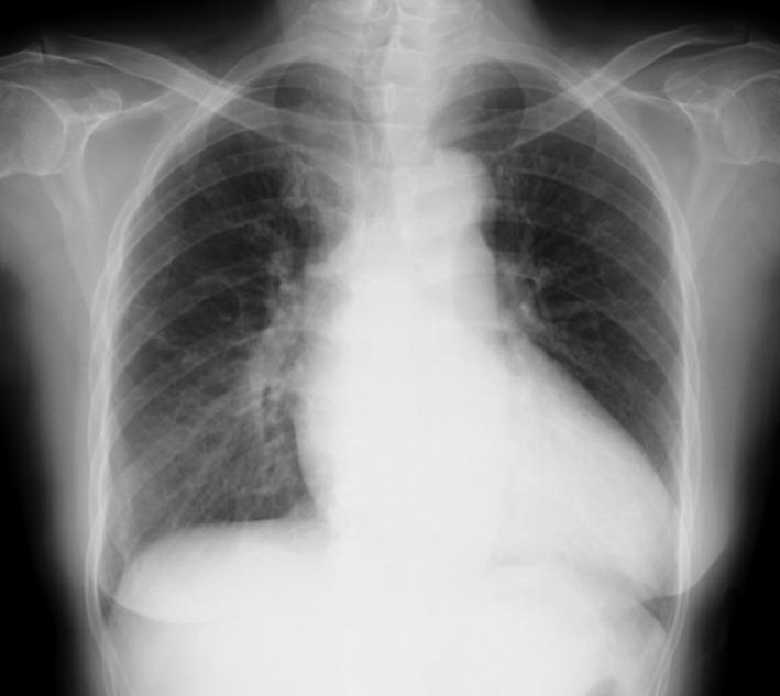
A posterior-anterior (PA) chest X-ray demonstrating a combination of thoracic findings. The image reveals prominent bilateral hilar lymphadenopathy (BHL), characterized by well-defined, bulky opacities in both hilar regions, consistent with Stage II sarcoidosis. Additionally, there is evidence of pulmonary edema, visualized as increased perihilar haziness and interstitial markings radiating from the central lung zones, which obscures the sharp margins of the pulmonary vasculature. The cardiac silhouette appears slightly enlarged, and there is a mild reduction in the clarity of the costophrenic angles. The musculoskeletal structures and overlying soft tissues are unremarkable. This radiographic presentation is significant for assessing systemic conditions such as cardiac sarcoidosis, where lymphadenopathy and acute congestive heart failure findings may coexist.
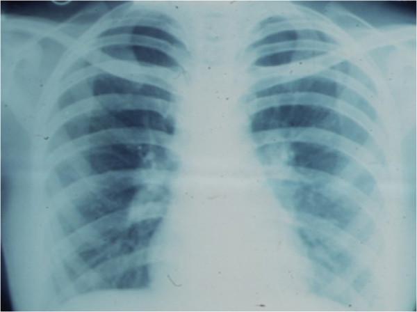
This diagnostic image is an anteroposterior chest X-ray demonstrating a classic presentation of bilateral hilar lymphadenopathy. The primary finding is the symmetrical enlargement of the hilar regions, appearing as lobulated, increased-density masses where the major bronchi and pulmonary vessels enter the lung parenchyma. This 'potato-node' appearance is a hallmark of pulmonary sarcoidosis. The lung fields are generally clear without obvious parenchymal infiltrates or consolidation, though the hilar margins are ill-defined due to the lymphatic enlargement. The mediastinal borders are slightly widened, but the cardiac silhouette and diaphragmatic contours remain distinguishable. The ribcage and clavicles are visualized and provide anatomical orientation. Clinically, this imaging is characteristic of Stage I sarcoidosis, often used to evaluate systemic granulomatous disease in both radiology and internal medicine education.
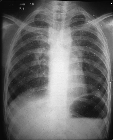
This diagnostic image is a posteroanterior (PA) chest X-ray illustrating classic features of stage II sarcoidosis. The primary finding is significant bilateral hilar lymphadenopathy, characterized by symmetrical enlargement and lobulated contours of both lung roots. Additionally, there is prominent soft tissue density in the left paratracheal region, indicating paratracheal lymphadenopathy. The lung parenchyma exhibits a diffuse reticulonodular pattern, with granular infiltrates most concentrated in the upper and middle lung zones bilaterally. These small, ill-defined opacities represent granulomatous inflammation within the interstitial spaces. The combination of symmetric hilar nodes and upper-lobe predominant parenchymal changes is highly characteristic of sarcoidosis. The image serves as a clinical reference for pulmonary manifestations of systemic granulomatous disease and the radiological assessment of mediastinal lymphadenopathy.
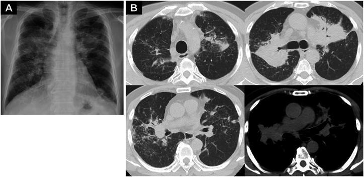
This composite diagnostic image features a posterior-anterior (PA) chest X-ray and axial chest computed tomography (CT) scans illustrating systemic sarcoidosis. (A) The chest X-ray shows bilateral, ill-defined hilar and mid-zone opacities, consistent with lymphadenopathy and pulmonary parenchymal involvement. (B) The CT scans (lung and mediastinal windows) provide detailed views of extensive mediastinal and bilateral hilar lymphadenopathy. Significant parenchymal findings include bilateral tumor-like consolidations and diffuse micronodular (granular) shadows with a perilymphatic distribution, characteristic of Stage II or III pulmonary sarcoidosis. Some lesions appear as large, dense masses mimicking malignancy, while others are small and scattered. The mediastinal window specifically highlights the conglomerate enlargement of the paratracheal and subcarinal lymph nodes. These findings, in conjunction with elevated ACE and sIL-2R levels mentioned in clinical context, demonstrate the multisystemic nature of the disease and its potential for varied radiological manifestations.
"bilateral hilar lymphadenopathy" diagnosis
Bilateral Hilar Lymphadenopathy (BHL)
Causes / Differential Diagnosis
Most Common
Other Causes (to exclude)
| Category | Specific Conditions |
|---|---|
| Infections | Tuberculosis, histoplasmosis, coccidioidomycosis, blastomycosis, Mycobacterium avium complex, EBV (infectious mononucleosis) |
| Malignancy | Lymphoma (Hodgkin's and non-Hodgkin's), lung carcinoma with nodal spread, leukemia, metastatic disease |
| Occupational/Inhaled | Berylliosis (chronic beryllium disease), silicosis, coal dust |
| Autoimmune/Inflammatory | Hypersensitivity pneumonitis, ANCA-associated vasculitis, IgG4-related disease, rheumatoid arthritis |
| Drug-induced | Immune checkpoint inhibitors, antiretroviral therapy, interferon, TNF-α antagonists |
| Other | Amyloidosis, Castleman disease, Löfgren syndrome |
Sarcoidosis in Detail (Most Common Cause)
Radiographic Staging
| Stage | Chest X-ray Finding | Spontaneous Remission |
|---|---|---|
| 0 | Normal | -- |
| I | BHL alone, clear lung fields | ~65-90% |
| II | BHL + parenchymal infiltrates | ~50-60% |
| III | Parenchymal infiltrates, no BHL | ~30% |
| IV | Pulmonary fibrosis | Rare |


Pathogenesis
- Intra-alveolar accumulation of CD4+ T cells (CD4:CD8 ratio 5:1 to 15:1)
- Elevated Th1 cytokines (IL-2, IFN-γ) leading to macrophage activation
- TNF, IL-8 drive ongoing granuloma formation
- Resultant non-caseating epithelioid granulomas - tightly formed, without central necrosis (distinguishing from TB/fungal infection)
- Polyclonal hypergammaglobulinemia and cutaneous anergy (e.g., negative tuberculin test)
Histology
Epidemiology
- Adults < 40 years; peak incidence 35-45 years
- Higher in women (up to 2:1)
- African Americans (35.5/100,000) and Northern Europeans (24/100,000) have highest rates
- African Americans have 10× higher rates than White Americans in the US
- Rare in Chinese and Southeast Asian populations
Clinical Presentations
- Asymptomatic - found incidentally on imaging
- Löfgren syndrome (acute sarcoidosis): BHL + erythema nodosum + bilateral ankle arthritis/periarthritis. Excellent prognosis, 70-80% remission rate.
- Heerfordt syndrome: uveoparotid fever (uveitis + parotid enlargement + facial nerve palsy + fever)
- Lupus pernio: indurated violaceous plaques of nose/cheeks - marker of severe, chronic disease
- Nonspecific: fevers, malaise, night sweats, weight loss, cough, dyspnea
- Extrapulmonary: ocular (uveitis), hepatic, cardiac (arrhythmias, heart block), neurologic, skin, musculoskeletal
Diagnosis of BHL/Sarcoidosis
- Compatible clinical/radiographic presentation
- Non-necrotizing granulomatous inflammation on biopsy
- Exclusion of alternative granulomatous causes
- CXR (initial), HRCT chest for detailed parenchymal assessment
- Biopsy - preferred via EBUS (endobronchial ultrasound)-guided lymph node sampling - high yield, low risk
- BAL: elevated lymphocyte %, CD4:CD8 ratio >3.5 supports diagnosis
- Serum ACE (elevated in ~60%, non-specific)
- Calcium (hypercalcemia from 1-alpha-hydroxylase activity in granulomas)
- LFTs, creatinine, urinalysis, ECG, ophthalmology exam
- 18F-FDG PET-CT for active inflammation mapping in difficult cases
Treatment
| Indication | Agent |
|---|---|
| Mild/asymptomatic Stage I-II | Observation (many remit spontaneously) |
| Symptomatic/organ-threatening | Prednisone 20-40 mg/day (first-line) |
| Steroid-sparing / steroid failure | Methotrexate (most evidence), azathioprine, mycophenolate, leflunomide |
| Skin/hypercalcemia | Hydroxychloroquine |
| Refractory disease | Infliximab or adalimumab (anti-TNF-α) - etanercept is ineffective |
- Duration: ~1 year for most; shorter courses = higher relapse
- Bisphosphonates to prevent steroid-induced osteoporosis
- Regular eye exams with steroids or hydroxychloroquine use
Key Points for Exams / Boards
- BHL = Sarcoidosis until proven otherwise - the most common cause
- Stage I sarcoidosis (BHL alone) has the best prognosis with highest spontaneous remission rate
- Granulomas are non-caseating (caseation points to TB/fungal)
- Löfgren syndrome = BHL + erythema nodosum + ankle arthritis - clinically secure diagnosis, no biopsy needed
- Serum ACE is elevated in ~60% but not diagnostic
- Hypercalcemia in sarcoidosis is due to extrarenal 1α-hydroxylase in granulomas converting 25-OH-vit D to calcitriol
- Etanercept does NOT work in sarcoidosis (unlike infliximab/adalimumab)
- CD4:CD8 ratio >3.5 on BAL supports diagnosis