


1000043304.jpg
1000043306.jpg
What is diagnosis
I can see images of a young infant/toddler with a widespread skin rash. Let me analyze both images carefully and consult the medical library.
analyse_media
analyse_media
Reading File
The image analysis is very clear. Now let me search the medical library for confirmation.
Finding Sources
Finding Sources
Reading File
Reading File
I now have all the information needed. Let me fetch a relevant textbook image for the answer.
Loading Image
Diagnosis: Molluscum Contagiosum (MC)
The clinical findings in these images are classic and consistent with a well-recognized diagnosis.
Key Findings Supporting This Diagnosis
| Feature | Seen in Images |
|---|---|
| Discrete, dome-shaped pearly papules | Yes - multiple lesions 2-6 mm |
| Central umbilication (dimple) | Yes - visible on many lesions |
| Widespread disseminated distribution | Yes - buttocks, lower back, thighs, arms, face/neck |
| Age group (infant/toddler) | Yes - highest incidence ages 1-4 years |
| Molluscum dermatitis (eczematous reaction around lesions) | Yes - confluent/scaly patches seen on buttocks |
What Is Molluscum Contagiosum?
Molluscum contagiosum is caused by MCV-1 (Molluscipoxvirus), the type responsible for virtually all infections in young children. The hallmark lesion is a small, firm, smooth-surfaced, dome-shaped pearly papule averaging 3-5 mm with a central umbilication or white core.
Textbook comparison image (Andrews' Diseases of the Skin - Fig. 19.34: Molluscum contagiosum in a child with atopic dermatitis):
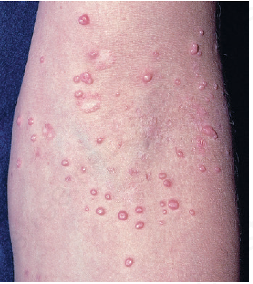
Clinical Notes for This Child
- Molluscum dermatitis is present (the eczematous, scaly, confluent patches around the buttock lesions) - this occurs in ~40% of affected children and is more common in atopic children
- The "BOTE" sign (Beginning Of The End) - inflamed/irritated lesions can signal the immune system is beginning to clear the infection
- The widespread distribution (buttocks, thighs, arms, face) is typical of childhood MC - in children, lesions are generalized and can number from a few to over 100
- Genital area involvement as part of widespread disease in a child does not automatically imply sexual contact - this is a recognized pattern
- Children with atopic dermatitis are 4x more likely to have >50 lesions and a longer course
Management
Most cases in immunocompetent children resolve spontaneously (typically within 6-18 months). Treatment options include:
- Watchful waiting - the standard approach in healthy children; most resolve without scarring
- Cantharidin (applied by clinician) - most commonly used office treatment
- 10% Potassium hydroxide solution - gentle topical option for children
- Curettage or cryotherapy - effective but painful, generally avoided in young children
- Dilute povidone-iodine or hydrogen peroxide 1% cream - gentle alternatives
- Treat co-existing molluscum dermatitis with low-potency topical corticosteroids (does not worsen MC)
Important: Check for underlying immunodeficiency if lesions are extremely numerous, giant (>1 cm), or not resolving - especially consider atopic dermatitis, HIV, or rare conditions like DOCK-8 mutation.
- Andrews' Diseases of the Skin: Clinical Dermatology, pp. 453-454
This is a shared conversation. Sign in to Orris to start your own chat.