
Typhoid
typhoid fever Salmonella typhi rose spots rash
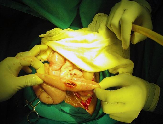
This clinical photograph shows an intraoperative view of an exploratory laparotomy, demonstrating a bowel perforation in the context of typhoid fever (Salmonella Typhi). The image focuses on a loop of small intestine held by a surgeon's gloved hands, revealing a discrete, longitudinal perforation with hyperemic and slightly everted edges. The surrounding serosa exhibits generalized inflammation and congestion. In the background, other segments of the intestine appear edematous and pale-pink, with visible white-yellowish peritoneal nodules and purulent exudate suggestive of concurrent miliary peritoneal tuberculosis and secondary peritonitis. Surgical instruments, suction tubing, and green sterile drapes define the surgical field. This image serves as an educational example of the severe gastrointestinal complications of typhoid fever, specifically intestinal perforation, and illustrates the challenging clinical scenario of co-infection with tuberculosis in an acute surgical setting.
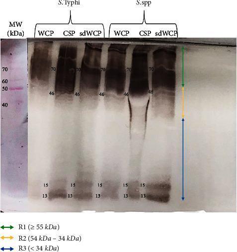
A diagnostic Western blot (WB) analysis image showing the immunoreactivity of antigens derived from Salmonella typhi and Salmonella spp. The image displays six experimental lanes representing three different protein extraction methods—Whole Cell Protein (WCP), Cell Surface Protein (CSP), and sodium deoxycholate-extracted WCP (sdWCP)—for each bacterial species. A molecular weight (MW) marker lane on the far left, stained with Ponceau S, shows reference bands from 40 kDa to 70 kDa. The WB profile is categorized into three vertical regions: R1 (≥ 55 kDa), R2 (54–34 kDa), and R3 (< 34 kDa). Prominent, high-intensity immunoreactive bands are observed in both species at approximately 70 kDa and 46 kDa, with a noticeable smear in the CSP lanes. Low molecular weight bands are consistently identified at 13 kDa and 15 kDa across all preparations. This visual provides comparative data on the antigenic profiles and antibody isotype reactivity against somatic and flagellar antigens, relevant for typhoid fever serodiagnosis research.
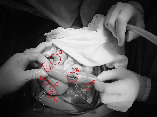
This monochrome clinical photograph captures an intraoperative view of the abdominal cavity during a surgical procedure for bowel perforation. The image illustrates two distinct infectious pathologies concurrently affecting the intestines and peritoneum. Label 'A' identifies a typhoid intestinal perforation, appearing as an irregular, longitudinal breach in the seromuscular layer of the small bowel, typical of advanced Salmonella typhi infection. Label 'B' shows a non-perforated typhoid lesion, characterized by localized inflammation and thickening without a full-thickness defect. Surrounding these intestinal segments, multiple small, discrete, whitish nodules are indicated by label 'C', representing miliary peritoneal tuberculosis. These nodules are scattered across the serosal surfaces and mesentery, demonstrating a grain-like distribution characteristic of disseminated mycobacterial infection. The image serves as a high-level educational example of acute surgical abdomen complications resulting from co-infection of typhoid fever and miliary tuberculosis, highlighting diagnostic landmarks such as transmural perforation and widespread peritoneal seeding.
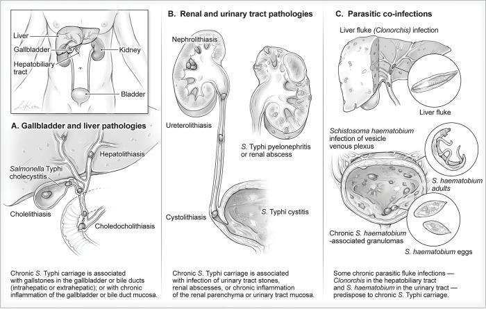
This medical illustration depicts anatomical locations and pathological conditions associated with chronic Salmonella Typhi carriage. The graphic is divided into three sections: (A) Gallbladder and liver pathologies, showing Hepatolithiasis, S. Typhi cholecystitis, Cholelithiasis, and Choledocholithiasis, illustrating how stones in the biliary system provide a niche for bacterial persistence. (B) Renal and urinary tract pathologies, highlighting Nephrolithiasis, Ureterolithiasis, Cystolithiasis, and S. Typhi-associated pyelonephritis or cystitis, where urinary stones or abscesses facilitate chronic carriage. (C) Parasitic co-infections, demonstrating how Liver flukes (Clonorchis) and Schistosoma haematobium (showing adults, eggs, and bladder granulomas) predispose patients to chronic infection. The diagram effectively connects infectious disease, pathology, and structural abnormalities of the hepatobiliary and urogenital systems, serving as an educational resource for understanding the reservoirs of chronic typhoid carriers.
Typhoid Fever (Enteric Fever)
Definition
- Harrison's Principles of Internal Medicine, 22nd Ed., p. 1359
Epidemiology
-
9.2-21 million cases of typhoid fever annually worldwide, causing 110,000-280,000 deaths per year
-
Highest incidence: Indian subcontinent (India, Pakistan, Bangladesh, Nepal), Eastern Mediterranean, and Africa - exceeding 1,000 cases per 100,000 children in some urban areas
-
Affects young children and adolescents disproportionately in endemic regions
-
In the US: ~5,700 cases/year; 78% are travel-associated (predominantly to India, Pakistan, Bangladesh)
-
Humans are the only reservoir - no animal hosts for S. Typhi or S. Paratyphi
-
Harrison's, p. 1359
Pathogen & Transmission
| Feature | Detail |
|---|---|
| Organism | Salmonella enterica serovar Typhi (Gram-negative rod, Enterobacteriaceae) |
| Transmission | Fecal-oral via contaminated food or water; fecal contamination by active cases or chronic carriers |
| Dose-dependent | Larger inoculum = shorter incubation & more severe disease |
| Risk factors | Contaminated drinking water/ice, street vendor food, raw produce fertilized with sewage, lack of hand hygiene, prior H. pylori infection (reduced gastric acidity) |
- Harrison's, p. 1359; Tintinalli's Emergency Medicine, p. 3379
Pathogenesis
- Ingestion - organism survives gastric acid and reaches the small intestine
- Invasion - penetrates intestinal mucosa at Peyer's patches (ileum)
- Lymphatic dissemination - travels to mesenteric lymph nodes
- Primary bacteremia - seeding of reticuloendothelial system (liver, spleen, bone marrow)
- Secondary bacteremia - massive multiplication in macrophages, re-entry into bloodstream causing the sustained febrile illness
- Unlike NTS gastroenteritis (PMN-driven), enteric fever produces mononuclear cell infiltration of the small-bowel mucosa
Clinical Course
Week-by-Week Progression
| Week | Features |
|---|---|
| Week 1 | Gradual onset fever (38.8-40.5°C), headache (80%), chills (35-45%), cough (30%), myalgias, malaise; relative bradycardia (Faget's sign) |
| Week 2 | High sustained fever, rose spots appear (~30% of fair-skinned patients), splenomegaly, abdominal distension, hepatomegaly, coated tongue |
| Week 3 | Complications peak: GI bleeding (6%), intestinal perforation (1%), neuropsychiatric features |
| Week 4 | Gradual defervescence if untreated; or complications/death |
Key Clinical Signs
- Rose spots - pale pink-red macular rash on the trunk and chest, 2-5 mm, fades in 2-5 days; culturable for S. Typhi; faint and easily missed in dark-skinned patients
- Relative bradycardia - pulse rate lower than expected for the degree of fever (Faget's sign)
- "Pea-soup" diarrhea OR constipation (30% present with constipation)
- "Muttering delirium" / coma vigil - classic neuropsychiatric sign with picking at bedclothes
- Harrison's, pp. 1360-1361; Tintinalli's, p. 3383
Complications (~27% of hospitalized patients)
- Intestinal perforation (1%) and GI bleeding (6%) - occur in 3rd-4th week from ulceration of Peyer's patches; life-threatening, require surgical intervention and broadened antibiotic coverage
- Hepatitis, pancreatitis, cholecystitis
- Meningitis, Guillain-Barre syndrome, neuritis, encephalopathy, psychosis, ataxia, seizures, deafness
- DIC, hemophagocytic syndrome, myocarditis, pericarditis, endocarditis, osteomyelitis, septic arthritis, glomerulonephritis, hemolytic-uremic syndrome, pneumonia
-
2-5% develop chronic asymptomatic carriage (shedding >1 year in urine or stool)
-
More common in women, infants, and those with biliary abnormalities or Schistosoma co-infection
-
Associated with increased risk of gallbladder cancer
-
Harrison's, pp. 1360-1361
Diagnosis
Culture (Gold Standard)
| Specimen | Sensitivity | Notes |
|---|---|---|
| Blood culture | ~40-60% | Lower yield in week 1; small inoculum (<15 organisms/mL) |
| Bone marrow culture | ~80% | Best single test; yield maintained even after 5 days of antibiotics |
| Stool culture | 30-40% week 1 → positive in week 3+ | |
| Combined (blood + BM + duodenal string test) | >90% |
Serology
- Widal test - detects agglutinating antibodies to O and H antigens; historically used but poor sensitivity/specificity
- Typhidot / Tubex - rapid IgM/IgG detection; sensitivity 70-80%, specificity 80-90%; not accurate enough to replace blood culture but useful at point of care
Molecular
-
PCR: sensitivity 40-100% (variable by gene target); increasingly available post-COVID pandemic
-
Harrison's, p. 1361
Treatment
Antibiotic Therapy (Adults)
| Indication | First-line Agent | Dose & Duration |
|---|---|---|
| Empirical (non-endemic area / susceptible strain) | Ciprofloxacin | 500 mg PO BID or 400 mg IV q12h x 10-14 days |
| Indian subcontinent / DSC/MDR strains | Azithromycin (mild-moderate) | 1 g PO day 1, then 500 mg/day x 5-7 days |
| Severe disease / MDR | Ceftriaxone | 2 g IV/IM OD x 10-14 days |
| Oral option | Cefixime | 400 mg PO BID x 10-14 days (higher clinical failure rate than FQs) |
| XDR strains | Carbapenems or azithromycin (per susceptibility) |
Key 2022 update: A systematic review of 27 RCTs found no significant difference between ceftriaxone, fluoroquinolone, or azithromycin in treatment failure, relapse, or adverse events. However, fluoroquinolones should no longer be used empirically in patients from the Indian subcontinent or Africa due to widespread decreased susceptibility (DSC) strains.
- Harrison's, p. 1361; Tintinalli's, p. 3387
Vaccines
| Vaccine | Type | Route | Protection | Notes |
|---|---|---|---|---|
| Ty21a | Live attenuated oral | Oral (4 capsules) | ~50-80% for 5-7 years | Avoid with antibiotics or immunosuppression |
| Vi-CPS (Typhim Vi) | Vi polysaccharide (inactivated) | IM injection | ~50-80% for 2-3 years | Not effective in children <2 years |
| Typhbar TCV / TCV | Typhoid conjugate vaccine (Vi-TT) | IM injection | ~80%+ for 5+ years | WHO preferred; effective in children >6 months; WHO SAGE recommended since 2018 |
-
Vaccines confer only partial immunity - vaccinated travelers must still avoid potentially contaminated food and fluids
-
Revaccination is required since protection wanes
-
Goodman & Gilman's Pharmacological Basis of Therapeutics; Harrison's
Prevention & Control
- Sanitation - safe drinking water, sewage treatment, food hygiene
- Hand hygiene - especially food handlers
- Case management - identify and treat chronic carriers (particularly food handlers); prolonged antibiotics +/- cholecystectomy for biliary carriers
- Vaccination - TCV (typhoid conjugate vaccine) campaigns in high-burden countries
- Carrier surveillance - 2-5% of patients become chronic carriers; cultures required to confirm clearance
- Park's Preventive & Social Medicine
Antimicrobial Resistance - Critical Alert
| Resistance Type | Drugs Resistant To | Origin/Spread |
|---|---|---|
| MDR S. Typhi | Chloramphenicol, ampicillin, TMP-SMX | Emerged 1980s; now widespread in Asia/Africa |
| DSC (decreased susceptibility to ciprofloxacin) | Ciprofloxacin (MIC ≥0.125 μg/mL) | Dominant on Indian subcontinent; ~66% of US travel-imported cases |
| XDR S. Typhi H58 | All of above + cephalosporins (ESBL) | Emerged Pakistan 2016; spreading globally via air travel; azithromycin is last oral option |
| Azithromycin resistance | Azithromycin | Emerging in multiple countries |
- Harrison's, pp. 1359-1361