
Whst is the association of sle of mother to baby
"neonatal lupus" AND "anti-Ro" AND outcomes
"neonatal lupus" AND "congenital heart block"
neonatal lupus rash skin manifestation
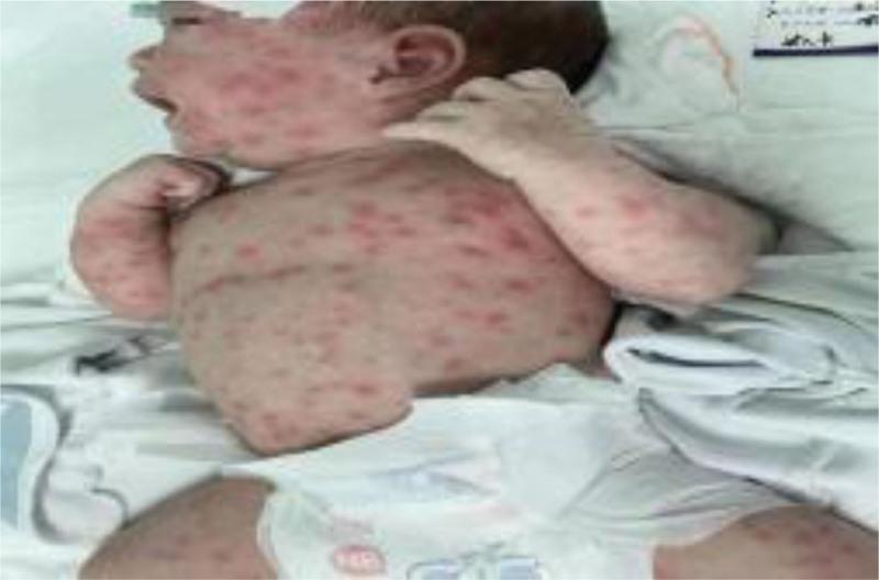
This clinical photograph displays a newborn infant in a neonatal care setting presenting with a widespread dermatological manifestation. The image depicts a diffuse, erythematous maculopapular rash consisting of individual, red, slightly raised lesions. The rash demonstrates extensive distribution, covering a high density on the face, scalp, and ears, while also involving the torso, upper extremities, and thighs. The clinical presentation is consistent with scarlet-like dermatitis or neonatal lupus erythematosus, particularly given the mention of positive ANA, SS-A, and SS-B antibodies in the context. The photograph serves as a visual reference for pediatric dermatology and neonatology, illustrating the morphology and generalized pattern of inflammatory skin conditions in a preterm infant. The background includes standard medical linens, emphasizing the clinical environment.
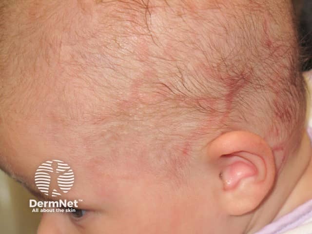
Clinical photograph with standard white-light illumination reveals a newborn scalp demonstrating erythematous, scaly patches along the hair-bearing skin. The lesions are modest in size, primarily clustered near the temporal and frontal scalp, with mild surface desquamation and surrounding mild perilesional erythema. This appearance is characteristic of cutaneous neonatal lupus erythematosus, a transient dermatologic manifestation caused by transplacental passage of maternal autoantibodies (SSA/Ro, SSB/La). The image emphasizes the typical distribution on the scalp in neonates, though other sites such as the face or neck may be involved. Although the cutaneous rash often resolves spontaneously by 6-12 months as maternal antibodies wane, neonatal lupus can be associated with congenital heart block; thus clinical correlation and cardiac surveillance are essential. Differential considerations include seborrheic dermatitis of infancy, atopic dermatitis, tinea capitis, cradle cap, and contact dermatitis. Histopathology (not shown) would demonstrate interface dermatitis with lymphocytic infiltrate and basal vacuolization in affected skin. Management is supportive, focusing on gentle cleansing, ceruminous moisturizers, and judicious topical corticosteroid therapy if inflammation is persistent. This image is valuable for dermatology, neonatal medicine, obstetric-pediatric collaboration, and educational case-based learning about lupus-related cutaneous disease in newborns. Clinically, early recognition supports targeted monitoring for cardiac rhythm abnormalities and long-term follow-up.
SLE in Mother: Effects on the Baby
Overview
1. Adverse Pregnancy Outcomes
- Pregnancy loss (miscarriage and stillbirth)
- Preterm birth
- Preeclampsia and pregnancy-induced hypertension
- Intrauterine growth restriction (IUGR)
- Cesarean section
2. Neonatal Lupus (NLE)
Manifestations:
| Feature | Details |
|---|---|
| Cutaneous rash | Most common and transient; erythematous, annular/maculopapular rash on face, scalp, and body. Resolves spontaneously. |
| Hematologic | Thrombocytopenia, neutropenia - seen in 10-25% of cases; usually self-limited |
| Hepatic | Asymptomatic transaminitis; dissipates after delivery |
| Congenital heart block | Most serious complication - see below |
3. Congenital Heart Block (CHB) - The Most Serious Complication
- CHB affects approximately 2% of infants exposed to maternal anti-Ro antibodies
- This risk rises to up to 20% in subsequent pregnancies after a previous infant was affected
- Risk is amplified by coexisting hypothyroidism and anti-La antibodies
- CHB typically develops between 18 and 24 weeks of gestation
- Survival is ~80% at 1 year, but the majority of affected infants will need a permanent pacemaker
- Premature atrial contractions and pericardial effusions in the fetus may foreshadow CHB
4. Monitoring During Pregnancy
- Anti-Ro/SSA antibody-positive mothers should be referred to high-risk obstetricians for serial fetal echocardiography between 18-24 weeks (ACR conditional recommendation)
- If first- or second-degree heart block is detected, treatment with dexamethasone may be initiated (some small series report reversal of early heart block)
- Prophylactic steroids are NOT recommended due to risk of IUGR and preterm birth
5. Protective Role of Hydroxychloroquine (HCQ)
- Reduces the risk of disease flares during pregnancy and postpartum
- Significantly reduces the risk of cardiac neonatal lupus / CHB
- A prospective open-label trial showed an odds ratio of 0.44 (p = 0.02) for CHB reduction in anti-Ro-positive women with a prior affected infant
- IVIG was NOT shown to be effective in preventing neonatal lupus
6. Key Antibody Summary
| Antibody | Association |
|---|---|
| Anti-Ro/SSA | Neonatal lupus, congenital heart block (primary antibody) |
| Anti-La/SSB | Neonatal lupus, congenital heart block (amplifies risk from anti-Ro) |
| Antiphospholipid antibodies (aPL) | Pregnancy loss, preterm birth, preeclampsia, IUGR |
| Lupus anticoagulant | Worst obstetric outcomes; use LMWH in pregnancy |