
Describe anatomy of heart
Reading File
Now I'll search the medical library for heart anatomy content in parallel.
Finding Sources
Finding Sources
Reading File
Finding Sources
Excellent - I found several strong sources. Let me read the most comprehensive ones in parallel.
Reading File
Reading File
Good content so far. Let me now fetch the detailed gross anatomy section from the anesthesia textbook and the key heart diagram image, in parallel.
Reading File
Loading Image
I now have rich content from multiple authoritative sources. Let me read more of the gross anatomy section and also get details on valves and conduction system.
Reading File
I now have very rich content from multiple textbooks. Let me also grab the valves and conduction system sections, and fetch the cardiac muscle histology image.
Reading File
Loading Image
I now have comprehensive content from multiple textbooks. Let me also get the valves section content that was partially cut off.
Reading File
I now have all the content needed for a comprehensive, well-structured answer. Let me compile it.
Anatomy of the Heart
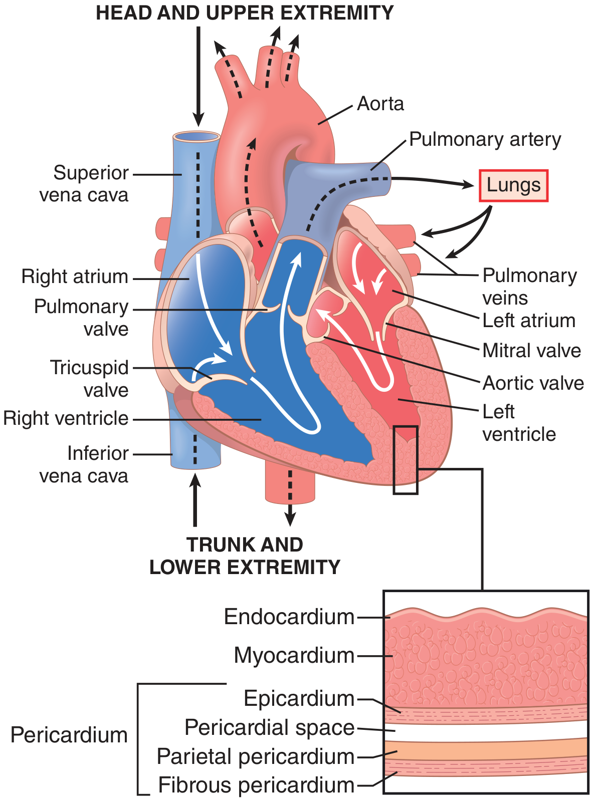
Figure: Structure of the heart and course of blood flow through the heart chambers and valves, showing all four layers of the heart wall (Guyton and Hall Textbook of Medical Physiology)
1. Overview
The heart is a phasic, electrically self-actuated, variable-speed hydraulic pump composed of two dual-component elastic muscular chambers - each consisting of an atrium and a ventricle. It simultaneously delivers equal quantities of blood to the pulmonary and systemic circulations. It lies in the mediastinum, with its base (superior aspect) facing upward and its apex pointing inferiorly and to the left, usually palpable at the left 5th or 6th intercostal space in the midclavicular line.
- Barash, Cullen, and Stoelting's Clinical Anesthesia, 9e, p. 842
2. Layers of the Heart Wall
The heart wall has four concentric layers (from outside in):
| Layer | Description |
|---|---|
| Fibrous pericardium | Outermost tough fibrous sac |
| Parietal pericardium | Lines the inner surface of fibrous pericardium |
| Pericardial space | Contains a small amount of serous fluid (~15-50 mL) for lubrication |
| Epicardium | Visceral pericardium; the outer surface of the heart itself; contains coronary vessels and fat |
| Myocardium | The thick muscular layer; composed of cardiomyocytes and cardiac fibroblasts |
| Endocardium | Innermost endothelial lining of all chambers and valves |
The heart is composed of 60-70% cardiac fibroblasts (not muscle cells). These produce the collagen extracellular matrix that surrounds cardiomyocytes and withstands the stress of constant pumping. - Goldman-Cecil Medicine, p. 412
3. Fibrocartilaginous Skeleton
The heart's skeleton is formed by:
- The annuli (rings) of all four valves
- The aortic and pulmonary arterial roots
- The central fibrous body
- The left and right fibrous trigones
This flexible cartilaginous structure at the base of the heart serves three functions:
- Supports the avascular valve leaflets
- Resists the forces of blood pressure and flow
- Provides a site of insertion for superficial subepicardial muscle
Critically, this skeleton also acts as an electrical insulator between the atria and ventricles, forcing all conduction through the AV node. - Barash, Cullen, and Stoelting's Clinical Anesthesia, 9e, p. 843
4. The Four Chambers
Right Atrium (RA)
- Thin-walled, low-pressure chamber
- Receives deoxygenated blood from the superior vena cava (from head/upper limbs) and inferior vena cava (from trunk/lower limbs)
- The coronary sinus also drains venous blood from the heart muscle here
- Composed of two thin sheets of myocardium oriented perpendicular to each other
Right Ventricle (RV)
- Located anterior and to the right of the LV within the mediastinum
- Crescent-shaped, thin-walled (~5 mm)
- Pumps deoxygenated blood into the low-pressure pulmonary arterial tree
- Contracts in a peristaltic "bellows-like" manner - the RV free wall moves toward the septum during contraction, with the LV providing a "splint"
- More compliant than the LV; accommodates volume more easily but is vulnerable to acute afterload increases
Left Atrium (LA)
- Receives oxygenated blood from four pulmonary veins
- Thin-walled; separated from the LV by the mitral (bicuspid) valve
- Serves three mechanical functions: conduit, reservoir, and contractile chamber
Left Ventricle (LV)
- The primary pumping chamber; ellipsoidal, thick-walled (~10 mm)
- Composed of billions of cardiomyocytes connected end-to-end through gap junctions
- Ejects oxygenated blood into the high-pressure systemic circulation
- The LV wall has three distinct muscle layers:
- Superficial bulbospiral (outer) - spirals leftward, rotates the apex counterclockwise
- Superficial sinospiral
- Deep sinospiral (inner) - spirals rightward, rotates the apex clockwise
- The net result of this double-helix fiber arrangement is a torsional "wringing" motion during systole (like wringing a cloth), which greatly increases ejection efficiency
- At end-systole, the LV acts like a loaded spring and recoils (untwists) during diastole to rapidly draw in blood
The LV generates 5-7x more pressure-volume work than the RV.
Interventricular septum: Shared wall between LV and RV; mostly derived from LV myocardium, so it normally thickens toward the LV during systole.
Trabeculae carneae: Irregular muscular ridges on the inner surface of the LV apex and RV ("meaty ridges" in Latin). The papillary muscles are specialized trabeculae that tether the AV valve leaflets via chordae tendineae.
- Barash, Cullen, and Stoelting's Clinical Anesthesia, 9e, p. 843-846
5. Heart Valves
Two pairs of valves ensure unidirectional blood flow:
Atrioventricular (AV) Valves
These separate the atria from the ventricles and are attached to papillary muscles via chordae tendineae:
| Valve | Location | Leaflets | Function |
|---|---|---|---|
| Tricuspid | Right AV junction | 3 leaflets | Opens when RV pressure < RA pressure; prevents backflow during systole |
| Mitral (Bicuspid) | Left AV junction | 2 leaflets | Opens when LV pressure < LA pressure; prevents regurgitation during systole |
The papillary muscles contract during systole to keep valve leaflets properly positioned and prevent regurgitation (they do not close the valves - pressure does).
Semilunar Valves
These separate the ventricles from the great arteries. They have no chordae tendineae:
| Valve | Location | Cusps | Function |
|---|---|---|---|
| Pulmonary | RV-pulmonary artery junction | 3 | Opens during RV systole; closes to prevent backflow |
| Aortic | LV-aorta junction | 3 | Opens during LV systole; closes to prevent backflow; coronary arteries arise just above it |
- Goldman-Cecil Medicine; Barash, Cullen, and Stoelting's Clinical Anesthesia, 9e, p. 847
6. Conduction System
The electrical activation sequence is tightly coupled to mechanical function:
| Structure | Role |
|---|---|
| SA node | Primary pacemaker; located in RA wall; initiates each heartbeat |
| Internodal pathways (anterior, middle/Wenckebach, posterior/Thorel) | Rapidly transmit SA impulse through RA to AV node |
| Bachmann's bundle | Transmits impulse from RA to LA across the atrial septum |
| AV node | Slow-conducting junction in the interatrial septum; introduces a delay allowing atrial contraction before ventricular contraction; only normal conduction path between atria and ventricles |
| Bundle of His | Pierces the fibrous skeleton; splits into left and right bundle branches |
| Left and Right bundle branches | Carry impulse down each side of the interventricular septum |
| Purkinje network | Rapidly distributes depolarization throughout the inner one-third of ventricular walls, ensuring fast, synchronized ventricular contraction |
Pathologic accessory pathways (e.g., Bundle of Kent in Wolff-Parkinson-White syndrome) bypass the AV node and can cause re-entrant tachyarrhythmias.
- Barash, Cullen, and Stoelting's Clinical Anesthesia, 9e, p. 848-849
7. Coronary Circulation
The coronary arteries arise from the aortic sinuses (just above the aortic valve) and travel through the epicardium:
| Artery | Territory Supplied |
|---|---|
| Left Anterior Descending (LAD) + septal/diagonal branches | Medial half of LV anterior wall, apex, anterior 2/3 of interventricular septum |
| Left Circumflex (LCCA) + obtuse marginal branches | Anterior and inferior aspects of the lateral wall |
| Right Coronary (RCA) + distal branches | Medial portions of inferior wall, posterior 1/3 of interventricular septum |
- Right-dominant circulation (RCA supplies posterior descending artery): ~80% of individuals
- Left-dominant circulation (LCCA supplies PDA): ~20%
Coronary flow occurs mainly during diastole (especially to the endocardium), because during systole the myocardium compresses the intramural coronary vessels.
- Barash, Cullen, and Stoelting's Clinical Anesthesia, 9e, p. 849-850; Goldman-Cecil Medicine, p. 413
8. Cardiac Muscle Microanatomy
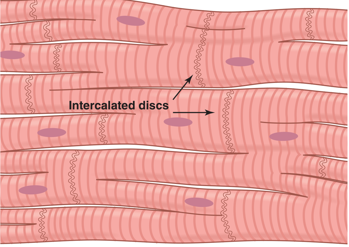
Figure: Syncytial interconnecting nature of cardiac muscle fibers, with prominent intercalated discs (Guyton and Hall Textbook of Medical Physiology)
-
Cardiac muscle fibers are striated (like skeletal muscle) and contain actin and myosin myofibrils
-
Cells are connected end-to-end by intercalated discs - specialized cell membranes containing gap junctions (low-resistance connections allowing rapid ion diffusion)
-
This makes the myocardium a functional syncytium: an action potential in one cell propagates to all connected cells
-
Cardiac muscle cells branch and interconnect in a latticework pattern
-
Nuclei are central (unlike skeletal muscle), and cells are rich in mitochondria (reflecting high oxidative energy demands)
-
Guyton and Hall Textbook of Medical Physiology, p. 122
Summary Table
| Structure | Key Fact |
|---|---|
| Location | Mediastinum; base up, apex down-left |
| Size | ~250-350 g in adults |
| LV wall thickness | ~10 mm |
| RV wall thickness | ~5 mm |
| Normal heart rate | 60-100 bpm (SA node driven) |
| Cardiac output | ~5 L/min at rest |
| Valves | 4 total: tricuspid, pulmonary, mitral, aortic |
| Dominant coronary circulation | Right dominant (~80%) |
Sources:
- Guyton and Hall Textbook of Medical Physiology
- Barash, Cullen, and Stoelting's Clinical Anesthesia, 9e
- Goldman-Cecil Medicine, International Edition
Describe mechanism of normal labour
Reading File
Searching sources...
Finding Sources
Finding Sources
Finding Sources
Finding Sources
Finding Sources
Reading File
Good content from Creasy & Resnik. Now let me read more of this section and also pull the Family Medicine / Anesthesiology sources in parallel for cardinal movements and physiology.
Reading File
Reading File
Excellent content. Now let me get the cardinal movements section and physiology of labour, plus fetch the Friedman curve image.
Reading File
Reading File
Excellent - very rich content from multiple sources. Now let me fetch the Friedman curve image and also search for the cardinal movements section specifically.
Loading Image
Finding Sources
Finding Sources
Reading File
I found the cardinal movements section in Roberts and Hedges. Let me read it.
Reading File
I now have comprehensive content from multiple authoritative textbooks. I have everything needed for a thorough, well-structured answer on the mechanism of normal labour. Let me compile it now.
Mechanism of Normal Labour
1. Definition and Initiation of Labour
Labour is defined as the onset of regular, painful uterine contractions associated with progressive effacement and dilation of the cervix. On average, term labour commences at 40 ± 2 weeks from the last menstrual period.
Initiating factors
The precise trigger is still incompletely understood but involves:
- Uterine distension - stretching of the myometrium stimulates contractility
- Increased myometrial oxytocin receptor density - circulating oxytocin levels may not rise initially, but the number of receptors increases sharply
- Prostaglandins - particularly PGE₂ and PGF₂α - produced by fetal membranes and decidual tissue drive myometrial contractions and cervical softening
- Fetal hypothalamo-pituitary-adrenal axis maturation - signals fetal readiness
Prodromal (pre-labour) signs (2-4 weeks before delivery)
-
Lightening - the fetal presenting part settles into the pelvis; the fundal height drops and pelvic pressure increases
-
Braxton Hicks contractions - irregular, painless contractions become more frequent
-
Cervical ripening - the cervix softens, effaces, and moves anteriorly (due to collagen breakdown mediated by prostaglandins and relaxin)
-
Bloody show - passage of blood-tinged mucous plug from the cervical os, usually within 1-3 days before true labour begins
-
Morgan & Mikhail's Clinical Anesthesiology, 7e, p. 1587; Textbook of Family Medicine 9e, p. 501
2. Stages of Labour
By convention, labour is divided into three (or four) stages:
| Stage | Onset | Completion |
|---|---|---|
| First stage (latent + active phases) | Onset of true labour | Complete cervical dilation (10 cm) |
| Second stage | Complete dilation | Delivery of the baby |
| Third stage | Delivery of the baby | Delivery of the placenta |
| Fourth stage (optional) | Delivery of placenta | 1 hour post-delivery; uterus contracted |
- Creasy & Resnik's Maternal-Fetal Medicine, p. 937; Textbook of Family Medicine 9e, p. 502
3. First Stage of Labour
The first stage is subdivided into the latent phase and the active phase - graphically depicted as a sigmoid (S-shaped) curve by Friedman (the "Friedman curve").
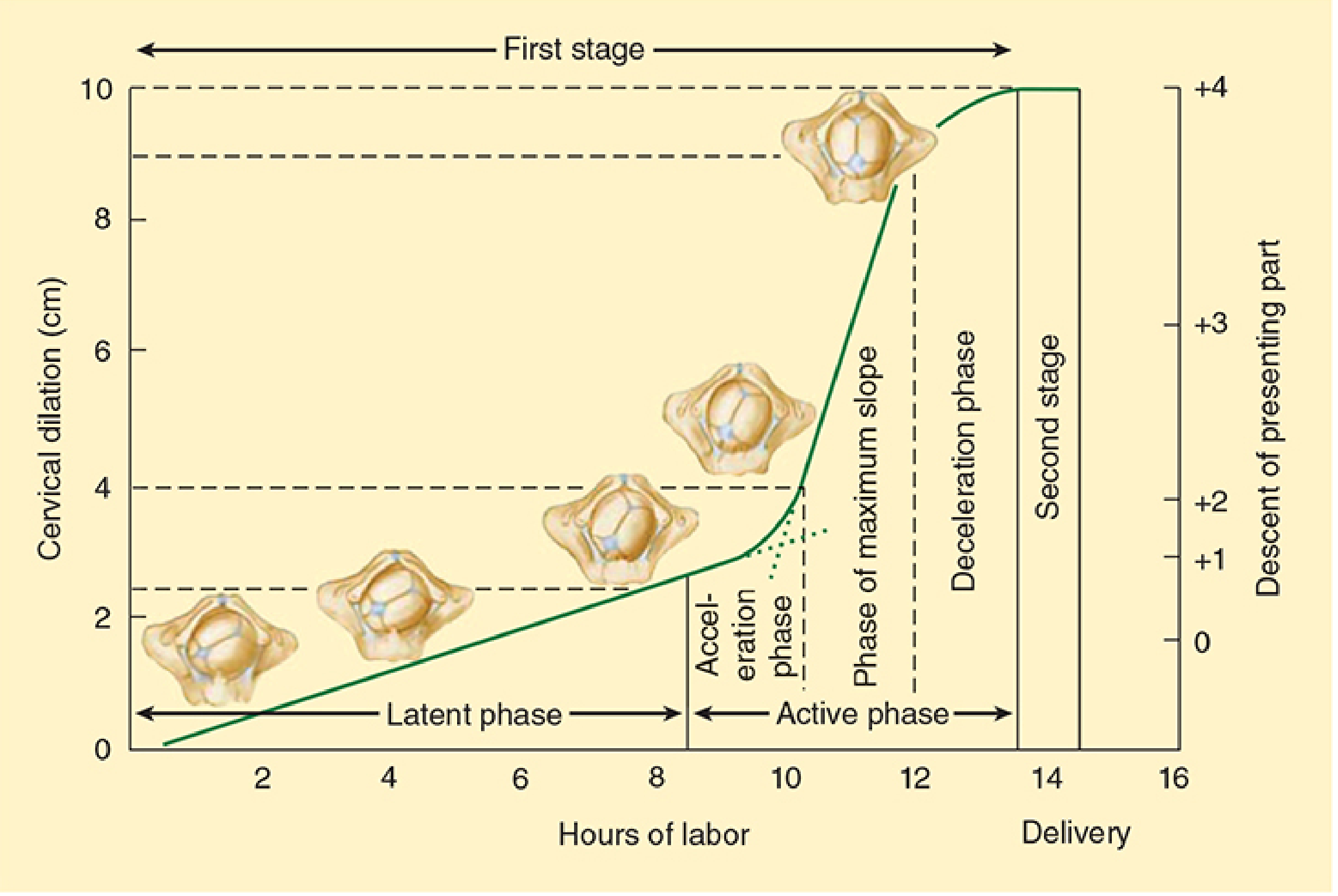
Figure: The course of normal labour. Cervical dilation (cm) on the left y-axis and descent of the presenting part on the right y-axis, plotted over hours of labour. The sigmoid Friedman curve shows the latent phase, acceleration phase, phase of maximum slope, and deceleration phase of the first stage. (Morgan & Mikhail's Clinical Anesthesiology, 7e)
Latent Phase
- From onset of regular contractions to the beginning of rapid dilation (~3-4 cm)
- Slow, minimal dilation - cervix effaces, softens, and moves anterior
- Collagen and connective tissue changes transform the cervix from firm to soft
- Normal upper limit: <20 hours in nulliparas; <14 hours in multiparas
- Contractions: irregular, becoming progressively more frequent
Active Phase
Friedman further divided the active phase into three sub-phases:
- Acceleration phase - rate of dilation begins to increase (~3-4 cm)
- Phase of maximum slope - most rapid dilation; normal minimum rate:
- Nulliparas: ≥ 1.2 cm/hr
- Multiparas: ≥ 1.5 cm/hr
- Deceleration phase - dilation slows as the cervix approaches 10 cm; fetal descent accelerates
Active phase contractions are stronger (25-60 mmHg), more coordinated, and occur every 3-5 minutes, lasting about 1 minute each.
Arrest of active phase is defined as no cervical dilation for ≥2 hours in the active phase.
Duration of First Stage (Friedman's data)
| Nulliparas | Multiparas | |
|---|---|---|
| Latent phase | 6.4 ± 5.1 hr | 4.8 ± 4.9 hr |
| Active phase | 4.6 ± 3.6 hr | 2.4 ± 2.2 hr |
| Total first stage | 11.0 ± 8.7 hr | 7.2 ± 7.1 hr |
- Creasy & Resnik's Maternal-Fetal Medicine, p. 937-938; Morgan & Mikhail's Clinical Anesthesiology, 7e, p. 1587
4. Cardinal Movements of Labour (Second Stage - Mechanism of Delivery)
The second stage begins at full cervical dilation (10 cm) and ends with delivery of the baby. During this stage, the fetus must navigate through the bony pelvis by a series of cardinal movements - passive mechanical adaptations of the fetal head to the pelvic dimensions. The fetus follows the path of least resistance by presenting its smallest diameter to the most favourable pelvic contour at each level.
The cardinal movements in vertex (cephalic) presentation are:
1. Engagement
- The biparietal diameter (BPD - the widest transverse diameter of the fetal head) passes through the pelvic inlet
- The fetal head is engaged when the presenting part is at station 0 (level of the ischial spines)
- In nulliparas: usually occurs 2 weeks before labour onset
- In multiparas: may occur only at the onset of labour
2. Flexion
- As the head descends and meets resistance from the pelvic floor and cervix, it flexes - the chin is brought to the chest
- This substitutes the larger occipitofrontal diameter (~12 cm) for the smaller suboccipitobregmatic diameter (~9.5 cm), greatly facilitating descent
- Occurs passively due to the lever-arm effect of the fetal body
3. Descent
- The downward passage of the presenting part through the pelvis
- Gradual and progressive but not necessarily continuous
- Driven by: uterine contractions, abdominal muscles (maternal pushing/Valsalva), straightening of the fetal body
- Descent rate accelerates in the late active phase and through the second stage
- Maximum descent rate: 3.3 cm/hr (nulliparas); 6.6 cm/hr (multiparas)
4. Internal Rotation
- Occurs as the head descends to the level of the ischial spines
- The occiput, initially in the transverse position (occiput transverse), rotates anteriorly toward the symphysis pubis → occiput anterior (OA) position - the most common and favourable
- Less commonly it rotates posteriorly → occiput posterior (OP) position
- This rotation aligns the longest diameter of the head with the longest diameter of the pelvic outlet (the anteroposterior diameter)
- The rotation is driven by the shape of the levator ani muscles forming the pelvic floor
5. Extension
- Once the occiput reaches and slides under the pubic symphysis, the head extends around the pubic arch
- The occiput, bregma, forehead, nose, mouth, and chin pass successively over the anterior perineum in that order
- Extension is due to the upward, anteriorly directed angle of the vaginal introitus - resistance from the perineum deflects the head upward
6. External Rotation (Restitution)
- Immediately after delivery of the head, it rotates back to its original transverse position - aligning with the fetal shoulders
- This is a passive movement (restitution) as the head realigns with the fetal body
- The occiput turns 45° to face the maternal thigh on the side of the fetal back
7. Expulsion
-
The anterior shoulder is delivered first with gentle downward traction, then the posterior shoulder with gentle upward traction
-
The remainder of the body follows rapidly
-
Roberts and Hedges' Clinical Procedures in Emergency Medicine, p. 1351-1352; Textbook of Family Medicine 9e, p. 400-401
5. Second Stage Duration
| Average | Upper limit | |
|---|---|---|
| Nulliparas | 50 minutes | 2 hr (no epidural), 3 hr (with epidural) |
| Multiparas | 20 minutes | 1 hr (no epidural), 2 hr (with epidural) |
Contractions: every 1.5-2 minutes, lasting 1-1.5 minutes - maternal bearing down can significantly augment intrauterine pressure.
6. Third Stage of Labour (Placental Delivery)
- Begins with delivery of the baby; ends with expulsion of the placenta
- Normal duration: 15-30 minutes
- Signs of placental separation:
- Lengthening of the umbilical cord
- A sudden gush of blood from the vagina
- The uterine fundus rises and changes shape (from discoid to globular)
- Mechanism: uterine contractions reduce the size of the placental implantation site, creating shearing forces that separate the placenta from the decidua
- Gentle cord traction (Brandt-Andrews manoeuvre) with uterine counter-pressure aids delivery
- After delivery, uterine contraction occludes open sinusoids at the placental bed - the primary haemostatic mechanism (the "living ligatures")
7. Fourth Stage of Labour
- The first hour after placental delivery - a period of physiological stabilisation
- The uterine fundus should be firm and contracted, palpable below the umbilicus
- Ongoing uterine atony (boggy, soft fundus) is the most common cause of primary postpartum haemorrhage
- Management: uterine fundal massage, oxytocin 20-40 units in IV fluids, or methylergonovine 0.2 mg IM
8. Uterine Contractions Throughout Labour
| Stage | Frequency | Duration | Intensity |
|---|---|---|---|
| Latent phase | Irregular, every 5-10 min | 30-45 sec | Mild (~20-25 mmHg) |
| Active phase | Every 3-5 min | ~60 sec | Moderate (25-50 mmHg) |
| Second stage | Every 1.5-2 min | 60-90 sec | Strong (50-80 mmHg) |
Adequate relaxation between contractions is essential to allow oxygenated blood to reach the intervillous space and transfer oxygen to the fetus. Contractions closer than every 2 minutes may compromise uteroplacental perfusion.
Summary: Cardinal Movements at a Glance
| Movement | What Happens | Why |
|---|---|---|
| Engagement | BPD through pelvic inlet; station 0 | Entry into pelvis |
| Flexion | Chin to chest; smaller diameter presented | Fit smallest head diameter to pelvis |
| Descent | Head moves downward | Propelled by contractions + maternal effort |
| Internal rotation | Occiput rotates to OA position | Align head with AP diameter of outlet |
| Extension | Head extends under pubic arch | Vaginal introitus directed anteriorly |
| External rotation | Head realigns with shoulders | Passive restitution |
| Expulsion | Anterior then posterior shoulder; body delivered | Completion of delivery |
Sources:
- Creasy & Resnik's Maternal-Fetal Medicine: Principles and Practice
- Morgan & Mikhail's Clinical Anesthesiology, 7e
- Textbook of Family Medicine, 9e
- Roberts and Hedges' Clinical Procedures in Emergency Medicine
This is a shared conversation. Sign in to Orris to start your own chat.