
Anti gbm disease from harrison 22 edition explain everything to know about md medicne resident
"anti-GBM disease" OR "Goodpasture syndrome"
anti-GBM disease Goodpasture syndrome immunofluorescence linear IgG glomerulus
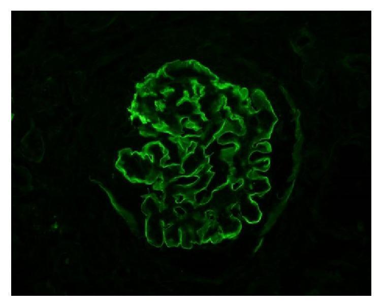
This diagnostic image is a high-power immunofluorescence (IF) microscopy of a human renal glomerulus. The image demonstrates a hallmark diagnostic pattern for anti-glomerular basement membrane (anti-GBM) disease. There is intense, continuous, and highly smooth linear staining for IgG (immunoglobulin G) along the glomerular capillary loops. Unlike the granular appearance typical of immune complex deposition (e.g., in lupus nephritis or post-infectious glomerulonephritis), this linear ribbon-like fluorescence highlights the entire circumference of the capillary basement membranes. The background is appropriately dark, emphasizing the bright green fluorescein isothiocyanate (FITC) signal. This visual finding is a key educational feature for identifying Goodpasture syndrome or anti-GBM mediated crescentic glomerulonephritis in a clinical pathology context. The pattern reflects the direct binding of autoantibodies to the non-collagenous domain of the alpha-3 chain of type IV collagen within the basement membrane.
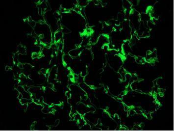
This diagnostic image is a direct immunofluorescence microscopy of a human renal biopsy. It shows a single glomerulus exhibiting intense, bright green fluorescent staining against a dark background. The staining pattern is classically linear, continuous, and smooth, outlining the glomerular basement membrane (GBM) along the capillary loops. This distinct visual feature indicates the diffuse deposition of IgG autoantibodies. Such findings are pathognomonic for anti-GBM disease (Goodpasture syndrome), demonstrating high specificity as evidenced by the lack of non-specific background staining in the surrounding mesangial or interstitial areas. This educational image is critical for distinguishing anti-GBM disease from other nephropathies like IgA nephropathy or post-infectious glomerulonephritis, which typically present with granular immunofluorescence patterns.
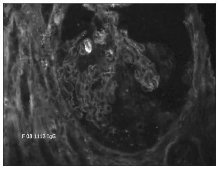
This diagnostic image shows a direct immunofluorescence (DIF) microscopy of a percutaneous kidney biopsy specimen at 20X magnification. The image demonstrates a hallmark pathognomonic finding: strong, continuous, linear immunofluorescence staining along the glomerular basement membrane (GBM). The staining outlines the capillary loops of the glomerulus with high intensity and uniformity, indicating the deposition of immunoglobulin G (IgG) antibodies directly against the basement membrane. The surrounding mesangial regions and Bowman's space show significantly less fluorescence, creating a sharp contrast that emphasizes the linear pattern. This visual presentation is characteristic of anti-glomerular basement membrane (anti-GBM) antibody disease, often associated with Goodpasture syndrome or rapidly progressive (crescentic) glomerulonephritis. The clinical significance of this finding is the confirmation of autoimmune-mediated renal damage where autoantibodies target Type IV collagen within the GBM.
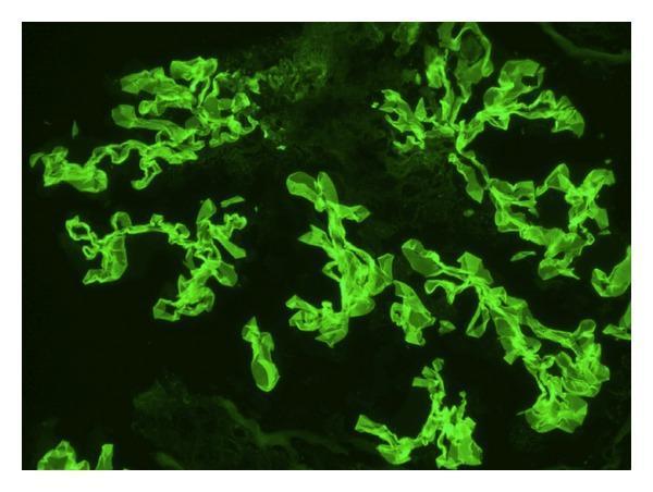
This diagnostic image is a direct immunofluorescence (DIF) micrograph of human renal tissue. The image demonstrates a characteristic linear staining pattern along the glomerular basement membrane (GBM). The fluorescent signal, appearing as a bright green (FITC) stain, reveals continuous, ribbon-like deposits of IgG that follow the complex, convoluted contours of the glomerular capillaries. This specific linear distribution is a hallmark of anti-GBM disease, such as Goodpasture syndrome, distinguishing it from the granular deposits typically seen in immune-complex-mediated glomerulonephritis. The clusters of highlighted structures represent multiple glomerular segments within the biopsy sample, showing uniform intensity of antibody binding. This finding is critical for the diagnosis of rapidly progressive glomerulonephritis (RPGN) and guides urgent clinical intervention like plasmapheresis and high-dose corticosteroid therapy.
Anti-GBM Disease (Goodpasture Syndrome)
Harrison's 22E - Complete Resident Review
1. DEFINITION & TERMINOLOGY
- Anti-GBM disease: Autoimmune glomerulonephritis caused by antibodies directed against the α3 NC1 domain of collagen IV in the glomerular basement membrane (GBM).
- Goodpasture's syndrome: When anti-GBM disease presents with both lung hemorrhage AND glomerulonephritis (pulmonary-renal syndrome).
- Renal-limited anti-GBM disease: Anti-GBM antibodies present, glomerulonephritis, but NO pulmonary involvement.
2. PATHOGENESIS
The Collagen IV Target
How Epitopes Are Exposed (Triggers)
- Infection (e.g., influenza, viral respiratory illness)
- Cigarette smoking (most common trigger for pulmonary involvement)
- Oxidants
- Hydrocarbon solvents / glue sniffing
Why the Lung is Involved
MHC Restriction
Mechanism of Kidney Injury
3. EPIDEMIOLOGY
- Rare disease - incidence ~1-2 per million per year
- Bimodal age distribution (key exam point):
- Young men in their late 20s - more likely full Goodpasture syndrome (renal + pulmonary)
- Older adults (60s-70s) - men AND women, more likely isolated glomerulonephritis
- Male predominance overall
- Majority of patients who develop pulmonary hemorrhage are active smokers
- A 2024 systematic review/meta-analysis (PMID: 38493958) confirmed these epidemiological features
4. CLINICAL PRESENTATION
Younger Patients (Full Goodpasture Syndrome)
- Hemoptysis (can be massive and life-threatening)
- Sudden fall in hemoglobin
- Fever
- Dyspnea
- Hematuria
- Better prognosis as a group because they present acutely
Older Patients (Renal-Limited Disease)
- Isolated glomerulonephritis - more insidious
- Prolonged, asymptomatic kidney injury
- Worse prognosis - often have significant fibrosis by the time of diagnosis
General Features of RPGN
- Nephritic syndrome features: hematuria, red cell casts, proteinuria (usually subnephrotic), hypertension, decreased GFR
- Rapidly declining kidney function over days to weeks
- Oliguria = particularly bad prognostic sign
Key Clinical Distinction from ANCA Vasculitis
| Feature | Anti-GBM Disease | ANCA Vasculitis |
|---|---|---|
| Disease course | Single acute fulminant episode | Can recur, even subclinically |
| Age | Bimodal (young men + elderly) | Older adults primarily |
| Pulmonary | Hemoptysis (smokers) | Hemoptysis, sinusitis, epistaxis |
| Serology | Anti-GBM Ab positive | ANCA (PR3 or MPO) positive |
| IF pattern | Linear IgG | Pauci-immune (no/scant deposits) |
| Recurrence | Rare after treatment | Common |
5. DIAGNOSIS
Serology
- Serum anti-GBM antibodies - the key test
- Critical technical point (Harrison's): The assay must use α3 NC1 domain of collagen IV alone as the target antigen
- Non-nephritic antibodies against α1 NC1 domain are seen in paraneoplastic syndromes and cannot be distinguished if whole basement membrane fragments are used as binding target
- ANCA testing (always do both anti-GBM AND ANCA)
- 10-15% of anti-GBM patients ALSO have ANCA (anti-MPO = p-ANCA)
- "Double positive" (anti-GBM + ANCA) = worse prognosis, behaves more like ANCA disease with tendency to relapse
Kidney Biopsy (Urgent - Do Not Delay)
- Focal or segmental necrotizing glomerulonephritis
- Crescent formation in Bowman's space (cellular crescents = parietal epithelial cell + monocyte/macrophage proliferation)
- Crescents compress and destroy the glomerular capillary tuft
- With progression: interstitial nephritis, fibrosis, and tubular atrophy
- Linear IgG deposition along the GBM - the hallmark finding
- Smooth, ribbon-like, continuous fluorescence along ALL capillary loops
- Contrast with granular deposits in immune-complex GN (lupus, post-strep, IgAN)
- With or without C3
- Rarely IgA instead of IgG
- No electron-dense immune deposits (unlike immune complex GN)
- GBM disruption and necrosis
Pulmonary Findings (if present)
- Diffuse alveolar hemorrhage on imaging (bilateral infiltrates)
- Lung biopsy: focal necrosis of alveolar walls, intraalveolar hemorrhage, hemosiderin-laden macrophages, fibrous thickening of septa, type II pneumocyte hypertrophy
- Linear IgG along alveolar septa (same pattern as kidney)
Lab Findings
- Elevated creatinine (often severely elevated at presentation)
- Urinalysis: hematuria, RBC casts, proteinuria
- Anemia (normocytic - due to hemorrhage or AKI)
- Complement normal (C3, C4 normal - unlike lupus nephritis or post-strep GN)
- Anti-GBM antibody titer elevated
6. PROGNOSIS DETERMINANTS
Poor Prognostic Factors at Presentation (Harrison's):
- >50% crescents on kidney biopsy with advanced fibrosis (fibrous/sclerotic crescents = irreversible)
- Serum creatinine >5-6 mg/dL at presentation
- Oliguria present
- Need for acute dialysis at presentation
- Older age with isolated renal disease (delayed diagnosis)
- Double-positive serology (anti-GBM + ANCA)
Better Prognosis:
- Younger patients with pulmonary presentation (caught earlier)
- Cellular crescents (potentially reversible) rather than fibrous crescents
- Lower creatinine at diagnosis
- Early aggressive treatment
7. TREATMENT
Step 1: Urgent Kidney Biopsy
- Confirms diagnosis and assesses % crescents and fibrosis
- Guides whether aggressive treatment is warranted (no benefit with >90% fibrous crescents + dialysis-dependent)
Step 2: Plasmapheresis (Plasma Exchange - PLEX)
- First-line treatment - removes circulating anti-GBM antibodies
- 8-10 treatments of plasmapheresis (typically daily or alternate day x 14 days)
- Especially indicated for pulmonary hemorrhage - lung hemorrhage responds to plasmapheresis
- Also used when dialysis-dependent (to save residual function) or with severe RPGN
Step 3: Immunosuppression (to prevent new antibody synthesis)
- Oral prednisone (1 mg/kg/day, max 60-80 mg) given concurrently with plasmapheresis
- Cyclophosphamide (oral 2-3 mg/kg/day OR IV pulse 500-1000 mg/m² monthly x 3-6 months)
- Duration: until anti-GBM antibody titers become negative
| Drug | Dose | Duration |
|---|---|---|
| Methylprednisolone IV pulse | 500-1000 mg/day x 3 days | Initial days |
| Oral prednisone | 1 mg/kg/day (taper) | Months |
| Cyclophosphamide | 2-3 mg/kg/day oral OR IV pulse | Until antibody negative |
| Plasmapheresis | 4L/session, 8-10 treatments | ~14 days |
Maintenance Therapy
- Low-dose immunosuppressants until antibody titers are negative
- Azathioprine, MMF (mycophenolate mofetil), or methotrexate used for maintenance
- Scarce data on rituximab or MMF as alternatives to cyclophosphamide (per Harrison's) - but rituximab is being used in refractory/anti-GBM + ANCA double-positive cases
Kidney Transplantation
- Wait at least 6 months after disease onset
- Serum anti-GBM antibodies must be undetectable before transplantation
- Alport syndrome patients who receive transplants typically develop anti-GBM antibodies directed against collagen IV epitopes absent in their native kidneys (important distinction)
When NOT to Treat Aggressively
- Dialysis-dependent at presentation + >50% fibrous crescents on biopsy + no pulmonary hemorrhage
- In these patients, the risk of cyclophosphamide toxicity outweighs the minimal chance of renal recovery
8. ANTI-GBM vs ANCA vs IMMUNE COMPLEX RPGN - Classification of RPGN
| RPGN Type | Immunofluorescence | Serology | Examples |
|---|---|---|---|
| Type I - Linear | Linear IgG along GBM | Anti-GBM Ab | Goodpasture syndrome, anti-GBM disease |
| Type II - Granular | Granular immune deposits | ANA, anti-dsDNA, low C3 | Lupus nephritis, post-strep GN, MPGN |
| Type III - Pauci-immune | No/scant deposits | ANCA (PR3 or MPO) | GPA, MPA, EGPA |
| Type IV - Double positive | Linear + scarce deposits | Anti-GBM + ANCA | Worse prognosis, may relapse |
9. SPECIAL SCENARIO: ANTI-GBM DISEASE IN ALPORT SYNDROME PATIENTS
- Patients with Alport syndrome have mutations in COL4A3/COL4A4/COL4A5 genes
- Their kidneys LACK normal α3·α4·α5(IV) collagen
- After kidney transplantation, they can develop de novo anti-GBM antibodies directed at the collagen IV epitopes present in the transplanted kidney but absent in their native kidneys
- This is a rare but recognized complication of renal transplantation in Alport patients
10. LUNG PATHOLOGY (Robbins/Harrison's)
- Focal necrosis of alveolar walls
- Intraalveolar hemorrhage
- Fibrous thickening of septa
- Hypertrophic type II pneumocytes
- Abundant hemosiderin-laden macrophages (residuum of prior hemorrhage)
- Linear IgG along alveolar septa on immunofluorescence (same as kidney)
11. KEY HIGH-YIELD EXAM POINTS
| Point | Detail |
|---|---|
| Antigen target | α3 NC1 domain of type IV collagen |
| "Conformeropathy" | Perturbation of α3·α4·α5 NC1 hexamer quaternary structure |
| IF pattern | Linear IgG (vs. granular in immune complex disease) |
| HLA association | HLA-DR15 (DRB1*15:01), HLA-DR4 |
| Triggers | Smoking, hydrocarbons, infections, oxidants |
| Double positive | Anti-GBM + ANCA (10-15%) = worse prognosis |
| Complement | Normal C3/C4 |
| Prognosis | Oliguria, Cr >6, >50% fibrous crescents = poor |
| Treatment | Plasmapheresis + cyclophosphamide + prednisone |
| Transplant timing | 6 months + antibody negative |
| Recurrence | Rare (unlike ANCA vasculitis) |
| Assay caution | Use α3-NC1 alone, not whole GBM - to avoid false positives from paraneoplastic α1-NC1 Ab |
Sources
- Harrison's Principles of Internal Medicine 22E (2025, McGraw Hill) - Sections "Antiglomerular Basement Membrane Disease" and "Basement Membrane Syndromes," pp. 2462-2469
- Brenner and Rector's The Kidney, 2-Volume Set - Anti-GBM chapter
- Robbins & Kumar Basic Pathology - Goodpasture Syndrome section
- Comprehensive Clinical Nephrology, 7th Edition - Goodpasture Disease chapter
- Recent systematic review: Kuang H et al., Autoimmun Rev 2024 - Epidemiology and clinical features meta-analysis (PMID: 38493958)