
Neurulation
Reading File
Neurulation is the embryological process by which the neural plate folds to form the neural tube, giving rise to the brain and spinal cord. It occurs in two phases: primary neurulation (folding and fusion of the neural folds) and secondary neurulation (cavitation of a solid cord of cells in the tail region). Defects in this process lead to neural tube defects such as spina bifida and anencephaly.
Searching sources...
Finding Sources
Finding Sources
Good - multiple strong sources found. Now let me read the key sections in parallel.
Reading File
Reading File
Reading File
Good content so far. Let me now read the deeper sections on molecular regulation, neural crest, secondary neurulation, and birth defects.
Reading File
Reading File
Reading File
Now let me get the sections on molecular regulation of neural induction, secondary neurulation, and neural tube defects.
Reading File
Reading File
Now let me get the molecular regulation and secondary neurulation sections from Langman's, and fetch a key diagram image.
Reading File
Loading Image
Loading Image
Excellent - I now have comprehensive content and images. Let me also get the NTD details from Langman's and then compile the full answer.
Reading File
I now have comprehensive content from multiple authoritative sources. Here is the full detailed answer:
Neurulation
Neurulation is the process by which the neural plate forms the neural tube - the primordium of the entire central nervous system. It occurs during weeks 3-4 of human embryogenesis and is one of the most critical events in early development.
1. Induction of the Neural Plate
The process begins after gastrulation. The notochord, once formed, releases inducing signals - principally soluble growth factors including FGF (fibroblast growth factor), WNT proteins, and BMP inhibitors (noggin, chordin) - that act on the overlying ectoderm.
- Upregulation of FGF signaling combined with inhibition of BMP (bone morphogenetic protein) activity induces the dorsal ectoderm to become neural ectoderm (neuroectoderm)
- High BMP concentrations → surface epidermis; very low BMP → neural ectoderm; intermediate BMP (at the border) → neural crest
- On approximately day 16, the overlying ectoderm thickens in response to notochordal induction, forming the neural plate - a slipper-shaped sheet of tall columnar epithelium
The neural plate appears rostral to the primitive node and dorsal to the notochord. As the notochord elongates, the neural plate broadens and extends cranially as far as the oropharyngeal membrane. - The Developing Human: Clinically Oriented Embryology
2. Primary Neurulation: Step-by-Step
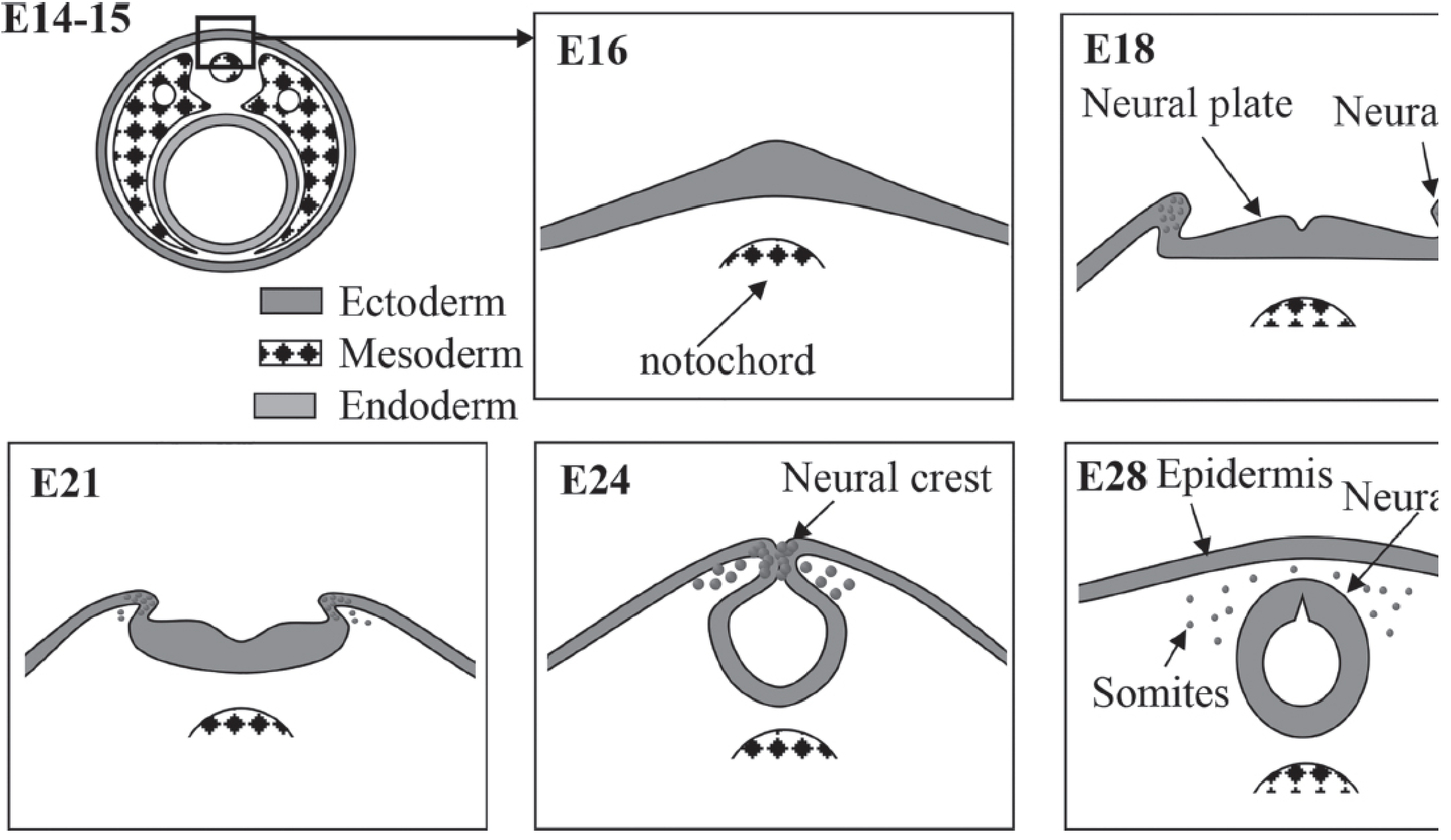
Cross-sections illustrating neurulation stages: notochord induction (E16), neural plate thickening (E18), neural fold elevation (E21), fold fusion with neural crest emergence (E24), and completed neural tube with surrounding epidermis and somites (E28). - Kaplan & Sadock's Comprehensive Textbook of Psychiatry
Step 1: Neural groove formation (~day 18)
- The neural plate invaginates along its central axis, forming a longitudinal neural groove flanked by neural folds on each side
- The neural folds become particularly prominent at the cranial end - these are the first signs of brain development
Step 2: Convergent extension (~day 19-20)
- The neural plate lengthens and the body axis extends via convergent extension: cells in the plane of the ectoderm and mesoderm undergo lateral-to-medial movement
- This process is regulated by the planar cell polarity (PCP) pathway (involving VANGL genes) and is essential for neural tube closure - Langman's Medical Embryology
Step 3: Neural fold elevation and fusion (~day 20-23)
- The lateral edges of the neural plate elevate as neural folds, which gradually approach each other in the midline
- Fusion begins in the cervical region (at the 5th somite / hindbrain level) and proceeds both cranially and caudally simultaneously
- This bidirectional "zipper-like" closure converts the neural plate into the neural tube
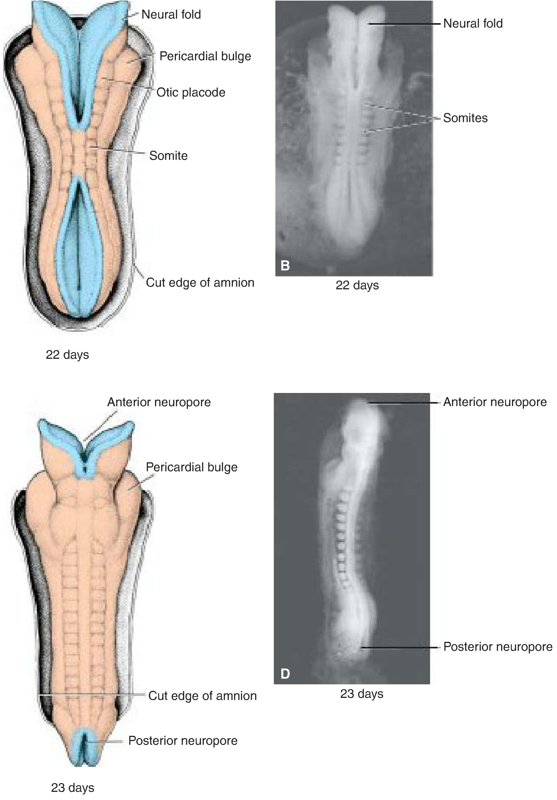
Dorsal views of human embryos showing the neural folds at day 22 and the nearly closed neural tube at day 23 with anterior and posterior neuropores. - Langman's Medical Embryology
Step 4: Neuropore closure
Until fusion is complete, the open ends of the neural tube communicate with the amniotic cavity through two temporary openings:
| Neuropore | Closure Day | Somite Stage |
|---|---|---|
| Anterior (cranial) neuropore | ~Day 25 | 18-20 somite stage |
| Posterior (caudal) neuropore | ~Day 28 | 25-somite stage |
Closure of the caudal neuropore marks the completion of primary neurulation by the end of the 4th week.
3. Secondary Neurulation
In the caudal-most part of the embryo (sacral and coccygeal segments), the neural tube forms differently:
- A solid cord of cells (the medullary cord/caudal cell mass) appears below the posterior neuropore
- This cord undergoes cavitation (hollowing out), and the resulting lumen connects with the canal of the primary neural tube
- Secondary neurulation produces the most distal part of the spinal cord (conus medullaris, filum terminale region)
4. Neural Crest Formation - A Key By-product of Neurulation
As the neural folds fuse, cells at the lateral borders (crests) of the neuroectoderm undergo an epithelial-to-mesenchymal transition (EMT) and dissociate from their neighbors. These are the neural crest cells (NCCs).
Molecular regulation of NCC specification:
- Intermediate BMP concentrations + FGF + WNT → induce PAX3 and other transcription factors specifying the neural plate border
- A second wave then induces SNAIL and FOXD3 to specify neural crest identity, and SLUG promotes migration
- Wnt/β-catenin signaling activates GBX2 homeobox gene, essential for neural crest development
Migration pathways from trunk NCCs:
- Dorsal pathway - through the dermis, then into the ectoderm to form melanocytes in skin and hair follicles
- Ventral pathway - through the anterior half of each somite to form sensory ganglia, sympathetic/parasympathetic neurons, Schwann cells, and enteric nervous system cells
Neural crest derivatives include:
- Bones and connective tissue of the face and skull
- Cranial nerve ganglia
- C cells of the thyroid
- Conotruncal septum of the heart
- Odontoblasts, dermis of face and neck
- Dorsal root (spinal) ganglia
- Sympathetic chain, preaortic, and parasympathetic ganglia
- Adrenal medulla
- Schwann cells, glial cells, meninges (forebrain)
- Melanocytes
- Smooth muscle of blood vessels in the face and forebrain
- Langman's Medical Embryology, Table 6.1
5. Cellular Mechanics of Neurulation
The shape changes that drive neural fold elevation involve:
- Apical constriction of neuroepithelial cells (mediated by actomyosin contractions) - cells become wedge-shaped
- Changes in intracellular cytoskeleton and cell-extracellular matrix attachment
- Differential cell adhesion - separating neural ectoderm from surface ectoderm
- After tube closure, surface ectoderm fuses over the neural tube and differentiates into the epidermis
6. Molecular Regulation Summary
| Signal | Effect |
|---|---|
| FGF (upregulated) | Promotes neural induction; essential for neural tube development and convergent extension |
| BMP inhibitors (Noggin, Chordin) | Block BMP activity → neural fate (low BMP) |
| Wnt/β-catenin | Neural crest specification (via GBX2) |
| PAX3 | Neural plate border specification |
| SNAIL, FOXD3 | Neural crest specification |
| SLUG | Promotes NCC migration |
| Planar Cell Polarity pathway / VANGL genes | Convergent extension → axis lengthening → tube closure |
| Retinoic acid (excess) | Teratogenic - disrupts neurulation |
| Valproic acid | Teratogenic NTD risk (especially folate-sensitive mechanism) |
7. Birth Defects from Abnormal Neurulation - Neural Tube Defects (NTDs)
Failure of neural fold fusion results in neural tube defects (NTDs), among the most common congenital anomalies:
| Defect | Region of Failure | Consequence |
|---|---|---|
| Anencephaly | Anterior neuropore fails to close | Most of the brain fails to form; lethal |
| Spina bifida | Posterior (cervical to sacral) failure | Variable neurological loss by level; most common site is lumbosacral |
Risk factors:
- Folate deficiency
- Retinoic acid (isotretinoin) exposure
- Valproic acid (antiepileptic)
- VANGL gene mutations (planar cell polarity pathway)
Prevention: 400 μg folic acid daily starting 3 months before conception and throughout pregnancy reduces NTD risk by 50-70%. Prior to flour fortification in the US, incidence was ~1/1,000; now approximately 1/1,500 births. - Langman's Medical Embryology
The neurocutaneous disorders (tuberous sclerosis, neurofibromatosis) also have their basis in the neural crest origin of both neural tissues and melanocytes at the border of neural and epidermal ectoderm. - Kaplan & Sadock's Comprehensive Textbook of Psychiatry
Quick Timeline Summary
| Day | Event |
|---|---|
| Day 16 | Neural plate induced by notochord; ectoderm thickens |
| Day 18 | Neural groove forms; neural folds appear |
| Day 19-20 | Neural folds elevate; convergent extension lengthens axis |
| Day 22 | Fusion begins at cervical region (5th somite) |
| Day 25 | Anterior neuropore closes |
| Day 28 | Posterior neuropore closes → primary neurulation complete |
| Week 4+ | Secondary neurulation (caudal cord) by cavitation |
This is a shared conversation. Sign in to Orris to start your own chat.