
Aast liver traima classification
AAST liver trauma injury classification grade scale
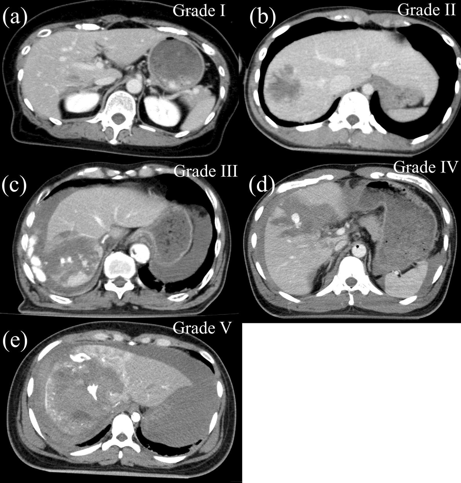
This composite diagnostic image presents five axial contrast-enhanced abdominal CT scans (labeled a-e) illustrating the AAST grading scale for blunt liver trauma. (a) Grade I: Shows a superficial laceration (<1 cm depth) or a small subcapsular hematoma involving <10% of the surface area. (b) Grade II: Demonstrates a deeper laceration (1–3 cm) or an intraparenchymal hematoma. (c) Grade III: Displays a large laceration (>3 cm depth) or a hematoma >10 cm in diameter, often with active intraparenchymal bleeding or extravasation of contrast. (d) Grade IV: Exhibits significant parenchymal disruption involving 25–75% of a hepatic lobe, showing extensive mottled density. (e) Grade V: Represents severe injury with parenchymal disruption exceeding 75% of a lobe, often accompanied by major venous injuries. The progression from Grade I to V is visually characterized by increasing heterogeneity of the liver parenchyma, loss of vascular architecture, larger areas of hypoattenuating lacerations/hematomas, and more extensive perihepatic fluid accumulation (hemoperitoneum).
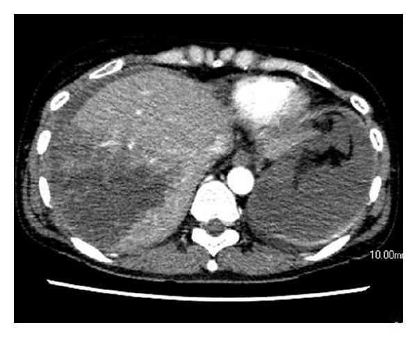
This diagnostic image is an axial contrast-enhanced CT scan of the upper abdomen showing acute traumatic liver injury. The primary finding is a deep, heterogeneous hypodense area within the right hepatic lobe, consistent with a Grade IV liver laceration according to the American Association for the Surgery of Trauma (AAST) Organ Injury Scale. There is notable disruption of the normal hepatic parenchyma architecture. Surrounding the liver and spleen, high-density fluid is visible, indicative of a significant hemoperitoneum (intra-abdominal hemorrhage). Despite the extensive laceration, there is no evidence of active contrast extravasation, pseudoaneurysm, or arterioportal fistula (APF) at this stage. The scan is a critical educational example of blunt abdominal trauma imaging, demonstrating the hallmarks of major solid organ injury and the typical appearance of associated peritoneal free fluid in an emergency radiology context.
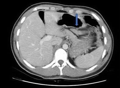
This diagnostic image is an axial contrast-enhanced CT scan of the upper abdomen demonstrating a grade II pancreatic injury. Key anatomical landmarks include the liver in the right upper quadrant, the spleen in the left upper quadrant, the gas-filled stomach centrally, and the vertebral column posteriorly. A blue arrow identifies a focal area of hypoattenuation and disruption within the pancreatic parenchyma, located posterior to the stomach. The injury represents a laceration that does not appear to involve the major pancreatic duct, consistent with the classification of a grade II injury according to the American Association for the Surgery of Trauma (AAST) scale. There is no evidence of large-volume hemoperitoneum or extensive peripancreatic fluid collection in this single slice. The scan is clinically significant for evaluating blunt abdominal trauma and determining the feasibility of non-operative management versus surgical intervention, such as laparoscopic debridement.
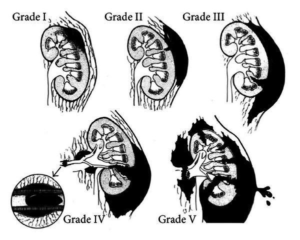
An anatomical illustration depicting the American Association for the Surgery of Trauma (AAST) Organ Injury Severity Scale for renal trauma, classified from Grade I to Grade V. The diagram uses cross-sectional views of the kidney to show progressive anatomical disruption. Grade I: Displays a subcapsular hematoma and renal contusion without laceration. Grade II: Shows a superficial parenchymal laceration (less than 1 cm depth) confined to the cortex. Grade III: Illustrates a deeper parenchymal laceration (greater than 1 cm) without involvement of the collecting system. Grade IV: Depicts a laceration extending through the cortex, medulla, and into the renal collecting system, accompanied by vascular injury and a segmental hematoma (an inset highlights the vascular thrombus). Grade V: Features a 'shattered kidney' with multiple major lacerations, devascularization, and complete avulsion of the renal hilum with extensive hemorrhage. This educational chart serves as a standardized reference for urological and trauma surgery to guide clinical management and surgical intervention.
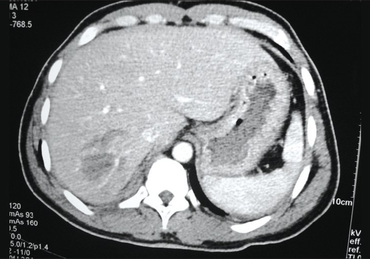
**Modality:** Axial contrast-enhanced computed tomography (CT) of the upper abdomen. **Anatomical Region:** Liver and surrounding upper abdominal structures. **Observed Pathology:** Grade II hepatic injury according to the AAST (American Association for the Surgery of Trauma) classification. **Visual Features:** - **Parenchymal Findings:** A localized, ill-defined hypodense area is visible within the posterior segment of the right hepatic lobe, consistent with an intraparenchymal hematoma or contusion. - **Injury Characteristics:** The lesion measures less than 10 cm in diameter with no evidence of major vascular disruption or active extravasation of contrast material in this section. - **Associated Findings:** A thin rim of hyperdense fluid is noted in the perihepatic and perisplenic spaces, suggestive of a small volume of hemoperitoneum. The spleen and visualized vasculature appear intact. **Diagnostic Context:** Findings are characteristic of blunt abdominal trauma resulting in a stable hepatic parenchymal injury. The absence of deep lacerations (>3 cm) or expanding hematomas supports the Grade II classification, making this image a primary reference for non-operative trauma management indexing.
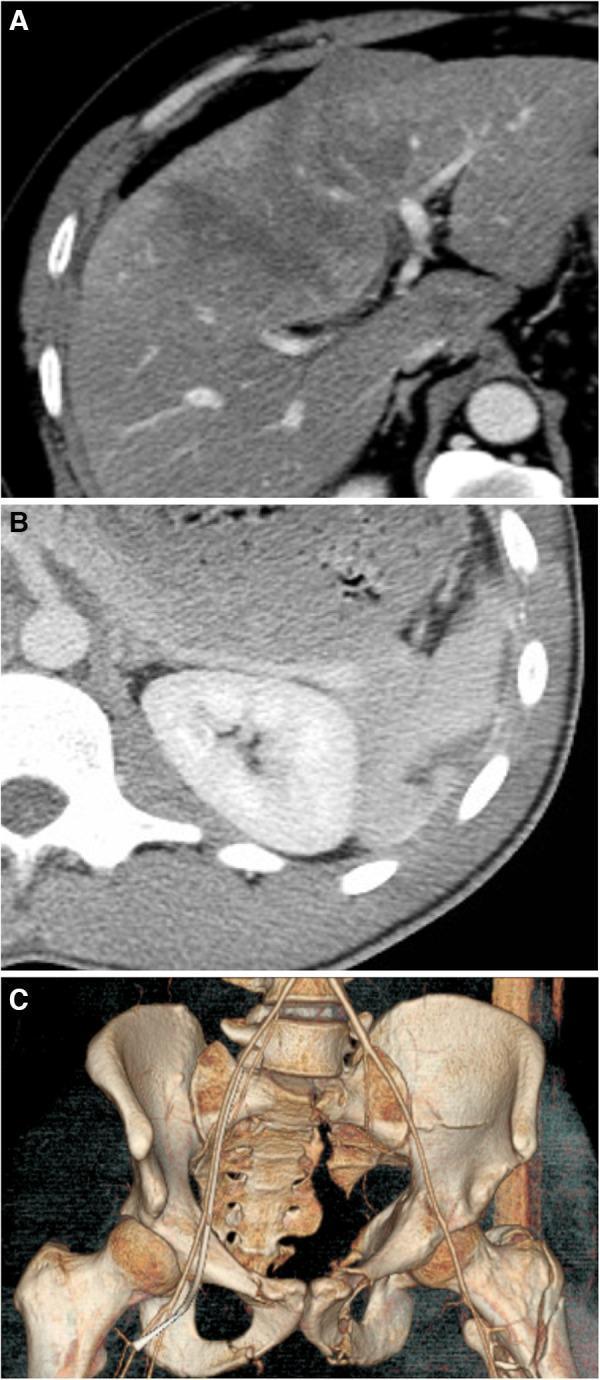
This composite diagnostic image illustrates traumatic solid organ and skeletal injuries using computed tomography (CT). Panel A shows an axial CT scan of the liver demonstrating an AAST Grade II liver injury, characterized by a low-density subcapsular hematoma and a focal intraparenchymal hematoma. Panel B displays a contrast-enhanced axial CT scan of the abdomen featuring a Grade II splenic laceration, visible as a linear hypodensity within the splenic parenchyma. Panel C presents a 3D CT reconstruction of the pelvis showing complex multisite fractures following high-energy trauma. Notable findings include displaced fractures of the left superior and inferior pubic rami, a left acetabular fracture, a left sacral fracture with moderate displacement, and a left intertrochanteric femur fracture. Non-displaced fractures of the right pubic rami are also suggested. The series serves as an educational resource for identifying visceral and pelvic girdle trauma on cross-sectional and reconstructed imaging, emphasizing injury grading systems and anatomical displacement.
AAST Liver Trauma Classification (OIS 2018)
Grade I - AIS 2 (Minor)
| Criteria | Description |
|---|---|
| CT/Imaging | Subcapsular hematoma <10% surface area; parenchymal laceration <1 cm depth |
| Operative | Same + capsular tear |
| Pathologic | Same + capsular tear |
Grade II - AIS 2 (Minor-Moderate)
| Criteria | Description |
|---|---|
| CT/Imaging | Subcapsular hematoma 10-50% surface area; intraparenchymal hematoma <10 cm; laceration 1-3 cm depth and ≤10 cm length |
| Operative | Same |
| Pathologic | Same |
Grade III - AIS 3 (Moderate-Severe)
| Criteria | Description |
|---|---|
| CT/Imaging | Subcapsular hematoma >50% surface area; ruptured subcapsular or parenchymal hematoma; intraparenchymal hematoma >10 cm; laceration >3 cm depth; any injury with liver vascular injury or active bleeding contained within parenchyma |
| Operative | Subcapsular hematoma >50% or expanding; ruptured hematoma; intraparenchymal hematoma >10 cm; laceration >3 cm depth |
| Pathologic | Same as operative |
Grade IV - AIS 4 (Severe)
| Criteria | Description |
|---|---|
| CT/Imaging | Parenchymal disruption 25-75% of a hepatic lobe; active bleeding extending beyond liver parenchyma into peritoneum |
| Operative | Parenchymal disruption 25-75% of a hepatic lobe |
| Pathologic | Same |
Grade V - AIS 5 (Critical)
| Criteria | Description |
|---|---|
| CT/Imaging | Parenchymal disruption >75% of hepatic lobe; juxtahepatic venous injury (retrohepatic vena cava and central major hepatic veins) |
| Operative | Same |
| Pathologic | Same |
Note: Grade VI (hepatic avulsion) existed in older versions but was removed in the 2018 revision.
Key Rules (2018 Update)
- Grading principle: Grade based on the highest-grade assessment - whether from imaging, operative findings, or pathologic specimen.
- Multiple injuries: When more than one grade is present, classify by the higher grade.
- Multiple injuries up-grading: Advance one grade for multiple injuries, up to Grade III maximum.
- Vascular injury definition: A pseudoaneurysm or arteriovenous fistula - appears as a focal collection of vascular contrast that decreases in attenuation on delayed imaging. Active bleeding increases in size or attenuation on delayed phase.
- Grade does not equal hemodynamic instability: Grades do not necessarily correlate with hemodynamic instability or need for emergent surgery.
CT Images of AAST Liver Grades (I-V)
Clinical Relevance
| Grade | Typical Management |
|---|---|
| I-II | Non-operative management (NOM) in hemodynamically stable patients |
| III | NOM if stable; angioembolization if active blush on CT |
| IV-V | High risk - often require angioembolization or operative intervention; damage control surgery |
New page