
Mm confirmed further treatment and evaluation
What does 'Mm confirmed further treatment and evaluation' refer to?
What aspect of post-diagnosis multiple myeloma management would you like to focus on?
"multiple myeloma" AND "staging" AND "diagnostic workup"
"multiple myeloma" AND diagnosis AND staging
multiple myeloma bone marrow plasma cells lytic lesions skull
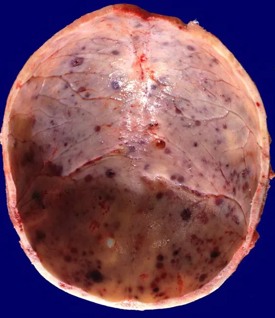
Gross pathology photograph of the cranial vault (calvaria) from a 60-year-old male with plasma cell myeloma. The image demonstrates multiple round, punched-out lytic lesions scattered across the skull with destructive osteolysis of the calvarial bone. Lesions are variably sized, ranging from 1 to 4 cm in diameter, and are largely devoid of surrounding sclerosis. The affected bone shows pale, creamy to tan-rose, gelatinous tumor tissue within the radiolucent areas; the interior is soft and hemorrhagic, with scattered dark red foci representing microhemorrhages. The periosteum is disrupted in focal regions, and there is minimal inflammatory reaction. This macroscopic appearance reflects clonal proliferation of immunoglobulin-producing plasma cells occupying medullary spaces and replacing normal bone marrow. Clinically, such skull lesions are highly characteristic of multiple myeloma and can lead to pain, pathologic fracture, and neurological compromise if the skull base or spine is involved. The image provides a visual correlate for the classic punched-out radiolucencies described in hematologic malignancies and should be correlated with serum protein electrophoresis, immunofixation, and bone marrow biopsy. Differential considerations include metastatic skull lesions, Langerhans cell histiocytosis, osteomyelitis, and eosinophilic granuloma. This photograph is often used in educational materials to illustrate gross features of monoclonal gammopathy with skeletal involvement and to aid in teaching macroscopic recognition of myeloma.
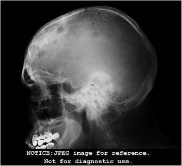
This lateral skull radiograph (X-ray) demonstrates the classic 'punched-out' or 'moth-eaten' appearance characteristic of multiple myeloma. The image reveals numerous well-circumscribed, round-to-oval radiolucent (lytic) lesions of varying sizes distributed across the calvarium, including the frontal, parietal, and occipital bones. These lesions show no evidence of peripheral sclerotic margins, indicating aggressive bone resorption without significant reactive bone formation. The calvarial diploe is notably involved, showing a generalized decrease in bone density and disruption of the normal trabecular pattern. Clinical significance of these findings includes the hallmark skeletal manifestations of plasma cell dyscrasias, where monoclonal plasma cells infiltrate the bone marrow, triggering osteoclast activity. This diagnostic image serves as an educational example of hematologic malignancy-associated osteolysis and is a critical component of a skeletal survey for staging conditions like multiple myeloma or metastatic disease.
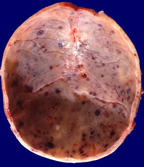
Gross pathology photograph of calvarial skull bones from a 60-year-old male with plasma cell myeloma showing multiple round lytic lesions. The skull base and vault are involved with discrete, punched-out defects ranging approximately 1 to 4 cm in diameter. The lesions are filled with soft, gelatinous red-brown tumor tissue, giving the inner table a slightly eroded appearance and thinning of the surrounding cortex. The overlying periosteum is variably preserved; surrounding diploic spaces may appear expanded and vascularized. The appearance is characteristic of osteolytic activity produced by clonal plasma cells that suppress normal bone formation and promote bone resorption, commonly involving the vertebral column, skull, ribs, pelvis, proximal long bones, and clavicles. In this specimen, the cranial vault bears multiple nodular foci with conspicuous dark hematopoietic plugs and subtle hemorrhagic foci. The gross features correlate with radiographic images of lytic skull lesions seen in multiple myeloma, or plasmacytomas, and reflect high tumor burden. Clinically, these findings support a diagnosis of multiple myeloma in the appropriate systemic context (anemia, hypercalcemia, renal dysfunction, monoclonal protein). Differential diagnoses include metastatic cancer and Langerhans cell histiocytosis. This image is useful for surgical pathology education, gross anatomy training, and radiology-pathology correlation exercises.
Multiple Myeloma: Post-Confirmation Diagnostic Workup & Staging
1. Diagnostic Criteria Recap (IMWG)
- ≥10% clonal plasma cells on bone marrow biopsy, AND
- M protein in serum or urine (except nonsecretory myeloma), AND
- ≥1 myeloma-defining event (MDE):
- Hypercalcemia (Ca >1 mg/dL above upper normal or >11 mg/dL)
- Renal insufficiency (creatinine >2 mg/dL or CrCl <40 mL/min)
- Anemia (Hb <10 g/dL or >2 g below normal)
- Bone lesions - ≥1 lytic lesion on skeletal survey, CT, or PET-CT
- Biomarker MDEs: BM plasma cells ≥60%, serum involved/uninvolved FLC ratio ≥100, or >1 focal lesion on MRI ≥5 mm
2. Laboratory Workup
Serum Studies
| Test | Purpose |
|---|---|
| Serum protein electrophoresis (SPEP) | Detect and quantify M spike |
| Serum immunofixation electrophoresis (IFE) | Identify immunoglobulin class (IgG, IgA, IgM, IgD, IgE) |
| Serum free light chains (kappa/lambda ratio) | Detect nonsecretory/light-chain myeloma; monitor response |
| Quantitative immunoglobulins | Assess immunosuppression of uninvolved Ig |
| CBC with differential | Anemia, cytopenias, rouleaux formation on smear |
| Comprehensive metabolic panel | Creatinine, calcium, albumin, LDH, uric acid |
| Beta-2 microglobulin (β2M) | Key staging marker (ISS) |
| Albumin | Paired with β2M for ISS staging |
| LDH | Elevated = high tumor burden, poor prognosis (R-ISS) |
| CRP | Indirect measure of IL-6 activity |
| Coagulation studies | Factor X deficiency in amyloid co-occurrence |
Serum immunofixation is more sensitive than SPEP - it can detect low M components not visible on electrophoresis. - Harrison's 22E
Urine Studies
- 24-hour urine protein electrophoresis + IFE - quantify and type Bence Jones protein (free light chains in urine)
- Spot urine total protein and albumin:creatinine ratio
Bone Marrow Examination
- Aspirate + trephine biopsy - mandatory; trephine (core) biopsy is preferred for accurate plasma cell percentage when there is discordance with aspirate (use the higher value)
- Immunohistochemistry with CD138 (syndecan-1) staining confirms clonal plasma cells
- Flow cytometry: kappa/lambda light chain restriction to establish clonality
- FISH cytogenetics (or DNA sequencing where available) - essential for risk stratification (see below)
3. Imaging
| Modality | Notes |
|---|---|
| Whole-body low-dose CT (WBLDCT) | Preferred first-line imaging; detects lytic lesions, vertebral collapse, pathologic fractures |
| Whole-body MRI | Best for detecting bone marrow infiltration and cord compression; particularly valuable in oligosecretory/nonsecretory myeloma |
| PET-CT (FDG) | Evaluates extramedullary disease, focal lesions, and is useful for response assessment; identifies metabolically active sites |
| Plain skeletal survey | Still performed in some centers but less sensitive; replaced by CT or MRI in modern practice |
4. Staging
International Staging System (ISS) - based on β2M and albumin
| Stage | Criteria | Median Survival |
|---|---|---|
| I | β2M <3.5 mg/L AND albumin ≥3.5 g/dL | ~62 months |
| II | Neither I nor III | ~44 months |
| III | β2M ≥5.5 mg/L | ~29 months |
β2M is the single most powerful predictor of survival and can substitute for staging. - Harrison's 22E
Revised ISS (R-ISS) - adds cytogenetics and LDH
| Stage | Criteria |
|---|---|
| R-ISS I | ISS Stage I + standard-risk FISH + normal LDH |
| R-ISS II | Neither I nor III |
| R-ISS III | ISS Stage III + high-risk FISH or elevated LDH |
5. Cytogenetic Risk Stratification (FISH/DNA Sequencing)
| Risk | Features |
|---|---|
| Standard risk (75-80%; expected survival 8-10+ years) | t(11;14), del(13q), hyperdiploidy |
| High risk (20-25%; expected survival 3-4 years) | del(17p) with ≥20% clonality, TP53 mutation, t(4;14), t(14;16), t(14;20) with 1q gain or 1p deletion, del(1p) + 1q gain, biallelic del(1p) |
- Current recommendation is DNA sequencing as the preferred method; FISH if sequencing is unavailable
- Chromosome 13q deletion and t(11;14) are not considered high-risk predictors
- β2M >5.5 with normal creatinine (suggesting tumor burden, not renal-related elevation) also signals high risk
6. Transplant Eligibility Assessment
- Performance status (ECOG/KPS)
- Cardiac evaluation (echo/ECG)
- Pulmonary function tests
- Renal function (affects high-dose melphalan conditioning)
- Absence of uncontrolled infection
7. Summary Workup Checklist
- SPEP, serum IFE, serum free light chains (FLC)
- 24-hr urine PEP + IFE
- Quantitative Ig levels
- CBC, CMP (Ca, Cr, albumin), LDH, CRP
- β2-microglobulin
- Bone marrow aspirate + core biopsy (with FISH or DNA sequencing panel)
- Whole-body low-dose CT (or MRI if CT negative/equivocal)
- PET-CT - extramedullary disease or nonsecretory myeloma
- Whole-body MRI - suspected cord compression, MRI-defined focal lesions
- Serum viscosity - IgM or IgA M component or M protein >7 g/dL
- Abdominal fat pad biopsy - suspected concurrent AL amyloidosis
- Myd88/CXCR4 mutation analysis - if IgM component present (to rule out Waldenstrom's)
Recent Evidence Note
- The IMS/IMWG consensus on high-risk MM definition (PMID 40489728, J Clin Oncol 2025) refines high-risk cytogenetic criteria
- The EHA-EMN evidence-based guidelines for diagnosis, treatment, and follow-up (PMID 40624367, Nat Rev Clin Oncol 2025) represent the most current European consensus