Flowchart of Pathogenesis of diebetic retinopaty . Take reference from datta , zia & khurana
pathogenesis diabetic retinopathy mechanisms Datta Khurana Zia
diabetic retinopathy biochemical pathways polyol pathway PKC VEGF pericyte loss
A detailed medical flowchart titled "Pathogenesis of Diabetic Retinopathy" showing the following sequence clearly with arrows and labeled boxes: Top: "HYPERGLYCEMIA (Chronic)" Four parallel biochemical pathways branching from hyperglycemia: 1. Sorbitol/Polyol Pathway → Osmotic stress, NADPH depletion, Oxidative stress 2. Advanced Glycation End-products (AGEs) → Crosslinking of proteins, Basement membrane thickening 3. Protein Kinase C (PKC) Activation → Increased vascular permeability, Neovascularization 4. Oxidative Stress → Free radical damage, Endothelial cell injury These four pathways converge into: "Retinal Microvascular Changes" - Pericyte loss - Basement membrane thickening - Endothelial cell damage - Increased vascular permeability Then arrow down to two parallel boxes: Left: "NON-PROLIFERATIVE DIABETIC RETINOPATHY (NPDR)" - Microaneurysms - Dot & blot hemorrhages - Hard exudates - Cotton wool spots - Venous beading - IRMA (Intraretinal Microvascular Abnormalities) Right: "Retinal Ischemia → VEGF Release (Vascular Endothelial Growth Factor)" Both converge to: "PROLIFERATIVE DIABETIC RETINOPATHY (PDR)" - Neovascularization (NVD / NVE) - Vitreous hemorrhage - Tractional retinal detachment - Rubeosis iridis Bottom box (complication): "DIABETIC MACULAR EDEMA (DME)" - Breakdown of Blood-Retinal Barrier - Macular thickening - Visual loss Clean clinical diagram style, white background, use color-coded boxes (blue for hyperglycemia, orange for biochemical pathways, red for complications, green for retinal changes), professional medical textbook appearance, clear readable fonts, well-organized layout
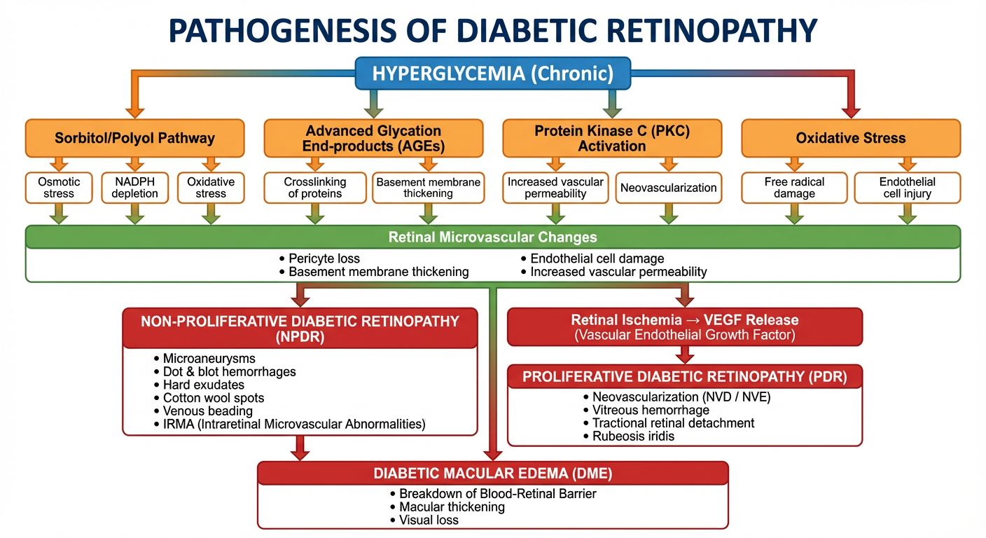
Pathogenesis of Diabetic Retinopathy
🔴 Starting Point: Chronic Hyperglycemia
1️⃣ Four Key Biochemical Pathways (Parallel)
| Pathway | Mechanism | Effect |
|---|---|---|
| Sorbitol / Polyol Pathway | Excess glucose → Sorbitol via Aldose Reductase | Osmotic stress, NADPH depletion, oxidative damage |
| Advanced Glycation End-products (AGEs) | Non-enzymatic glycation of proteins | BM thickening, crosslinking, pericyte dysfunction |
| Protein Kinase C (PKC) Activation | Diacylglycerol accumulation activates PKC | ↑ Vascular permeability, VEGF upregulation |
| Oxidative Stress | Free radical overproduction | Endothelial cell injury, mitochondrial dysfunction |
2️⃣ Retinal Microvascular Changes
- Pericyte loss (earliest histological change)
- Basement membrane thickening
- Endothelial cell damage
- Breakdown of the Blood-Retinal Barrier (BRB)
- Increased vascular permeability
3️⃣ Non-Proliferative Diabetic Retinopathy (NPDR)
- Microaneurysms (earliest clinical sign)
- Dot & blot hemorrhages
- Hard exudates (lipid leakage)
- Cotton-wool spots (nerve fiber layer infarcts)
- Venous beading
- IRMA (Intraretinal Microvascular Abnormalities)
4️⃣ Retinal Ischemia → VEGF Release
- Capillary non-perfusion → retinal hypoxia
- VEGF secreted → drives neovascularization
5️⃣ Proliferative Diabetic Retinopathy (PDR)
- NVD (Neovascularization at Disc) / NVE (elsewhere)
- Vitreous hemorrhage
- Tractional retinal detachment
- Rubeosis iridis (iris neovascularization) → Neovascular glaucoma
6️⃣ Diabetic Macular Edema (DME) (Can occur at any stage)
- BRB breakdown → fluid accumulation in macula
- Macular thickening → central visual loss
References: Khurana AK – Comprehensive Ophthalmology; Datta & Zia – Eye Care of the Patient with Diabetes Mellitus (p. 34); supported by established biochemical pathway literature (sorbitol pathway, AGEs, PKC, VEGF).