
Anatomy of larynx for anesthesia resident
Reading File
Finding Sources
Finding Sources
Searching Images
larynx anatomy cartilages muscles nerves recurrent laryngeal nerve diagram
Searching PubMed
laryngeal anatomy anesthesia
Searching the Web
larynx anatomy anesthesia airway recurrent laryngeal nerve external branch superior laryngeal nerve
Reading File
Reading File
Reading File
Reading File
query_images_db
Larynx anatomy for the anesthesia resident
The larynx extends from approximately C3 to C6, connecting the laryngopharynx to the trachea. It functions in phonation, airway patency, aspiration prevention, cough, and pressure generation during Valsalva. During swallowing it elevates and moves anteriorly, helping close the inlet and open the upper esophageal sphincter. Gray's Anatomy for Students, p. 1192.
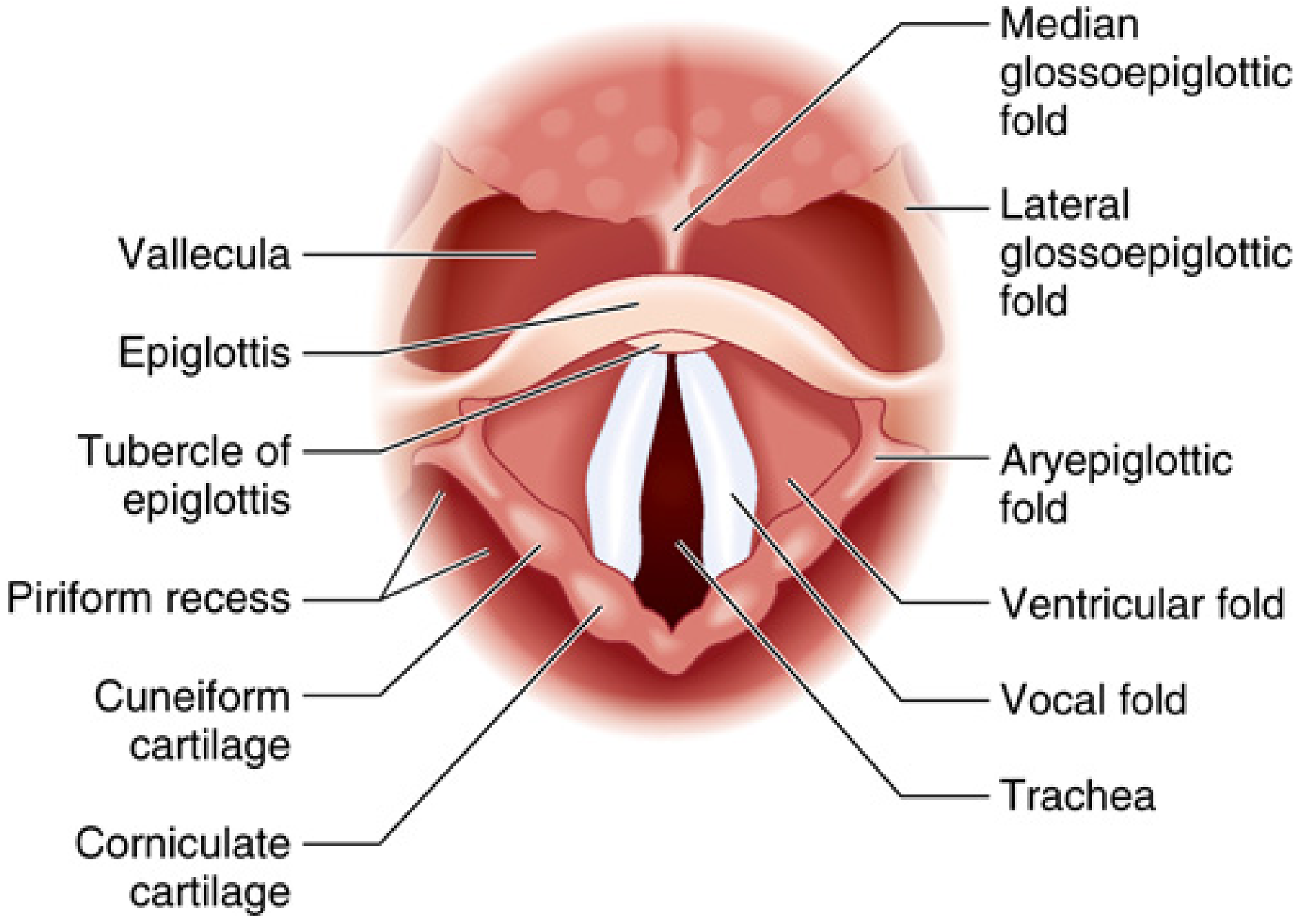
Laryngoscopic view: recognize epiglottis, aryepiglottic folds, false cords, true vocal cords, arytenoids, and the glottic opening. Miller's Anesthesia, 10e, p. 5843.
1. Cartilaginous framework
There are nine cartilages:
- Unpaired: thyroid, cricoid, epiglottis
- Paired: arytenoid, corniculate, cuneiform
| Structure | High-yield anatomy | Relevance to anesthesia |
|---|---|---|
| Hyoid bone | Suspends the larynx superiorly | Relevant surface landmark for superior laryngeal nerve block |
| Thyroid cartilage | Largest cartilage; laryngeal prominence is palpable | Its superior notch is a useful anterior-neck landmark |
| Epiglottis | Leaf-shaped, attached to thyroid cartilage by thyroepiglottic ligament | Key landmark in direct/video laryngoscopy; its shape and position may make exposure difficult |
| Cricoid cartilage | At about C6; signet-ring shaped, narrow arch anteriorly and broad lamina posteriorly; only complete cartilaginous ring | Landmark for cricothyrotomy orientation and cricoid pressure. The posterior lamina is adjacent to the esophagus |
| Arytenoids | Sit on the posterior cricoid lamina; vocal folds attach anteriorly to thyroid cartilage and posteriorly to vocal processes | Posterior structures visible during laryngoscopy. Arytenoid trauma/dislocation may mimic vocal-fold palsy |
| Corniculate and cuneiform | Small cartilages within the aryepiglottic folds | Produce the posterior and lateral contours of the laryngeal inlet |
The cricothyroid membrane (CTM) lies between the inferior thyroid cartilage and superior cricoid arch. It is the target for emergency front-of-neck access. The cricoid is the only complete cartilaginous airway ring. Miller's Anesthesia, 10e, p. 5841-5842.
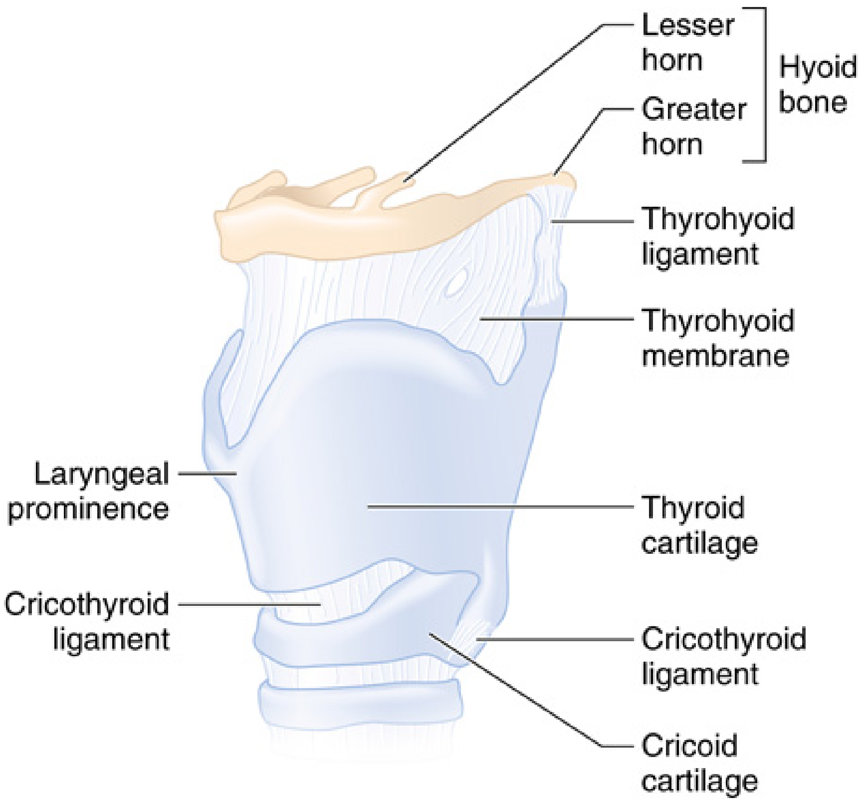
Cartilaginous and membranous framework. Miller's Anesthesia, 10e, p. 5843.
2. Cavities and laryngoscopic divisions
From superior to inferior:
-
Laryngeal inlet (aditus)
- Anterior: epiglottis
- Lateral: aryepiglottic folds
- Posterior: interarytenoid region and arytenoids
-
Supraglottis
- Epiglottis, aryepiglottic folds, false vocal folds, laryngeal ventricles
- In adults, supraglottic structures commonly obstruct dynamically during induction or deep sedation.
-
Glottis
- True vocal folds plus the rima glottidis.
- Narrowest part of the adult larynx at the level of the vocal cords in a functional sense.
-
Subglottis
- From lower margin of the vocal folds to inferior border of cricoid.
- A fixed, non-distensible region due to the complete cricoid ring. Important in edema and acquired subglottic stenosis.
Pediatric distinction: Traditionally the cricoid was described as the narrowest region. Modern imaging indicates the glottis is often the narrowest transverse level, but the subglottic cricoid remains clinically important because it is less distensible and susceptible to pressure injury from an oversized tube.
3. Vocal folds and intrinsic muscles
Vocal folds
- True vocal folds: mucosa over the vocal ligament and vocalis muscle.
- Attach from the inner thyroid cartilage anteriorly to the vocal processes of arytenoids posteriorly.
- False vocal folds lie superiorly and do not normally contribute to phonation.
Intrinsic muscle actions
| Muscle | Principal action | Airway relevance |
|---|---|---|
| Posterior cricoarytenoid (PCA) | Only abductor of the vocal folds | Opens glottis during inspiration. Bilateral weakness causes a dangerously narrow airway |
| Lateral cricoarytenoid (LCA) | Adducts anterior vocal folds | Closure for phonation and airway protection |
| Interarytenoid, transverse and oblique | Adduct arytenoid cartilages, close posterior glottis | Improves glottic closure |
| Thyroarytenoid/vocalis | Relaxes/shortens and fine-tunes vocal folds | Voice control |
| Cricothyroid (CT) | Tenses and elongates vocal folds by tilting thyroid cartilage forward | Only intrinsic muscle not supplied by RLN |
Exam pearl:
PCA is the only vocal-fold abductor. If both recurrent laryngeal nerves are injured, the folds may rest near the midline, causing stridor and possible inability to ventilate or extubate safely.
PCA is the only vocal-fold abductor. If both recurrent laryngeal nerves are injured, the folds may rest near the midline, causing stridor and possible inability to ventilate or extubate safely.
4. Innervation: the anesthesia-critical section
All motor and sensory laryngeal innervation is via the vagus nerve (CN X).
| Nerve | Motor supply | Sensory supply | Key clinical implication |
|---|---|---|---|
| Internal branch of superior laryngeal nerve, iSLN | None | Supraglottic larynx: epiglottis, laryngeal vestibule, aryepiglottic folds, superior surface of vocal folds | Main afferent limb of laryngeal protective reflexes including laryngospasm. Block supports awake airway instrumentation |
| External branch of superior laryngeal nerve, eSLN | Cricothyroid | None of major importance | Injury produces impaired pitch control and vocal fatigue |
| Recurrent laryngeal nerve, RLN | All other intrinsic muscles: PCA, LCA, interarytenoids, thyroarytenoid | Infraglottic larynx and upper trachea | Damage causes vocal-fold paresis/paralysis. Block reduces cough response below cords |
| Sympathetic fibers | Vasomotor and secretomotor influence | - | Limited relevance in routine airway management |
This is commonly summarized as:
“All intrinsic laryngeal muscles are recurrent laryngeal, except cricothyroid.”
Recurrent laryngeal nerve course
- Right RLN: loops under the right subclavian artery
- Left RLN: loops under the aortic arch, posterior to ligamentum arteriosum
- Both ascend in or near the tracheoesophageal groove, pass near the thyroid gland, and enter the larynx posterior to the cricothyroid joint.
The longer left-sided intrathoracic course makes left RLN palsy relevant in mediastinal, cardiac, and aortic pathology. The OpenAnesthesia laryngeal anatomy review gives a useful anesthesia-oriented overview.
Reflex arcs
- Laryngospasm: supraglottic stimulation -> iSLN afferent -> brainstem -> vagal/RLN-mediated adductor response.
- Cough: laryngeal/tracheal sensory input, especially below the cords via RLN, produces expiratory and glottic responses.
5. Blood supply
- Superior laryngeal artery, from the superior thyroid artery, accompanies the iSLN through the thyrohyoid membrane.
- Inferior laryngeal artery, from the inferior thyroid artery, accompanies the RLN.
Procedural implication: During an iSLN block, the needle is advanced through the thyrohyoid membrane. Aspirate carefully because the superior laryngeal vessels travel in this area.
6. External landmarks and airway procedures
| Landmark | Clinical use |
|---|---|
| Hyoid bone | Identify superior laryngeal region |
| Thyroid notch and laryngeal prominence | Surface orientation, especially in front-of-neck access |
| Thyrohyoid membrane | iSLN block access |
| Thyroid cartilage | Superior boundary when identifying CTM |
| Cricothyroid membrane | Emergency cricothyrotomy target |
| Cricoid cartilage | Inferior boundary of CTM; complete ring at C6 |
| Tracheal rings | Tracheostomy orientation |
Cricothyrotomy: Palpate thyroid cartilage, move caudad to the depression of the CTM, then identify the firm cricoid cartilage below it. Avoid excessively cephalad puncture, which risks laryngeal injury, and excessively caudad placement, which enters trachea rather than the membrane.
7. Anesthesia applications
Direct and video laryngoscopy
- Identify: tongue base -> epiglottis -> vallecula -> aryepiglottic folds -> posterior arytenoids -> cords.
- In Macintosh laryngoscopy, blade tip in the vallecula tensions the hyoepiglottic ligament and indirectly lifts the epiglottis.
- A straight blade directly lifts the epiglottis.
Intubation trauma
Potential injuries include:
- Edema or hematoma of vocal folds/arytenoids
- Arytenoid dislocation
- Mucosal ulceration and granuloma
- RLN neuropraxia from cuff pressure or surgical factors
- Subglottic injury from an oversized or repeatedly manipulated tube
Use a tube that passes without force, minimize repeated attempts, monitor cuff pressure, and investigate persistent postoperative hoarseness, stridor, aspiration, or weak cough.
Laryngeal blocks for awake intubation
- iSLN block: anesthetizes supraglottic mucosa, decreases gagging and laryngospasm provoked by supraglottic contact.
- Transtracheal injection through CTM: anesthetizes infraglottic tracheal mucosa through cough-mediated distribution of local anesthetic.
- Topical local anesthetic addresses oral, pharyngeal, and laryngeal surfaces but requires dose accounting.
Extubation
High-risk anatomy or pathology includes:
- Supraglottic edema
- Post-intubation laryngeal edema
- Subglottic stenosis
- Bilateral vocal-fold immobility
- Head and neck surgery or thyroid surgery
- Prior neck radiation, tumor, or airway trauma
A cuff-leak test evaluates risk indirectly and cannot exclude post-extubation obstruction. Extubate only when oxygenation, ventilation, airway patency, neuromuscular recovery, and a rescue plan are satisfactory.
Rapid recall
- Level: C3-C6
- Only complete airway ring: cricoid
- Only vocal-fold abductor: posterior cricoarytenoid
- Only intrinsic muscle not supplied by RLN: cricothyroid
- Supraglottic sensation: internal SLN
- Infraglottic sensation: RLN
- Right RLN loop: subclavian artery
- Left RLN loop: aortic arch
- Emergency front-of-neck target: cricothyroid membrane
- Laryngospasm afferent limb: internal SLN
The cartilaginous structure, cricoid anatomy, and key surface landmarks are described in Miller's Anesthesia, 10e, pp. 5841-5843. The broader anatomical organization and laryngeal mobility during swallowing are described in Gray's Anatomy for Students, p. 1192.
This is a shared conversation. Sign in to Orris to start your own chat.