
About Axilla anatomy
Reading File
Now let me search the medical library for axilla anatomy content.
Finding Sources
Finding Sources
Gray's Anatomy for Students has a dedicated AXILLA section. Let me read it along with the Thieme Atlas content.
Reading File
Reading File
Now let me read more of Gray's Anatomy content on the axilla walls, contents, and nerves:
Reading File
Reading File
Now let me get the remaining walls and the key content (axillary vessels, brachial plexus, lymph nodes) from Gray's:
Reading File
Reading File
Now let me get the brachial plexus, axillary lymph nodes, and the quadrangular/triangular spaces from Gray's:
Reading File
Reading File
Reading File
Now let me get the lymph nodes and the axillary artery parts section:
Reading File
Let me look for the axillary artery parts and lymph node sections:
Reading File
Now let me get the lymph node section:
Reading File
Now let me fetch a few key diagrams to include in the answer:
Loading Image
Loading Image
Loading Image
I now have comprehensive content from Gray's Anatomy for Students. Let me compile the complete answer.
Axilla - Anatomy
The axilla (armpit) is the gateway to the upper limb - an irregularly shaped pyramidal space forming a transition between the neck and arm. All major vessels, nerves, and lymphatics passing to and from the upper limb travel through this region.
Shape and General Structure
The axilla is shaped like a four-sided pyramid, having:
- An apex (inlet) - directed superomedially into the neck
- A floor (base) - the skin of the armpit, opens laterally into the arm
- Four walls - anterior, posterior, medial, lateral
Boundaries
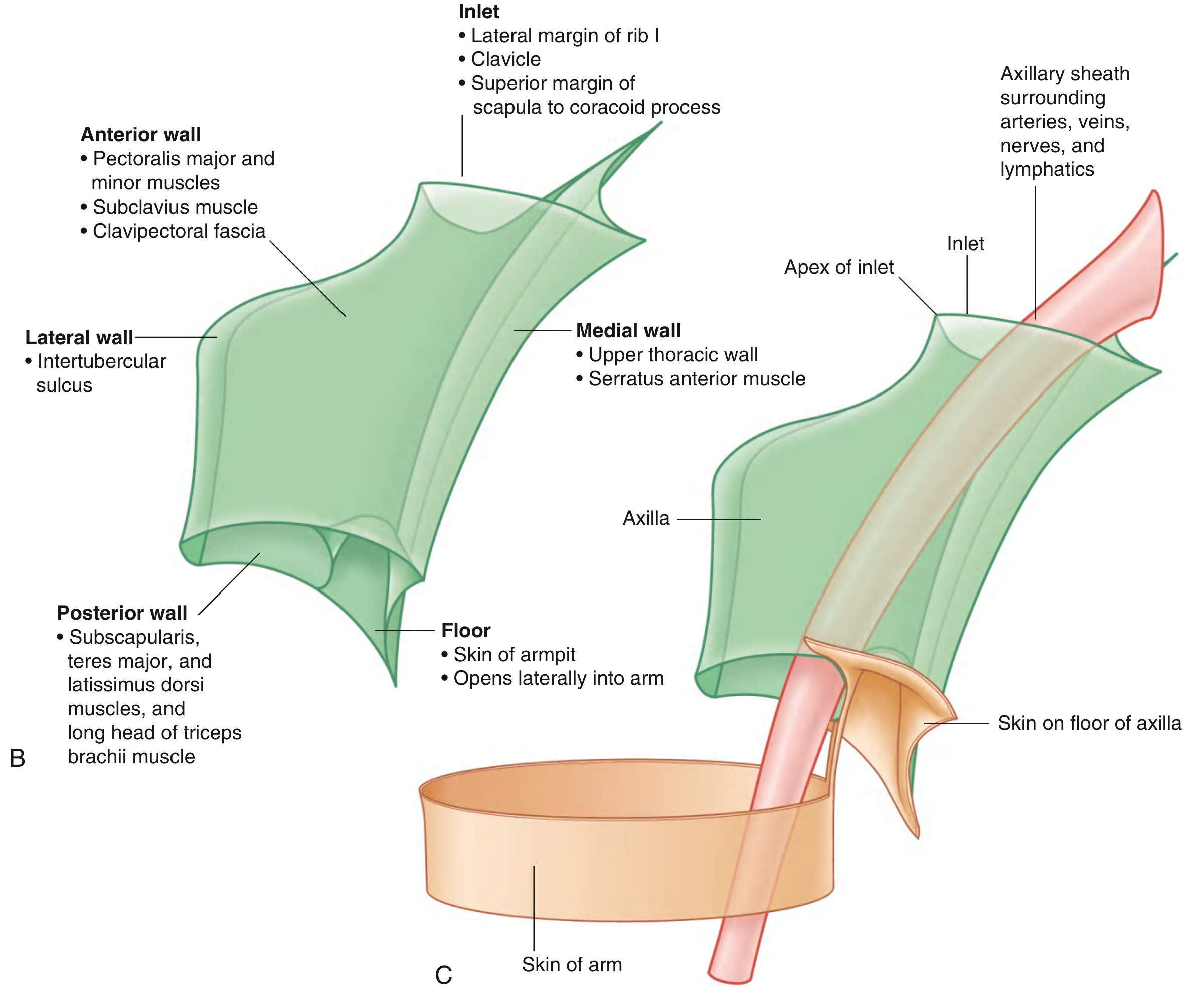
Axillary Inlet (Apex)
The inlet is triangular, oriented in the horizontal plane, apex pointing laterally. Its bony margins are:
- Medial - lateral border of rib I
- Anterior - posterior surface of the clavicle
- Posterior - superior border of the scapula up to the coracoid process
- Apex - medial aspect of the coracoid process
The subclavian artery becomes the axillary artery as it crosses the lateral border of rib I. At the inlet, the axillary vein is anterior to the artery, which is anterior to the trunks of the brachial plexus.
Walls
Anterior Wall
Formed by:
- Pectoralis major (most superficial; forms the anterior axillary fold)
- Pectoralis minor (deep)
- Subclavius
- Clavipectoral fascia
| Muscle | Origin | Insertion | Nerve | Function |
|---|---|---|---|---|
| Pectoralis major | Medial clavicle, sternum, costal cartilages 1-7 | Lateral lip of intertubercular sulcus | Medial + lateral pectoral nerves (C5-T1) | Flexion, adduction, medial rotation |
| Pectoralis minor | Ribs III-V (ant. surface) | Coracoid process | Medial + lateral pectoral nerves (C5-T1) | Depresses shoulder tip, protracts scapula |
| Subclavius | Rib I at rib-costal cartilage junction | Inferior groove of clavicle (middle 1/3) | Nerve to subclavius (C5,C6) | Pulls clavicle medially, stabilizes SC joint |
The clavipectoral fascia spans the gap between the subclavius and pectoralis minor. The cephalic vein, lateral pectoral nerve, and thoraco-acromial artery all pierce this fascia.
Posterior Wall
Formed by:
- Subscapularis (largest component - fills the subscapular fossa)
- Teres major
- Latissimus dorsi
- Long head of triceps brachii
| Muscle | Origin | Insertion | Nerve | Function |
|---|---|---|---|---|
| Subscapularis | Subscapular fossa (medial 2/3) | Lesser tubercle of humerus | Upper + lower subscapular nerves (C5,C6) | Medial rotation (rotator cuff) |
| Teres major | Inferior angle of scapula | Medial lip of intertubercular sulcus | Lower subscapular nerve (C5-C7) | Medial rotation + extension of arm |
| Latissimus dorsi | Lower 6 thoracic spinous processes, thoracolumbar fascia, iliac crest, lower 3-4 ribs | Floor of intertubercular sulcus | Thoracodorsal nerve (C6,C7,C8) | Adduction, medial rotation, extension |
Between these muscles lie two important apertures in the posterior wall:
- Quadrangular space - bounded by subscapularis/teres minor (above), teres major (below), long head of triceps (medial), surgical neck of humerus (lateral). Transmits the axillary nerve and posterior circumflex humeral artery.
- Triangular space - bounded by teres minor (above), teres major (below), long head of triceps (lateral). Transmits the circumflex scapular artery.
Medial Wall
Formed by:
- Serratus anterior overlying ribs 1-4 and intercostal muscles
- The long thoracic nerve (of Bell, C5,C6,C7) runs on the outer surface of serratus anterior
Lateral Wall (Narrowest)
Formed by:
- Intertubercular sulcus (bicipital groove) of the humerus
Floor (Base)
- Skin and superficial fascia of the armpit
- Opens laterally into the arm through the axillary fascia
Contents of the Axilla
The axilla contains all major structures passing into and out of the upper limb:
- Axillary artery (and branches)
- Axillary vein (and tributaries)
- Cords and branches of the brachial plexus
- Axillary lymph nodes and lymphatics
- Proximal parts of biceps brachii and coracobrachialis muscles
- Axillary process (tail) of the breast
- Axillary fat
Axillary Artery
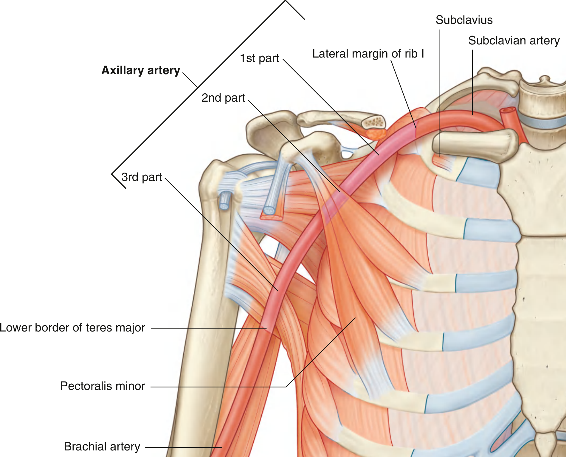
The pectoralis minor divides the axillary artery into 3 parts:
| Part | Relation to Pectoralis Minor | Branches (mnemonic: "1-2-3") |
|---|---|---|
| 1st | Proximal (medial) to it | 1 branch - Superior thoracic artery |
| 2nd | Posterior to it | 2 branches - Thoraco-acromial artery (4 branches: pectoral, deltoid, clavicular, acromial) + Lateral thoracic artery |
| 3rd | Distal (lateral) to it | 3 branches - Subscapular artery (largest branch, divides into circumflex scapular + thoracodorsal), Anterior circumflex humeral artery, Posterior circumflex humeral artery |
The axillary artery begins at the lateral border of rib I (continuation of subclavian artery) and ends at the lower border of teres major (continues as the brachial artery).
Axillary Vein
- Begins at the lower margin of teres major as the continuation of the basilic vein
- Passes medial and anterior to the axillary artery
- Receives: brachial veins, cephalic vein (which pierces clavipectoral fascia), and tributaries following arterial branches
- Becomes the subclavian vein as it crosses the lateral border of rib I
Brachial Plexus
The brachial plexus (C5-T1) enters the axilla from the neck, crossing over rib I. Its organization: Roots → Trunks → Divisions → Cords → Branches.
Three cords (named by their relation to the axillary artery) give off the major terminal branches in the axilla:
Lateral Cord branches:
- Lateral pectoral nerve - innervates pectoralis major
- Musculocutaneous nerve - pierces coracobrachialis, innervates anterior arm muscles
- Lateral root of median nerve - joins medial root to form the median nerve
Medial Cord branches:
- Medial pectoral nerve - innervates pectoralis minor (+ major)
- Medial cutaneous nerve of the arm
- Medial cutaneous nerve of the forearm
- Ulnar nerve
- Medial root of median nerve
Posterior Cord branches:
- Superior subscapular nerve - subscapularis
- Thoracodorsal nerve (C6,C7,C8) - latissimus dorsi
- Inferior subscapular nerve (C5,C6) - subscapularis + teres major
- Axillary nerve - exits via quadrangular space; innervates deltoid + teres minor
- Radial nerve - largest branch; passes into posterior compartment of arm
Key landmark: the musculocutaneous nerve, lateral root of median nerve, median nerve, medial root of median nerve, and ulnar nerve form an "M" configuration over the third part of the axillary artery.
Only trunk-level branches (from the superior trunk): suprascapular nerve and nerve to subclavius.
Axillary Lymph Nodes
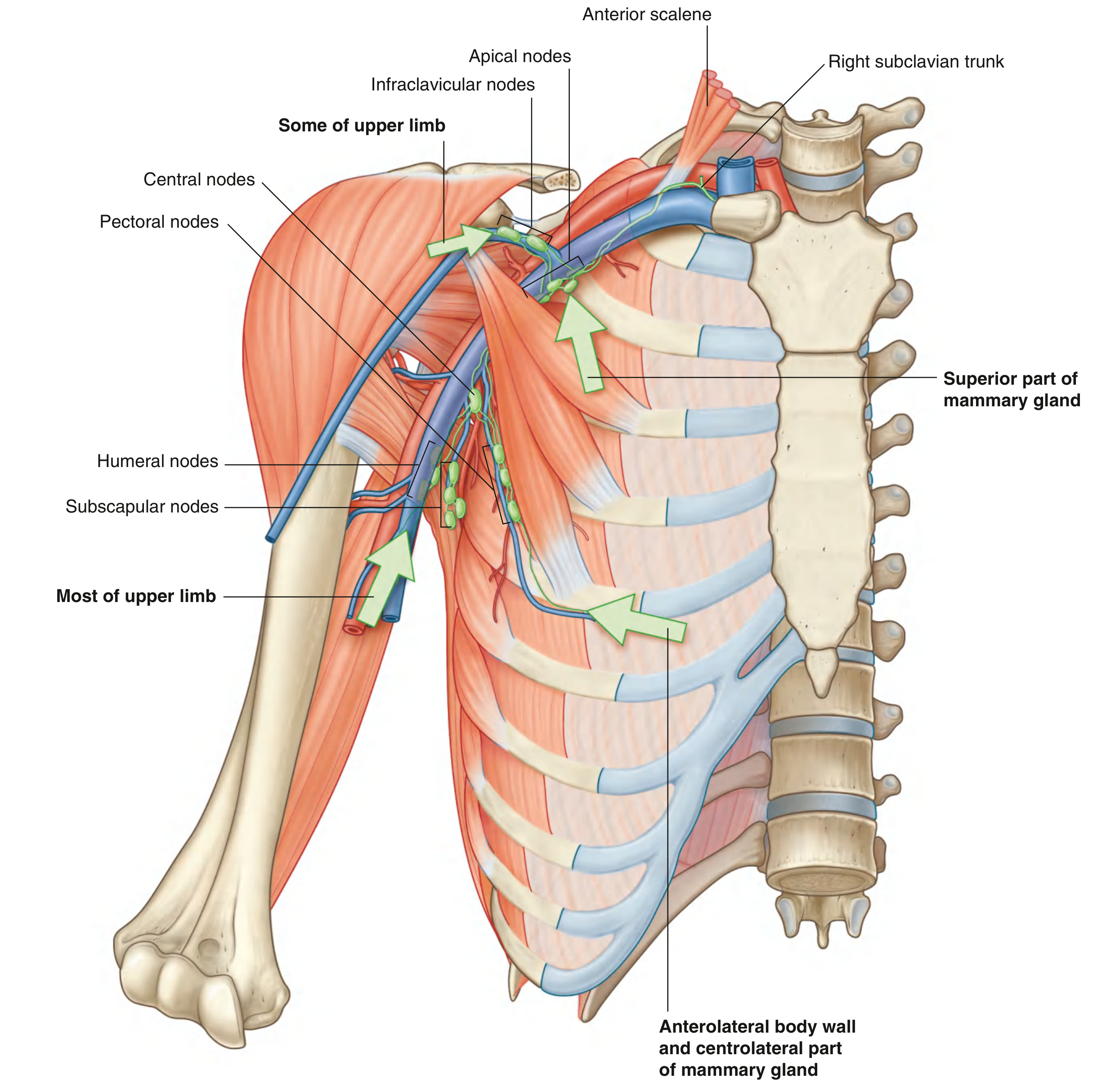
There are 20-30 axillary lymph nodes arranged in five groups. They drain the upper limb, chest wall, back, and approximately 75% of the mammary gland:
| Group | Location | Drainage |
|---|---|---|
| Humeral (lateral) | Posteromedial to axillary vein | Most of the upper limb |
| Pectoral (anterior) | Along inferior border of pectoralis minor, lateral thoracic vessels | Abdominal wall, chest wall, most of breast |
| Subscapular (posterior) | Posterior axillary wall, subscapular vessels | Posterior axillary wall, back, shoulder, neck |
| Central | Embedded in axillary fat | Receives from humeral, subscapular, pectoral groups |
| Apical | Apex of axilla, medial to axillary vein | Receives all other groups; drains to subclavian lymphatic trunk |
Infraclavicular (deltopectoral) nodes lie in the clavipectoral triangle and drain to apical nodes.
Important Gaps in the Posterior Wall
| Space | Boundaries | Contents |
|---|---|---|
| Quadrangular space | Subscapularis/teres minor (sup.), teres major (inf.), long head triceps (med.), surgical neck humerus (lat.) | Axillary nerve, posterior circumflex humeral artery |
| Triangular space | Teres minor (sup.), teres major (inf.), long head triceps (lat.) | Circumflex scapular artery |
| Triangular interval (lower triangular space) | Teres major (sup.), long head triceps (med.), humerus (lat.) | Radial nerve, profunda brachii artery |
Clinical Correlations
- Axillary nerve injury (e.g. anterior shoulder dislocation, proximal humerus fracture) - loss of deltoid function (abduction) and loss of sensation over the "regimental badge" area (lateral deltoid region)
- Breast cancer spreads primarily via the pectoral nodes and ultimately to apical nodes
- Long thoracic nerve injury (serratus anterior palsy) - causes "winging of the scapula"
- Axillary vein access (subclavian line) - should be done lateral to the midclavicular line to avoid catheter kinking by the subclavius muscle
- Quadrangular space syndrome - compression of the axillary nerve and posterior circumflex humeral artery in the quadrangular space
Sources: Gray's Anatomy for Students, pp. 841-868; General Anatomy and Musculoskeletal System (Thieme Atlas of Anatomy), pp. 401-402
This is a shared conversation. Sign in to Orris to start your own chat.