
Pulmonary tuberculosis radiology pg answer from standard radiology textbooks high yield everything i need to know for radiology pg Military tb 10 mark
Pulmonary tuberculosis radiology pg answer from standard radiology textbooks high yield everything i need to know for radiology pg Military tb 10 mark
Pulmonary Tuberculosis - Radiology PG High-Yield Answer
CLASSIFICATION OF PULMONARY TB (Radiological Perspective)
| Type | Synonyms | Timing | Key Radiological Pattern |
|---|---|---|---|
| Primary TB | Ghon focus disease | First infection | Lower/mid zone opacity + hilar adenopathy |
| Progressive Primary TB | Post-primary in children | Persistent primary | Cavitation, lobar consolidation |
| Reactivation TB | Post-primary, secondary TB | Remote reinfection | Apical/upper lobe cavitation + fibrosis |
| Miliary TB | Disseminated TB | Hematogenous spread | Innumerable 1-3 mm random nodules |
1. PRIMARY TUBERCULOSIS - Radiological Features
Chest X-Ray Findings
- Ghon focus: Focal parenchymal opacity, typically middle or lower lung zone (subpleural)
- Hilar/mediastinal lymphadenopathy: Ipsilateral hilar enlargement is characteristic (right > left)
- Pleural effusion: May be present on the affected side
- Combination of parenchymal opacity + lymphadenopathy = Ghon complex (or primary complex)
- Atelectasis may occur if enlarged nodes compress adjacent bronchi
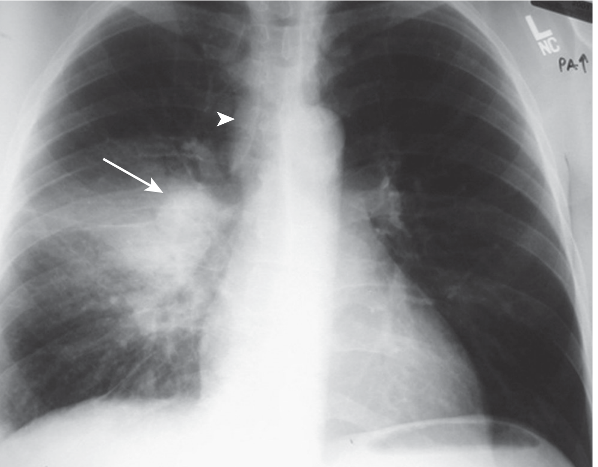
CT Findings in Primary TB
- Ghon focus: Focal parenchymal opacity with ipsilateral lymphadenopathy
- CT clearly demonstrates lymph node enlargement (more sensitive than CXR)
- Central necrosis in nodes appears as low-density center with rim enhancement
- Air bronchograms visible in areas of consolidation
2. REACTIVATION (POST-PRIMARY) TUBERCULOSIS
Classic Radiological Features - "The Upper Lobe Disease"
- Apical and posterior segments of RIGHT upper lobe (most common)
- Apical-posterior segment of LEFT upper lobe
- Superior segments of lower lobes (less common)
Key rule: Anterior segments of upper lobes are rarely involved in reactivation TB - this is a classic exam point.
CXR Pattern Progression
| Stage | Findings |
|---|---|
| Early | Patchy, poorly defined opacity in upper lobes |
| Active/Progressive | Coalescent consolidation, cavity formation |
| Spreading | Tree-in-bud opacities (bronchogenic spread), satellite nodules |
| Healing | Fibrosis, volume loss, calcification |
| Healed | Fibrocavitary scarring, calcified nodules, pleural thickening |
Cavitation in Reactivation TB
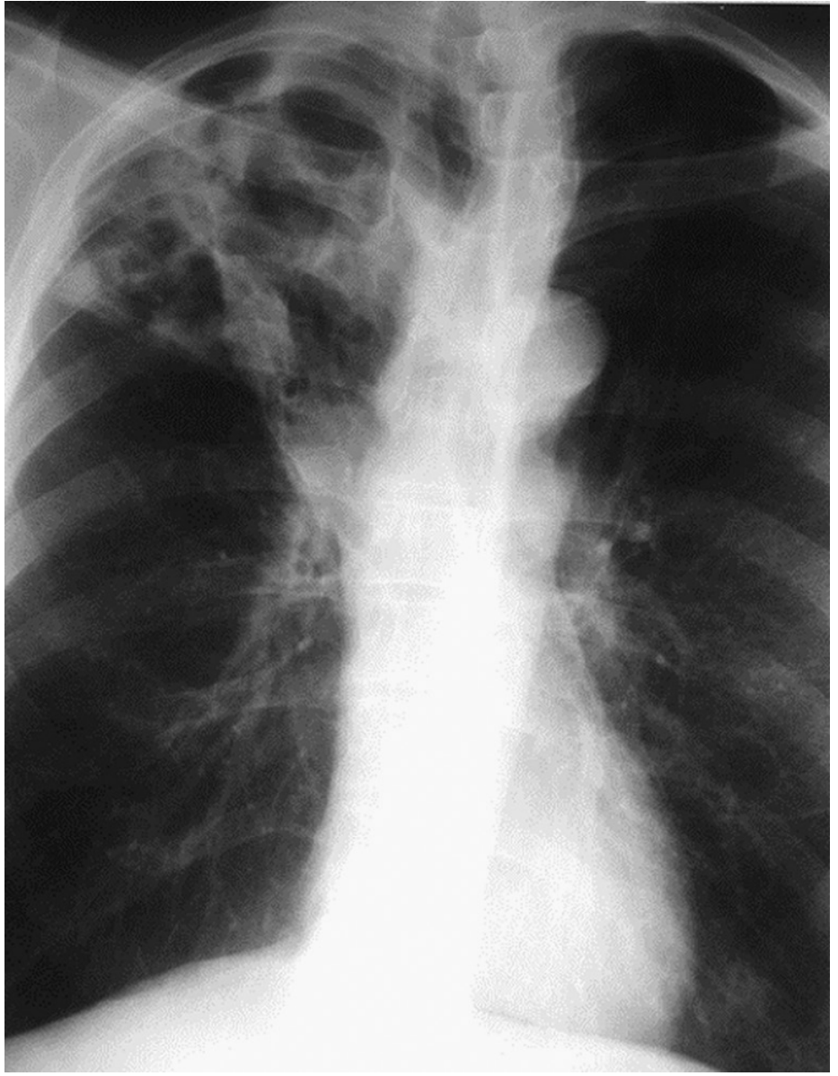
- Cavities form by liquefaction necrosis and drainage into airways
- Typically thick-walled initially, thin-walled in chronic/healed state
- Air-fluid levels may be present (superimposed infection)
- Rasmussen aneurysm: Dilated vessels in cavity wall - cause of hemoptysis
- Aspergilloma (fungus ball) may colonize old cavities - "crescent sign" / air crescent sign
CT Features of Reactivation TB (HIGH YIELD)
- Tree-in-bud pattern: Centrilobular nodules with branching linear densities = bronchogenic spread (HIGHLY specific sign)
- Nodular opacities in upper lobes
- Cavities with thick or thin walls
- Air bronchograms within consolidation
- CT is superior to CXR: sensitivity 96% vs 48% for active TB
- CT better demonstrates bronchogenic spread characteristic of reactivation TB
3. MILIARY TUBERCULOSIS (10-Mark Answer)
Definition and Pathogenesis
- The term "miliary" comes from the resemblance of lesions to millet seeds
- Results from hematogenous dissemination of M. tuberculosis
- Occurs when a tuberculous focus (primary or reactivation) erodes into a blood vessel or lymphatic, seeding the entire pulmonary vasculature simultaneously
- Grossly: 1-2 mm yellowish nodules that are granulomas on histology
- Can occur in both primary and reactivation settings; more common in immunocompromised
Classic CXR Features of Miliary TB
| Feature | Details |
|---|---|
| Size | 1-3 mm nodules (size of millet seeds) |
| Distribution | Bilateral, diffuse, symmetric - all lung zones equally affected |
| Pattern | Random distribution - no zonal or bronchovascular predilection |
| Number | Innumerable (too numerous to count) |
| Margins | Well-defined, sharply marginated |
| Density | Uniform soft tissue density |
- CXR abnormal in 85-90% of miliary TB cases at diagnosis
- Classic miliary pattern seen in 50-90% of disseminated TB patients
- Nodules appear 2-3 weeks after hematogenous seeding (CXR may initially be normal!)
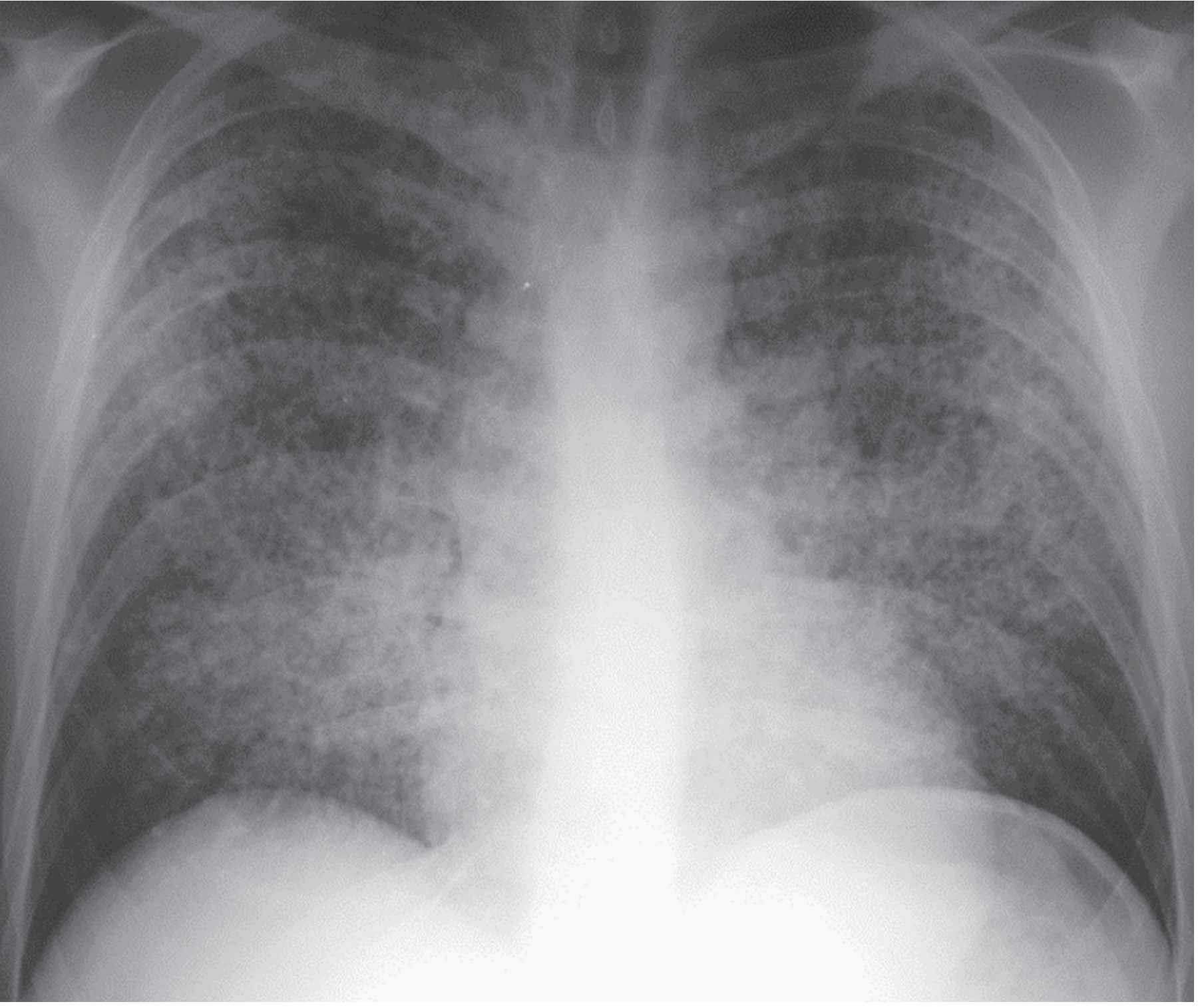
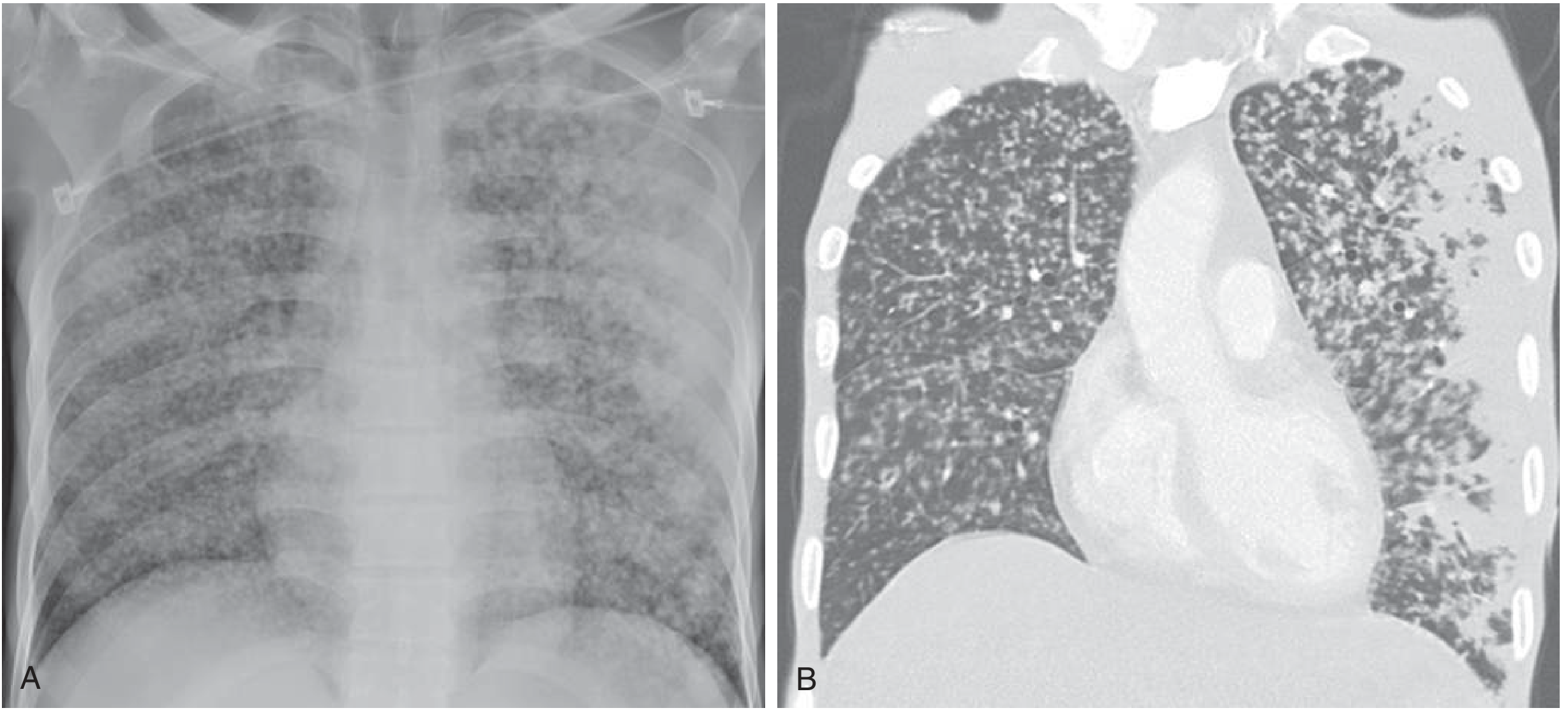
CT Features of Miliary TB
- Random distribution (not perilymphatic, not centrilobular) - this is the KEY CT pattern
- Nodules 1-3 mm, bilateral, diffuse
- Equal distribution in upper, middle, and lower zones
- Nodules are distributed randomly relative to secondary pulmonary lobule structures
- No zonal predilection - unlike reactivation TB which favors upper zones
- May see ground-glass opacity surrounding nodules ("halo sign")
- Additional findings: upper lobe opacities with/without cavitation, pleural effusion, pericardial effusion
CT Nodule Distribution Patterns - PG Exam Comparison Table
| Pattern | Distribution | Diseases |
|---|---|---|
| Random | Uniform, all zones | Miliary TB, hematogenous metastases, fungal infection |
| Perilymphatic | Septal/bronchovascular/subpleural | Sarcoidosis, lymphangitic carcinomatosis, pulmonary edema |
| Centrilobular | Around bronchioles, spares subpleura | Endobronchial TB (tree-in-bud), hypersensitivity pneumonitis |
Exam Tip: Miliary TB = RANDOM distribution. This is THE classic teaching point separating it from sarcoidosis (perilymphatic) and endobronchial TB (centrilobular tree-in-bud).
Atypical Presentations (in HIV/Immunosuppression)
- CXR may appear normal early in disease
- Granuloma formation is impaired; instead of discrete nodules, may show diffuse uniform opacification
- Intrathoracic lymphadenopathy more prominent (unlike immunocompetent patients)
- Cavitation less common with advanced HIV (CD4 < 200)
- Lower lobe/diffuse opacities instead of upper lobe predominance
- Pattern may resemble lobar pneumonia (Klebsiella-like appearance)
Miliary TB - Differential Diagnosis
| Condition | Key Differentiating Feature |
|---|---|
| Miliary fungal infection (histoplasma, cryptococcus) | Clinically indistinguishable; geographic history important |
| Hematogenous metastases | Known primary malignancy; nodules may be larger, non-uniform sizes |
| Sarcoidosis | Perilymphatic distribution (not random); upper lobe > lower |
| Pneumoconiosis (silicosis/CWP) | Occupational history; upper lobe predominance; egg-shell calcification of nodes |
| Langerhans cell histiocytosis | Cystic + nodular; upper > lower; spares costophrenic angles |
Organs Involved in Miliary TB (Autopsy Data)
4. TB-RELATED COMPLICATIONS - Radiological Findings
Pleural TB
- Unilateral exudative pleural effusion (lymphocyte predominant)
- Rarely bilateral; rarely large
- Empyema necessitans - TB empyema eroding through chest wall
- Bronchopleural fistula with hydropneumothorax
Endobronchial TB
- Bronchial stenosis leading to lobar/segmental atelectasis
- Obstructive pneumonitis distal to stenosis
- Air trapping on expiratory CT
Bronchogenic Spread
- Tree-in-bud pattern on CT: centrilobular branching opacities
- Ill-defined centrilobular nodules
- Patchy consolidation in dependent portions of lung
- Sign of active, highly infectious disease
Calcified Ghon Complex / Ranke Complex
- Calcified Ghon focus + calcified ipsilateral hilar node = Ranke complex
- Indicates healed primary TB
5. TB IN HIV - Radiological Spectrum (High-Yield Table)
| CD4 Count | Typical Radiological Pattern |
|---|---|
| CD4 > 350 (early HIV) | Similar to immunocompetent: upper lobe cavitation, typical reactivation pattern |
| CD4 200-350 | Mixed pattern |
| CD4 < 200 (advanced HIV) | Lower zone/diffuse opacities, lymphadenopathy, no cavitation, miliary pattern |
| CD4 < 50 | Miliary pattern, lobar consolidation mimicking bacterial pneumonia, normal CXR possible |
6. IMAGING MODALITY COMPARISON
| Modality | Role | Sensitivity for Active TB |
|---|---|---|
| CXR | Screening, initial evaluation | 48% |
| CT Chest | Characterization, bronchogenic spread, lymphadenopathy | 96% |
| MRI | Pleural assessment, caseous necrosis; pregnant/children | Comparable to CT for consolidation/adenopathy; lower for nodules |
| PET-CT | Monitoring treatment response, differentiating active vs inactive | High; increasing evidence |
QUICK-FIRE HIGH-YIELD POINTS FOR PG EXAM
- Primary TB: Lower/mid zone opacity + ipsilateral hilar adenopathy
- Reactivation TB: Apical and posterior segments of RUL (most common site)
- Anterior segment of upper lobe is spared in TB (unlike most other conditions)
- Miliary TB: 1-3 mm nodules, bilateral, symmetric, RANDOM distribution, all zones equal
- Tree-in-bud = bronchogenic spread = centrilobular distribution = active endobronchial TB
- Ranke complex = calcified Ghon focus + calcified hilar node = healed primary TB
- Rasmussen aneurysm: Dilated vessel in cavity wall - cause of massive hemoptysis
- Aspergilloma in old TB cavity: "crescent sign" (Monod sign)
- HIV + TB: More adenopathy, less cavitation, more lower zone/diffuse disease
- CXR may be normal in miliary TB for first 2-3 weeks (nodules too small)
- CT has 96% sensitivity vs 48% for CXR in active TB
- Miliary TB = RANDOM nodule pattern (differential: sarcoid = perilymphatic)
- 85-90% of miliary TB patients have abnormal CXR at diagnosis
- Snowstorm appearance on CXR = miliary TB
Pulmonary sequestration crisp radiology pg Rguhs ans for 10 marks From now on all my answers shud be based on graingers Sutton or aiims pg textbooks on any other standard radiology textbooks Double cross all the answers for any mistakes Let the answers be crisp and sweet All answers for 10 marks From now on
PULMONARY SEQUESTRATION
RGUHS Radiology PG - 10 Marks
DEFINITION
- Has no normal bronchial tree connection
- Has no pulmonary arterial supply
- Receives its blood supply from a systemic artery (usually thoracic or abdominal aorta)
CLASSIFICATION - COMPARISON TABLE (Most Important for PG)
| Feature | Intralobar (ILS) | Extralobar (ELS) |
|---|---|---|
| Frequency | 75-85% (more common) | 15-25% |
| Pleural investment | Shares pleura with normal lung | Has its own separate pleural covering |
| Location | Left lower lobe, posterior basal segment | Left base (77%), between lower lobe and diaphragm |
| Laterality | Left > right | Left >> right |
| Blood supply | Thoracic/abdominal aorta | Thoracic/abdominal aorta |
| Venous drainage | Pulmonary veins (left heart) | Systemic veins - azygos or portal system |
| Presentation | Adults - recurrent pneumonia or incidental | Infants/neonates |
| Infection | Common (frequently becomes infected) | Rare |
| Associated anomalies | Uncommon (~50% with scimitar syndrome variant) | Common - 50-65% (CDH, CPAM, cardiac defects, TEF) |
| Air bronchogram | May be present (collateral ventilation) | Absent |
Key exam point: ILS drains to pulmonary veins (left-to-right shunt). ELS drains to systemic veins (no shunt, smaller lesion).
PATHOGENESIS
CLINICAL PRESENTATION
- ILS: Often presents in adults as incidental finding on CXR or recurrent lower lobe pneumonia that fails to resolve
- ELS: Detected prenatally on US or in neonates; may present as respiratory distress
- ELS subdiaphragmatic: Can mimic neuroblastoma or adrenal hemorrhage
- Hemoptysis may occur from the aberrant vascular supply
IMAGING
1. Chest X-Ray
- ILS: Homogeneous opacity at the left lung base (posterior basal segment). May appear as solid mass or consolidation. Air-fluid level if infected.
- ELS: Well-defined homogeneous opacity at left base, silhouetting the posterior medial hemidiaphragm. Usually no air bronchogram.
- Both may be confused with pneumonia, mass, or diaphragmatic hernia.
2. Ultrasound (especially in neonates/prenatal)
- Uniformly echogenic mass with a hyperechoic rim
- Doppler demonstrates the aberrant feeding artery and venous drainage
- Prenatal US can diagnose ELS - hyperechoic mass near left diaphragm
3. CECT / MDCT - Investigation of Choice
- Demonstrates the systemic feeding artery arising from the aorta (the PATHOGNOMONIC finding)
- Shows venous drainage pattern (pulmonary vs systemic)
- ILS appears as:
- Solid homogeneous soft tissue mass, OR
- Air-containing single/multicystic lesion (from collateral ventilation), OR
- Mass with air-fluid level (if infected)
- ELS: Homogeneous, well-defined soft tissue density mass, occasionally with adjacent emphysema
- MDCT is the technique of choice - higher spatial/temporal resolution detects small systemic vessels better than MRI, especially in children; also evaluates lung parenchyma simultaneously
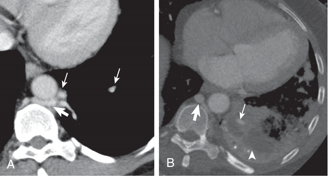
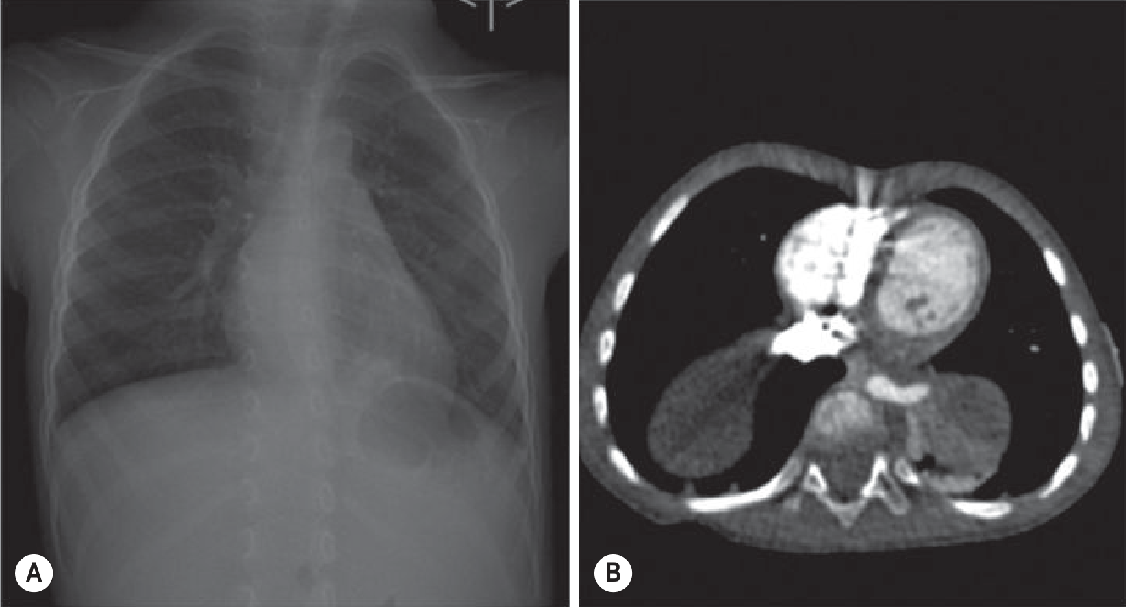
4. MRI
- Well-defined, solid mass - hyperintense on T2WI
- Systemic feeding artery identifiable
- Useful in pregnant patients and children (no radiation)
- Comparable to CT for demonstrating the vascular anatomy; lower sensitivity for small vessels than MDCT
5. Catheter Angiography
- Gold standard historically; now rarely needed
- Reserved for cases where CT/MRI fails to demonstrate the feeding artery
- Shows the aberrant systemic artery arising from the aorta
DIFFERENTIAL DIAGNOSIS
| Condition | Key Differentiator |
|---|---|
| Consolidation/Pneumonia | No persistent feeding artery; responds to antibiotics |
| CPAM (CCAM) | No systemic feeding artery; involves pulmonary circulation |
| Bronchogenic cyst | No systemic artery; thin-walled cyst; posterior mediastinum/subcarinal |
| Diaphragmatic hernia | Contains bowel; contrast swallow helps |
| Neuroblastoma | ELS subdiaphragmatic; mass calcification; elevated catecholamines |
TREATMENT
- Surgery (lobectomy or segmentectomy) is treatment of choice
- ~50% of adults with ILS are asymptomatic at diagnosis
- Preoperative selective systemic artery embolization useful in hemoptysis cases (reduces intraoperative bleeding)
- Perinatal management in a center equipped for pulmonary hypoplasia
HIGH-YIELD EXAM BULLETS
- Sequestration = lung tissue with no bronchial connection + systemic arterial supply (Pryce, 1946)
- ILS = 75-85%; ELS = 15-25%
- ILS location: left lower lobe, posterior basal segment
- ELS location: left base between lower lobe and diaphragm (77%)
- ILS venous drainage: pulmonary veins | ELS drainage: azygos/portal veins
- MDCT = investigation of choice - demonstrates systemic feeding artery
- MRI: T2 hyperintense mass with systemic artery
- ELS associated anomalies: 50-65% (CDH most common)
- ILS - recurrent lower lobe pneumonia in adults = classic presentation
- Pathognomonic CT finding: systemic artery from aorta supplying the mass
Same way bronchogenic cyst
BRONCHOGENIC CYST
RGUHS Radiology PG - 10 Marks
DEFINITION
- Timing of defect determines location: Early embryonic defect = mediastinal cyst; Later defect = intrapulmonary cyst
- Male predilection; incidence ~1 in 42,000-68,000 hospital admissions
PATHOLOGY
- Thin fibrous capsule lined with respiratory ciliated pseudostratified columnar epithelium
- Wall contains: cartilage, smooth muscle, bronchial glands
- Contents: usually thick mucoid material or clear serous fluid
- May contain proteinaceous fluid, blood, or milk of calcium
LOCATION
| Type | Frequency | Specific Sites |
|---|---|---|
| Mediastinal | 65-90% | Subcarinal (most common), paratracheal, paraesophageal, hilar |
| Intrapulmonary | 10-35% | Lower lobes predominantly |
| Rare sites | Uncommon | Pleura, diaphragm, neck, pericardium |
Key point: Subcarinal is the single most common location - classic exam answer.
CLINICAL FEATURES
- Usually asymptomatic (detected incidentally on imaging)
- Symptoms occur due to:
- Compression of airways - dyspnea, wheeze, stridor (especially in infants)
- Compression of SVC - SVC syndrome
- Compression of oesophagus - dysphagia
- Superinfection - fever, cough, purulent expectoration, hemoptysis (82% of symptomatic cases)
- Haemorrhage into cyst - rapid painful enlargement
IMAGING
1. Chest X-Ray (CXR)
- Well-defined, solitary, spherical or oval mass with homogeneous opacity
- Located just inferior to the carina, often projecting slightly to the right of midline into middle mediastinum
- Usually unilocular; non-lobulated outline
- Wall calcification is rare
- Milk of calcium: cyst-liquid calcium level within cyst (rare but classic finding)
- Characteristic displacement: carina pushed anteriorly, oesophagus pushed posteriorly (almost pathognomonic; only thyroid masses and aberrant left pulmonary artery can do the same)
- Intrapulmonary: ovoid parenchymal mass with sharp border; air-fluid level if infected (resembles lung abscess)
2. CT - Investigation of Choice
- Thin-walled cystic mass with no internal enhancing component (no enhancement post-contrast - key diagnostic criterion)
- CT attenuation varies:
- Water density (0-20 HU) - classic simple cyst appearance
- Soft-tissue density (up to 80+ HU) - due to high protein content, haemorrhage, or milk of calcium (can mimic solid mass - important pitfall)
- Rarely, uniformly high density (proteinaceous)
- Calcification in wall or contents - occasional
- If infected: thick irregular wall, increased cyst attenuation, surrounding mediastinal stranding/mediastinitis
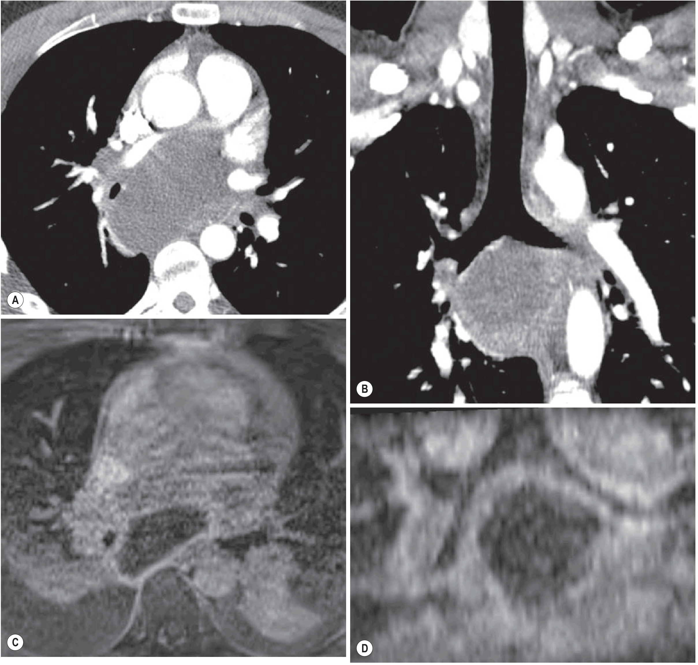
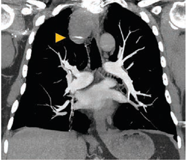
3. MRI
| Sequence | Signal |
|---|---|
| T1WI | Variable (low to high) - depends on protein/haemorrhage content |
| T2WI | High signal (characteristic) |
| Post-contrast | No enhancement (key diagnostic feature) |
- Fat-suppressed pre- and post-contrast subtraction MRI confirms absence of enhancement
- MRI superior for surgical planning - defines relation to surrounding structures
- Solid component + enhancement = consider malignancy
4. Prenatal / Fetal US
- Echogenic or anechoic cystic mass
- Most regress after birth; some persist
CT ATTENUATION - IMPORTANT PITFALL
A bronchogenic cyst with high CT attenuation (soft-tissue density) due to proteinaceous or haemorrhagic contents can be mistaken for a solid mediastinal mass (thymoma, lymphoma). MRI resolves this - T2 hyperintensity + no enhancement confirms cystic nature.
DIFFERENTIAL DIAGNOSIS
| Condition | Key Differentiator |
|---|---|
| Oesophageal duplication cyst | Identical imaging; thicker wall, more tubular, intimate contact with oesophagus; no cartilage on histology |
| Pericardial cyst | Anterior cardiophrenic angle; right > left |
| Hydatid cyst | Septations, daughter cysts; endemic area |
| Cystic teratoma | Anterior mediastinum; fat/calcium; inhomogeneous |
| Lung abscess | Thick wall; clinical sepsis; responds to antibiotics |
| Neurenteric cyst | Posterior mediastinum; vertebral anomalies; intraspinal component |
| Thymic cyst | Anterior mediastinum |
TREATMENT
- Asymptomatic: Conservative management (observation)
- Symptomatic / complicated: Surgical excision
- VATS (Video-Assisted Thoracoscopic Surgery) - treatment of choice
- Posterolateral thoracotomy for large/complicated cysts
- Intrapulmonary cysts: VATS cystectomy, wedge resection, segmentectomy, or lobectomy depending on size
- Recurrence: Low if complete resection; recurs if residual mucosa remains
- Mediastinoscopy / TBNA for diagnosis in selected cases
HIGH-YIELD EXAM BULLETS
- Bronchogenic cyst = congenital foregut duplication cyst; lined by respiratory epithelium + cartilage
- Most common location: subcarinal (middle mediastinum)
- CXR: well-defined oval mass inferior to carina, pushing carina anteriorly + oesophagus posteriorly
- CT: No enhancement post-contrast - defining feature
- CT attenuation is variable - can mimic solid mass (pitfall!) - use MRI to confirm
- MRI: T2 hyperintense, no enhancement
- Milk of calcium = calcium-fluid level within cyst - pathognomonic when present
- Infected cyst: thick wall + surrounding mediastinitis on MRI
- Treatment: VATS is modality of choice
- Differential from oesophageal duplication cyst: cartilage in wall = bronchogenic; smooth muscle + enteric mucosa = oesophageal duplication
Unilateral hyperlucent lung
UNILATERAL HYPERLUCENT LUNG
RGUHS Radiology PG - 10 Marks
DEFINITION
- Increased air (obstructive/check-valve, pneumothorax)
- Decreased blood flow (vascular)
- Decreased tissue (compensatory hyperinflation, congenital hypoplasia)
- Technical artefact
CAUSES - SYSTEMATIC CLASSIFICATION (Exam Table)
A. TECHNICAL / POSITIONAL
| Cause | Clue |
|---|---|
| Patient rotation | Spine not central; ribs asymmetric |
| Soft-tissue asymmetry (mastectomy, Poland syndrome) | No chest wall soft tissue on one side; lung markings normal |
| Grid/exposure artefact | Unusually uniform hyperlucency |
Mastectomy/Poland syndrome - vessels and lung markings present but no soft tissue shadow = true unilateral hyperlucency without lung pathology.
B. PLEURAL
| Cause | CXR Features |
|---|---|
| Pneumothorax | Visible pleural edge; absent lung markings beyond; mediastinum central (unless tension) |
| Tension pneumothorax | Mediastinal shift to opposite side; depressed ipsilateral diaphragm; emergency |
C. AIRWAY / OBSTRUCTIVE (Air Trapping)
| Cause | Key Features |
|---|---|
| Inhaled Foreign Body | Children; sudden onset; hyperinflation on ipsilateral side; expiratory film/fluoroscopy shows air trapping and mediastinal shift away from affected side on expiration |
| Congenital lobar overinflation | Neonates; check-valve bronchial lesion; most common in left upper lobe (42%), right middle lobe (35%); progressive hyperlucency of affected lobe |
| Endobronchial tumour/stricture | Adults; obstructing lesion on CT; post-obstructive hyperinflation early, then collapse |
Classic CXR sign for foreign body: On expiration, mediastinum swings AWAY from the obstructed (hyperinflated) side - "mediastinal swing" on fluoroscopy.
D. VASCULAR
| Cause | Key Features |
|---|---|
| Pulmonary embolism | Westermark sign - focal oligaemia; Hampton's hump (peripheral wedge consolidation); elevated hemidiaphragm |
| Congenital absence/hypoplasia of pulmonary artery | Small ipsilateral lung; small hilum; no air trapping on expiratory CT |
| Pulmonary artery stenosis | Post-stenotic lung oligaemia |
E. PARENCHYMAL / POST-INFECTIOUS
SWYER-JAMES-MACLEOD (SJM) SYNDROME - Detailed (10 Marks Focus)
Definition
- Also called: Unilateral hyperlucent lung syndrome / Unilateral emphysema
- First described by Swyer & James (1953) and independently by MacLeod (1954)
Pathogenesis
Radiological Features
CXR
| Feature | Description |
|---|---|
| Hyperlucent lung | Unilateral (or lobar) - blacker than contralateral side |
| Small hilum | Small ipsilateral pulmonary artery (key differentiator from other causes) |
| Decreased lung markings | Pruned vasculature throughout affected lung |
| Normal or small lung volume | Unlike compensatory emphysema where volume is increased |
| Mediastinum central | No shift at rest |
CT / HRCT (Most Important)
- Mosaic attenuation pattern: Areas of hyperlucency alternating with normal/denser areas
- Air trapping on expiratory CT: Affected lung remains lucent on expiration; does NOT deflate (hallmark finding)
- Decreased calibre and number of pulmonary vessels in hyperlucent areas
- Bronchial wall thickening and bronchiectasis (cylindrical) in affected lobe
- Normal contralateral lung
- The hyperlucent zones correspond to areas of obliterative bronchiolitis
Hallmark of SJM on CT: Hyperlucent lung with decreased vessel calibre that PERSISTS on expiratory CT = air trapping. This confirms small airways disease. (Grainger & Allison)
Expiratory CT / Fluoroscopy
- On expiration: affected lung remains hyperlucent and fails to deflate
- Mediastinum may shift TOWARD the normal (deflating) side on expiration
- This air trapping on expiratory imaging is pathognomonic of obliterative bronchiolitis
Ventilation-Perfusion (V/Q) Scan
- Reduced V and Q on affected side (matched defect - unlike PE which is mismatched)
- Confirms both ventilatory and perfusion defects in affected lung
DIFFERENTIATING KEY CAUSES - EXAM TABLE
| Feature | SJM Syndrome | Pneumothorax | Foreign Body | Mastectomy | Congenital absent PA |
|---|---|---|---|---|---|
| Hilum | Small | Normal | Normal | Normal | Absent/small |
| Lung markings | Decreased | Absent beyond pleural edge | Present | Present (no soft tissue) | Absent |
| Air trapping on expiry | YES | No | YES (acute) | No | No |
| Mediastinal shift | None at rest | Toward normal (if tension: away) | Away from obstructed side on expiry | None | None |
| Pleural line | Absent | Visible | Absent | Absent | Absent |
| Volume | Normal or small | Increased | Increased | Normal | Small |
| CT | Mosaic + bronchiectasis | Air outside lung | Obstructing FB | No lung abnormality | No vessels |
APPROACH TO UNILATERAL HYPERLUCENCY - ALGORITHM (PG Exam)
Unilateral Hyperlucent Lung
|
├── Is there a pleural line?
│ YES → Pneumothorax (check tension features)
│
├── Is there soft-tissue asymmetry?
│ YES → Mastectomy / Poland syndrome (technical)
│
├── What is the HILUM like?
│ Absent/small → Vascular (SJM, absent PA, PE)
│ Normal → Airway (foreign body, lobar overinflation, endobronchial)
│
├── Air trapping on expiratory CT/fluoroscopy?
│ YES → SJM syndrome / obliterative bronchiolitis / foreign body
│ NO → Compensatory emphysema / vascular cause
│
└── Is it a child? → Think: Foreign body, Congenital lobar overinflation, SJM
Is it an adult? → Think: SJM, Mastectomy, PE (Westermark), Endobronchial tumor
HIGH-YIELD BULLETS
- First step: Check for pleural line (pneumothorax) and soft tissue (mastectomy)
- Small hilum + hyperlucent lung = SJM syndrome or absent/hypoplastic PA
- SJM = post-adenovirus bronchiolitis obliterans in childhood
- SJM hallmark: Air trapping on expiratory CT; hyperlucent lung persists despite expiration
- Foreign body: Expiratory film shows mediastinal swing AWAY from obstructed lung
- Westermark sign = focal oligaemia due to PE (hyperlucent segment, not whole lung)
- Mastectomy/Poland syndrome: No soft tissue on one side; lung markings NORMAL
- SJM: mosaic attenuation + bronchiectasis + reduced vessels on HRCT
- V/Q scan in SJM: matched V and Q defects (not mismatched as in PE)
- Compensatory hyperinflation: Contralateral to atelectasis/collapse; larger lung volume; normal vessels
Unilateral radiolucent lung
- All causes (Technical, Pleural, Airway, Vascular, Parenchymal)
- Swyer-James-MacLeod Syndrome in detail
- Differentiating table
- Diagnostic algorithm
- High-yield bullets
| Category | Key Causes |
|---|---|
| Technical | Rotation, mastectomy, Poland syndrome |
| Pleural | Pneumothorax, tension pneumothorax |
| Airway/Obstructive | Inhaled foreign body, congenital lobar overinflation, endobronchial tumour |
| Vascular | PE (Westermark sign), absent/hypoplastic pulmonary artery |
| Parenchymal | Swyer-James-MacLeod syndrome (post-adenovirus bronchiolitis obliterans) |
| Compensatory | Contralateral collapse/lobectomy/agenesis |
Round atelectasis
ROUND ATELECTASIS (Folded Lung / Blesovsky Syndrome)
RGUHS Radiology PG - 10 Marks
DEFINITION
- Always associated with visceral pleural fibrosis / thickening
- Most common cause: Asbestos exposure (most important association)
PATHOGENESIS
- Exudative pleural effusion develops (commonly asbestos-related)
- As effusion resolves, visceral pleura thickens and fibroses
- Underlying peripheral lung is trapped and folds/invaginates
- Bronchi and vessels are dragged inward as the lung collapses
- Result: rounded subpleural mass with curved bronchovascular bundle = "comet tail"
- Pleural fibrosis overlying collapsed parenchyma
- Invaginations of fibrotic pleura into the region of collapse
- Folded atelectatic lung entrapped within fibrotic pleura
CAUSES / ASSOCIATIONS
| Cause | Frequency |
|---|---|
| Asbestos-related pleural disease | Most common (classic association) |
| Diffuse pleural thickening (DPT) | Common; follows asbestos pleuritis / benign asbestos effusion |
| Congestive cardiac failure | Resolving pleural effusion |
| TB pleuritis (post-effusion) | After resolving TB empyema |
| Parapneumonic effusion | Post-infectious pleural thickening |
| Uremia, Dressler syndrome | Any cause of fibrinous pleuritis |
| Coronary artery bypass surgery | Post-pericardiotomy effusion |
Key exam point: Round atelectasis is essentially ALWAYS associated with adjacent pleural thickening. In the context of asbestos exposure, it is a benign entity - must not be mistaken for mesothelioma or lung cancer.
LOCATION
- Dorsal regions of lower lobes - most common (dependent regions)
- Always subpleural / peripheral
- Left lower lobe > right lower lobe
- Can be bilateral
IMAGING
CXR Features
- Rounded, oval, or wedge-shaped opacity abutting the pleural surface
- Size: usually 2.5-5 cm (can be up to 7 cm)
- Adjacent pleural thickening always present
- Volume loss of affected lobe - key sign
- Bronchovascular markings swept toward the mass (comet tail visible on careful inspection)
- Relatively stable on serial films (unlike tumour which grows)
CT - Investigation of Choice
| CT Sign | Description |
|---|---|
| 1. Subpleural location | Mass always touches the pleura; inseparable from thickened visceral pleura |
| 2. Adjacent pleural thickening | Pleural fibrosis directly adjacent to and extending from the mass |
| 3. Comet tail sign | Curved/swirling bronchovascular bundle sweeping into the mass toward the hilum; pathognomonic |
| 4. Strong, homogeneous enhancement | After IV contrast - indicates viable atelectatic lung (not necrotic tumour) |
- Volume loss of affected lobe
- Obtuse angle with the pleura (broad pleural contact)
- Air bronchograms may be present within the mass
- Rounded or oval shape; well-defined margins
- No internal necrosis or cavitation
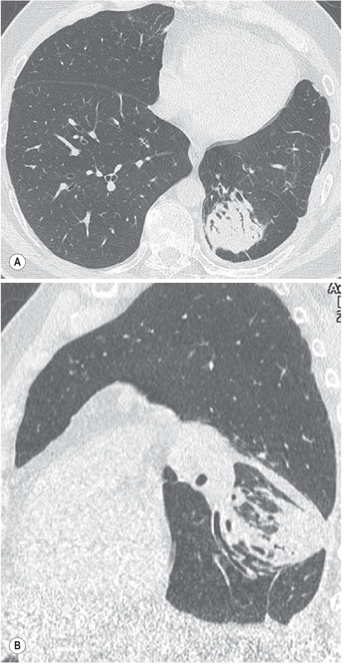
MRI Features
- T1WI: intermediate signal
- T2WI: high signal (akin to atelectatic lung)
- Homogeneous enhancement post-gadolinium (same rationale as CT enhancement - confirms atelectasis)
- MRI useful when CT enhancement ambiguous
Nuclear Medicine
- V/Q scan: matched ventilation-perfusion defect (both reduced in atelectatic lung)
DIFFERENTIAL DIAGNOSIS - KEY EXAM TABLE
| Feature | Round Atelectasis | Peripheral Lung Carcinoma | Mesothelioma |
|---|---|---|---|
| Shape | Round/oval | Round/irregular | Rindlike/nodular |
| Pleural thickening | Always present, adjacent | May/may not | Circumferential pleural thickening |
| Comet tail sign | Present | Absent | Absent |
| Enhancement (CT/MRI) | Strong, homogeneous | Variable, heterogeneous | Variable |
| Volume loss | Present (ipsilateral) | Absent | May be present |
| Growth on serial CT | Stable | Grows | Grows |
| Asbestos history | Yes (usually) | Possibly | Yes (usually) |
| Air bronchograms | May be present | Absent (usually) | Absent |
| PET-FDG | Low uptake | High uptake | High uptake |
Critical point: Strong homogeneous CT enhancement + comet tail sign + stability on serial imaging = confidently diagnose round atelectasis and avoid unnecessary biopsy.
CLINICAL FEATURES
- Usually asymptomatic - incidental finding
- Symptoms from associated pleural disease: dyspnea (restrictive defect from DPT)
- History of asbestos exposure (mesothelioma must be excluded in same patient)
- Lung function: restrictive pattern if diffuse pleural thickening is extensive
MANAGEMENT
- No treatment required for round atelectasis itself
- Recognition is key to avoid unnecessary biopsy / surgery
- Monitor with serial CT (6-12 months) to confirm stability
- Address underlying cause (asbestos exposure, pleural disease management)
- If diagnostic uncertainty persists → PET-CT (low FDG uptake in round atelectasis vs high in malignancy) or biopsy
HIGH-YIELD EXAM BULLETS
- Round atelectasis = folded lung = Blesovsky syndrome
- Always associated with pleural thickening (always subpleural)
- Most common cause: asbestos exposure (exam favourite)
- Location: dorsal lower lobes (most common)
- Comet tail sign = curved bronchovascular bundle sweeping into mass = PATHOGNOMONIC on CT
- CT: subpleural + pleural thickening + comet tail + strong homogeneous enhancement = diagnostic tetrad
- Stable on serial imaging = key to differentiate from lung carcinoma
- PET-CT: low FDG uptake (vs high in carcinoma / mesothelioma)
- No biopsy needed if all 4 CT criteria satisfied
- Murray & Nadel: folded lung is seen in diffuse pleural thickening from asbestos; develops in 4-5% of asbestos-exposed individuals
Bronchopleural fistula 10 marks Bronchopleural fistula vs lung abscess 10 mark
Bronchopleural fistula 10 marks Bronchopleural fistula vs lung abscess 10 mark
PART 1: BRONCHOPLEURAL FISTULA (BPF)
RGUHS Radiology PG - 10 Marks
DEFINITION
Differs from pneumothorax in that the communication is via airways (bronchus), NOT via distal air spaces (alveoli). - Grainger & Allison
CAUSES / ETIOLOGY
| Category | Specific Causes |
|---|---|
| Post-surgical (most common overall) | Pneumonectomy (most dangerous), lobectomy, segmentectomy, sleeve resection |
| Necrotising infections | Staphylococcal pneumonia, Klebsiella, TB empyema, anaerobic lung abscess, actinomycosis, fungal (coccidioidomycosis) |
| Trauma | Penetrating chest injury, barotrauma (ventilator-associated) |
| Tumour | Carcinoma eroding into pleural space |
| Radiation necrosis | Post-radiotherapy |
| Spontaneous | Rupture of peripheral cavity/bulla into pleura |
Post-pneumonectomy BPF is the most dangerous because the persistent pleural space cannot be obliterated by lung re-expansion, and the liquid contents of the pneumonectomy space can flood the remaining lung. - Pye's Surgery
PATHOPHYSIOLOGY
- Bronchus fails to heal (poor blood supply, infection, malignancy)
- Communication opens between bronchial lumen and pleural space
- Air enters pleural space via airways on each breath (not via alveolar rupture)
- If pleural space contains fluid (empyema / post-pneumonectomy fluid) → hydropneumothorax or pyopneumothorax
- Risk of aspiration of pleural fluid into remaining lung → acute respiratory failure
CLINICAL FEATURES
- Sudden cough productive of large volumes of brown/purulent fluid (post-pneumonectomy - pathognomonic)
- Progressive dyspnea, fever, sepsis
- Persistent air leak via chest drain
- Ventilator patients: inability to maintain tidal volume, persistent air leak through drain
IMAGING
CXR - KEY SIGNS
- Normal post-pneumonectomy space fills with fluid progressively → opacified hemithorax
- Cardinal sign: Appearance of a new air-fluid level in the previously opacified hemithorax
- Or: New air space (positive pressure) in operated side - semicircular in shape (instead of expected triangular negative space)
- Fall in fluid level on serial CXRs (fluid escaping via fistula into airways)
- Tracheal/mediastinal shift may or may not be present
- Air-fluid level in post-op space that increases in size = BPF until proven otherwise
- Hydropneumothorax or pyopneumothorax: air + fluid in pleural space = horizontal air-fluid level
- Air-fluid level occupies the ENTIRE width of the hemithorax (distinguishes from lung abscess)
- Pleural thickening, loculation
- Underlying pneumonia/consolidation with cavity
CT - Investigation of Choice
- Directly visualises the fistulous tract (thin linear air track connecting bronchus to pleural space)
- Air within pleural space with fluid = hydropneumothorax
- Split pleura sign: Enhancement of both visceral and parietal pleura (empyema component)
- Pleural thickening with smooth inner walls
- Underlying parenchymal disease (necrotising pneumonia, cavity)
- Loculated air pockets within empyema (multilocular)
- CT can identify level of fistula - helpful for bronchoscopic treatment
Classic CT/CXR Sign in Post-Pneumonectomy BPF:
"The cardinal sign is the appearance of a positive air space on the operated side - semicircular in shape. A fall in the fluid level with a new air-fluid level indicates BPF." - Pye's Surgery
INVESTIGATION SUMMARY
| Investigation | Findings |
|---|---|
| CXR | New air-fluid level; fall in post-pneumonectomy fluid level; hydropneumothorax |
| CT Chest | Fistulous tract; split pleura sign; loculated empyema + air |
| Bronchoscopy | Identifies proximal fistula; balloon occlusion identifies segment; guides treatment |
| Sinography | Contrast injected into pleural drain shows communication |
TREATMENT
- Minimise air leak: Low tidal volumes, PEEP, high frequency oscillatory ventilation
- Dependent positioning: Fistula side down (reduces aspiration of fluid into good lung)
- Bronchoscopic closure: Silver nitrate, cyanoacrylate, fibrin glue, endobronchial valves - balloon occlusion identifies the segment
- Surgical: Pleurodesis, surgical closure of bronchial stump, thoracoplasty - treatment of choice for post-pneumonectomy BPF
- Persistent BPF > 7 days refractory to medical therapy → surgery
PART 2: BPF vs LUNG ABSCESS - RADIOLOGY DIFFERENTIATION
RGUHS Radiology PG - 10 Marks
OVERVIEW
KEY DIFFERENCE TABLE (Murray & Nadel Table 109.1)
| Feature | Empyema / BPF | Lung Abscess |
|---|---|---|
| Shape | Lenticular (biconvex, D-shaped) | Rounded / spherical |
| Wall | Thick smooth wall | Thick irregular wall |
| Lung-fluid boundary | Surrounding lung compressed (sharp margin) | Boundary indistinct - necrosis of adjacent lung |
| Angle with chest wall | Obtuse angle (broad, tapering margins) | Acute angle |
| Lung vessels | No vessels close to collection | Vessels seen passing through or near collection |
| Air-fluid level length | Extends full width of hemithorax | Shorter - limited to cavity diameter |
| CT split pleura sign | Present (both pleural layers enhance) | Absent |
| Mediastinal shift | May be present (mass effect of large empyema) | Absent (lung destroyed, no mass effect) |
| Underlying lung | Compressed but preserved | Destroyed/necrotic |
CXR DIFFERENTIATION IN DETAIL
BPF / Empyema with Air-Fluid Level
- Air-fluid level horizontal and extends edge-to-edge (full width of pleural space)
- Lenticular/D-shaped opacity conforming to pleural space
- Medial border is concave (follows pleura)
- Smooth, tapering margins that form obtuse angles with chest wall
- On lateral view: air-fluid level extends further posteriorly (pleural space geometry)
- Associated pleural thickening
Lung Abscess
- Air-fluid level limited to cavity size (rounded/oval)
- Walls are ragged and irregular (necrotic lung)
- Located within the lung parenchyma
- Surrounding consolidation, satellite nodules
- Acute angle with chest wall (invades lung, not along pleura)
- Can cross fissures (empyema cannot)
CT DIFFERENTIATION - MOST IMPORTANT
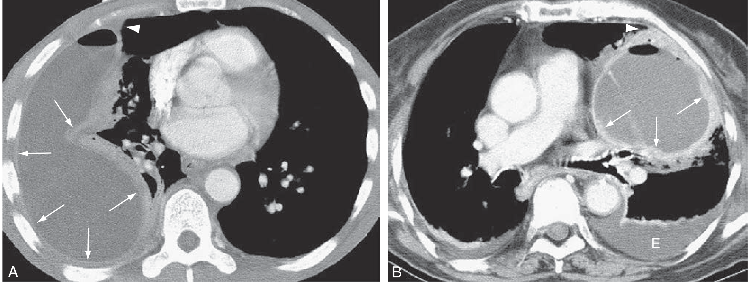
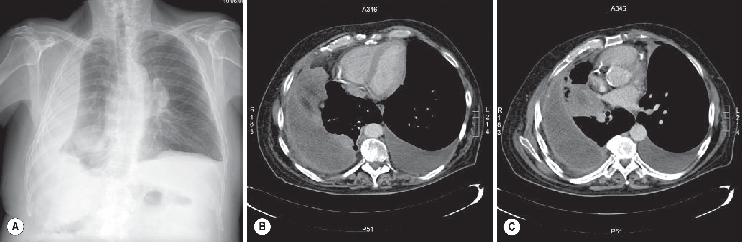
CT Split Pleura Sign (Empyema/BPF)
- Both visceral AND parietal pleura enhance separately after IV contrast
- Creates the appearance of two enhancing layers separated by fluid
- Pathognomonic of empyema (not seen in lung abscess)
- Represents fibrinous/inflammatory thickening of both pleural surfaces
COMPLETE COMPARISON TABLE
| Feature | BPF / Empyema | Lung Abscess |
|---|---|---|
| Shape | Lenticular / D-shaped | Rounded / spherical |
| Wall | Smooth, uniform thickness | Irregular, non-uniform |
| Chest wall angle | Obtuse | Acute |
| Split pleura sign | Present | Absent |
| Compressed lung | Yes | No (lung necrosed) |
| Crossing fissures | No (pleural space limited by fissures) | Yes (parenchymal, can cross) |
| Vessels within collection | No | Yes (traversing) |
| Air-fluid level | Full width of hemithorax | Short (cavity size only) |
| Mediastinal shift | Possible | Not typical |
| Enhancement post-contrast | Pleural layers enhance (split pleura) | Cavity wall enhances (ring) |
| Satellite lesions | Absent | Present (aspiration nodules) |
HIGH-YIELD EXAM BULLETS
- BPF = airway-to-pleural communication (NOT alveolar rupture like pneumothorax)
- Most common cause: post-pneumonectomy (most dangerous)
- Infections: staph, TB, necrotising pneumonia
- Cardinal CXR sign in post-pneumonectomy BPF: new air-fluid level / fall in fluid level on operated side
- Split pleura sign on CT = pathognomonic of empyema + BPF
- BPF: air-fluid level spans full width of hemithorax; lung abscess: short air-fluid level
- BPF/empyema: lenticular shape + obtuse angle; abscess: round + acute angle
- BPF: lung compressed; abscess: lung destroyed/necrotic (indistinct margin)
- Vessels traverse a lung abscess but are absent near empyema collection
- CT is the investigation of choice to differentiate and to detect the fistulous tract
Pleural tumours radiology. Malignant pleural mesothelioma in detail
PLEURAL TUMOURS - RADIOLOGY
Malignant Pleural Mesothelioma in Detail
RGUHS Radiology PG - 10 Marks
CLASSIFICATION OF PLEURAL TUMOURS
| Type | Examples |
|---|---|
| Primary Malignant | Malignant pleural mesothelioma (most common primary), Synovial sarcoma, Primary pleural lymphoma |
| Primary Benign | Solitary fibrous tumour of pleura (SFTP), Lipoma, Fibroma |
| Secondary / Metastatic | Most common overall - adenocarcinoma from lung, breast, ovary, GI tract |
| Tumour-like | Erdheim-Chester disease, Diffuse pulmonary lymphangiomatosis |
Most common pleural neoplasm overall = Metastatic (not mesothelioma). Most common primary pleural malignancy = Malignant Mesothelioma.
MALIGNANT PLEURAL MESOTHELIOMA (MPM)
DEFINITION & EPIDEMIOLOGY
- Primary malignant tumour of mesothelial cells lining the pleura
- 80% of all mesothelioma cases are pleural (20% peritoneal, rare pericardial/tunica vaginalis)
- Strongly linked to asbestos exposure (~70% of cases)
- Latency period: 30-40 years from asbestos exposure to disease development
- Peak incidence: 5th-7th decade; Male > Female
- Most common asbestos type: Amphiboles (crocidolite > amosite) > chrysotile
- 60% right-sided (due to gravitational predilection of asbestos fibers to right lower lobe)
HISTOLOGICAL SUBTYPES (WHO 2021 Classification)
| Subtype | Frequency | Prognosis |
|---|---|---|
| Epithelioid | 60% | Best prognosis |
| Sarcomatoid | 20% | Worst prognosis |
| Biphasic | 20% | Intermediate |
- Desmoplastic = subtype of sarcomatoid
- Well-differentiated papillary mesothelial tumour = now a separate entity (NOT mesothelioma)
GROSS PATHOLOGY / SPREAD
- Begins as multiple discrete nodules on parietal pleura preferentially
- Nodules coalesce → visceral + parietal fusion
- Progressive lung encasement by thick rind of tumor (several cm thick)
- Extends into interlobar fissures
- Advanced: chest wall invasion, diaphragmatic invasion, mediastinal involvement, pericardium
- 70% have mediastinal nodal involvement at autopsy
- Hematogenous mets: contralateral lung, liver, adrenals, bone, brain
CLINICAL FEATURES
- Chest pain (non-pleuritic): 60-70%
- Dyspnoea: 25%
- Cough: 20%
- Unilateral pleural effusion (may be only finding early)
- Palpable chest wall mass (advanced)
- Only 10% bilateral at presentation
IMAGING IN DETAIL
1. Chest X-Ray (CXR)
- Large unilateral pleural effusion (most common initial finding)
- Often with contralateral mediastinal shift (large effusion)
- Associated pleural plaques (evidence of asbestos exposure) - only 20% have radiographic asbestosis
- Ipsilateral mediastinal shift despite large effusion = CLASSIC SIGN of mesothelioma (lung encased by tumour - cannot expand to cause contralateral shift)
- Nodular / irregular / lobulated pleural thickening (rind)
- Thickening of interlobar fissures
- Intercostal space narrowing (volume loss)
- Elevated diaphragm (volume loss)
- Mediastinal widening (nodal/direct invasion)
- Rib destruction / chest wall soft-tissue mass (advanced)
- Pleural plaques or calcifications elsewhere (marker of asbestos exposure)
Classic exam sign: Massive pleural effusion + IPSILATERAL (not contralateral) mediastinal shift = mesothelioma until proven otherwise (lung is trapped and cannot re-expand).
2. CT Chest - Investigation of Choice for Staging
| Feature | Description |
|---|---|
| Circumferential pleural thickening | Rind-like, encasing lung; may be nodular or smooth |
| Nodular pleural thickening | Irregular nodules on pleural surface |
| Pleural thickening > 1 cm | Suggests malignancy |
| Mediastinal pleural involvement | Extends across mediastinum |
| Fissure thickening | Tumour extending into interlobar fissures |
| Pleural effusion | Usually large, often obscures underlying thickening |
| Volume loss | Ipsilateral hemithorax shrinkage |
| Chest wall invasion | Rib destruction, intercostal soft-tissue mass |
| Diaphragm invasion | Irregular/nodular diaphragm |
| Mediastinal nodes | Enlarged in up to 50% |
| Hypodense areas | Areas of necrosis within tumour |
- Circumferential pleural thickening
- Pleural nodules
- Parietal thickening > 1 cm
- Mediastinal pleural involvement
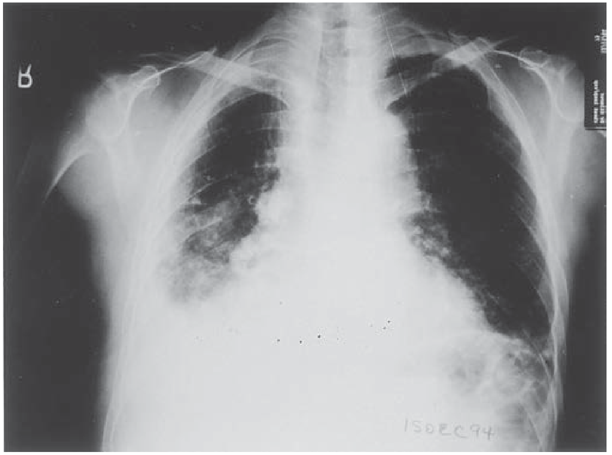
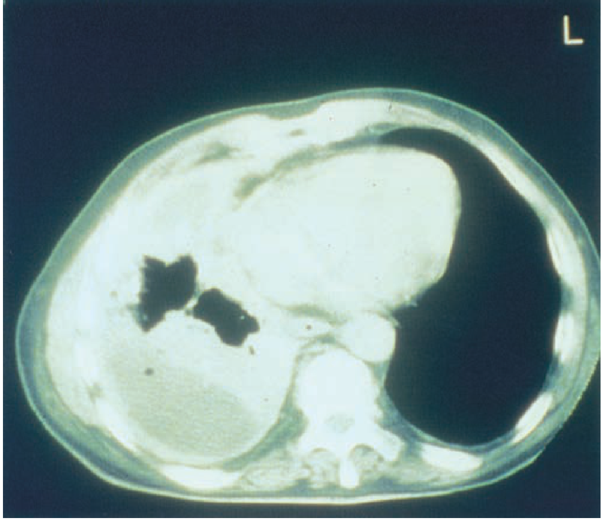
3. MRI
- Signal: minimally increased T1, moderately increased T2
- Superior to CT for:
- Diaphragmatic invasion (82% vs 55% accuracy for CT)
- Chest wall and endothoracic fascial invasion (69% vs 46% for CT)
- Overall mediastinal nodal accuracy: ~50% (both similar)
- MRI superior for relationship of tumour to chest wall, mediastinum and diaphragm
- Useful in surgical planning (extrapleural pneumonectomy candidates)
- Coronal MRI best for diaphragm assessment
4. PET-CT / FDG-PET
- Differentiates malignant from benign pleural disease: sensitivity 91%, specificity 100%
- FDG uptake is significantly higher in malignant vs benign lesions
- Staging: extent of pleural disease, mediastinal nodes, chest wall invasion, extrathoracic mets
- More sensitive than CT for extrathoracic metastases
- Treatment response assessment (chemo/radiotherapy)
- Radiation therapy planning
- Limitation: Limited sensitivity for locoregional staging (resectability)
- High SUV associated with worse prognosis
5. Ultrasound
- Detects pleural effusion and guides thoracocentesis/biopsy
- Identifies loculated effusions
- Supplementary for biopsy and surgical planning
CT vs MRI - COMPARISON TABLE
| Assessment | CT | MRI |
|---|---|---|
| Pleural thickening | Excellent | Good |
| Pleural calcifications | Superior | Poor |
| Fissure extension | Excellent | Good |
| Chest wall invasion | 46% accuracy | 69% |
| Diaphragm invasion | 55% accuracy | 82% |
| Mediastinal nodes | ~50% | ~50% |
| Bone involvement | Superior | Good |
DIFFERENTIAL DIAGNOSIS OF MPM
| Condition | Differentiating Features |
|---|---|
| Pleural metastases | Most common pleural malignancy; known primary (lung, breast, ovary); less volume loss; hilar nodes more common |
| Diffuse pleural thickening (benign) | No nodularity; no mediastinal pleural involvement; FDG-PET negative |
| Empyema | Clinical sepsis; split pleura sign; air-fluid level; resolves with drainage |
| Lymphoma | Systemic features; anterior mediastinal mass; responds to chemo |
| Pleural plaques (asbestos) | Parietal pleura; calcified; discrete; no mass effect |
Mesothelioma vs Metastases on CT: Hilar adenopathy more common in metastases. Mesothelioma more often circumferential, ipsilateral volume loss.
BENIGN PLEURAL TUMOURS - BRIEF
Solitary Fibrous Tumour of Pleura (SFTP)
- Previously called "benign fibrous mesothelioma" - misnomer (not from mesothelium)
- Origin: subpleural fibroblasts / CD34+ cells
- Usually benign; 10-20% malignant
- CXR: well-defined, lobulated, pedunculated pleural mass; changes position with posture (if pedunculated)
- CT: heterogeneous solid mass with areas of necrosis; enhances avidly
- Associated with: hypertrophic osteoarthropathy (Marie-Bamberger) and hypoglycaemia (Doege-Potter syndrome)
- Treatment: surgical resection
Pleural Metastases
- Most common pleural neoplasm
- Origin: lung > breast > ovary > GI
- CXR: pleural effusion (often only sign); multiple nodules; diffuse thickening
- CT: nodular pleural thickening + effusion; hilar/mediastinal nodes common
- Indistinguishable from mesothelioma on imaging alone → biopsy required
HIGH-YIELD EXAM BULLETS
- MPM = most common primary pleural malignancy; metastases = most common overall
- Asbestos exposure in 70%; latency 30-40 years; amphiboles most carcinogenic
- 60% right-sided mesothelioma
- Classic CXR: massive effusion + ipsilateral mediastinal shift (trapped lung)
- CT: circumferential nodular pleural thickening > 1 cm + mediastinal pleural involvement = malignant
- Epithelioid = best prognosis; sarcomatoid = worst
- MRI superior for diaphragm invasion (82% vs CT 55%) and chest wall invasion
- PET-CT: sensitivity 91%, specificity 100% for differentiating benign vs malignant pleural disease
- 70% have mediastinal nodal involvement at autopsy
- SFTP (solitary fibrous tumour): pedunculated mass; hypertrophic osteoarthropathy + hypoglycaemia (Doege-Potter syndrome)
- Mesothelioma indistinguishable from pleural metastases on imaging → biopsy required
- FDG-PET: high uptake in malignant mesothelioma; no uptake in benign (fibroma, TB pleuritis)
I didn’t get the ans yet
Causes of neonatal acute respiratory distress syndrome. Discuss imaging features in detail
Discuss imaging features of neonatal distress
Create a comprehensive study guide PDF on neonatal respiratory distress imaging
Causes of acute neonatal respiratorydistress. Describe their imaging
CAUSES OF ACUTE NEONATAL RESPIRATORY DISTRESS - IMAGING
RGUHS Radiology PG - 10 Marks
CAUSES - CLASSIFICATION
By Lung Volume on CXR (Exam Approach)
| Lung Volume | Causes |
|---|---|
| Low volume (small lungs) | RDS/HMD, Neonatal pneumonia |
| Normal volume | Transient Tachypnoea of Newborn (TTN), Neonatal pneumonia |
| High volume (hyperinflated) | Meconium Aspiration Syndrome (MAS), Air leak (pneumothorax) |
| Asymmetric | Diaphragmatic hernia, Pneumothorax, Lobar overinflation |
1. RESPIRATORY DISTRESS SYNDROME (RDS) / HYALINE MEMBRANE DISEASE (HMD)
Background
- Caused by surfactant deficiency in premature infants
- Commonest cause of respiratory distress in premature neonates (<34 weeks)
- Risk increases with decreasing gestational age
- Surfactant produced by Type II pneumocytes from ~28 weeks
CXR - Classic Features (Gradings I-IV)
| Grade | CXR Appearance |
|---|---|
| I | Fine reticulogranular / ground-glass pattern (diffuse bilateral) |
| II | Grade I + air bronchograms extending to periphery |
| III | Grade II + heart borders becoming obscured ("white-out beginning") |
| IV | Complete "white-out" - lungs completely opacified; heart borders invisible |
- Bilateral symmetrical involvement
- Low lung volumes (bell-shaped chest)
- Fine granular / reticulogranular opacification bilaterally
- Air bronchograms (air in bronchi contrasted against opaque alveoli)
- Heart border partially or completely obscured
- No pleural effusion (unlike TTN)
- Rapid resolution after surfactant therapy (dramatic improvement within hours)
- Normal CXR in first hour; findings develop over 4-6 hours
- After surfactant: patchy asymmetric clearing ("salt and pepper" pattern)
2. TRANSIENT TACHYPNOEA OF THE NEWBORN (TTN)
Background
- Delayed clearance of fetal lung fluid (wet lung)
- Commoner in: term or near-term infants, caesarean section births, maternal diabetes
- Self-limiting; resolves in 48-72 hours
CXR Features
- Normal or mildly hyperinflated lung volumes (opposite of RDS)
- Bilateral perihilar streakiness / prominent bronchovascular markings
- Fluid in fissures (horizontal fissure thickening - classic)
- Small pleural effusions bilaterally (often bilateral)
- Mild cardiomegaly may be present
- Clears rapidly (within 24-48 hours = key differentiating feature on serial CXR)
Exam Tip: TTN = fluid in fissures + pleural effusion + normal/high lung volume = resolves fast. RDS = granular opacification + low volume in preterm infant.
3. MECONIUM ASPIRATION SYNDROME (MAS)
Background
- Aspiration of meconium-stained amniotic fluid in utero or at delivery
- Full-term or post-term infants (fetal distress)
- Meconium causes: chemical pneumonitis, airway obstruction (check-valve), surfactant inactivation
CXR Features
- Hyperinflated lungs (overexpanded due to air-trapping)
- Coarse, irregular, asymmetric opacities bilaterally (patchy consolidation)
- Rope-like densities and streaky infiltrates
- Pneumothorax (10-20%) - common complication of ball-valve obstruction
- Pneumomediastinum
- Asymmetric; unilateral involvement possible
- No pleural effusions typically
Key: Post-term + hyperinflated + coarse irregular patches + pneumothorax = MAS
4. NEONATAL PNEUMONIA (Group B Streptococcus most common)
Background
- GBS (Group B Streptococcus) = most common cause of early-onset neonatal pneumonia
- Presentation: birth or within 6 hours (early onset) or after 7 days (late onset)
- Difficult to distinguish from RDS on CXR
CXR Features
- Indistinguishable from RDS in early stages (same granular pattern)
- Diffuse bilateral opacification (mimics HMD exactly)
- May show segmental/lobar consolidation (more focal than RDS)
- Pleural effusions more common than in RDS
- May have asymmetric opacification
- Normal or decreased lung volumes
- Air bronchograms present
Differentiating from RDS: pleural effusion + asymmetric + term infant = pneumonia more likely than RDS (RDS is symmetric, bilateral, low volume, premature).
5. NEONATAL PNEUMOTHORAX
Background
- Spontaneous or secondary to MAS, RDS, PPV (positive pressure ventilation)
- Life-threatening emergency
CXR Features
- Hyperlucent hemithorax (affected side)
- Visible pleural line with absent lung markings beyond
- Mediastinal shift to contralateral side (tension pneumothorax)
- Depressed ipsilateral diaphragm
- "Spinnaker sail sign" = pneumomediastinum (elevated thymic lobes on either side of mediastinum)
- In supine neonate: air rises anteriorly → anteromedial lucency, "deep sulcus sign"
Deep sulcus sign in supine neonate: lucency extending into the costophrenic angle (air anteriorly pools at base). Classic neonatal pneumothorax sign on AP film.
6. CONGENITAL DIAPHRAGMATIC HERNIA (CDH - Bochdalek)
Background
- Left-sided in 85% (Bochdalek hernia - posterior)
- Herniation of abdominal contents into chest
- Associated with pulmonary hypoplasia
CXR Features (Classic)
- Loops of bowel / gas-filled structures in left hemithorax
- Mediastinal shift to contralateral (right) side
- Small or absent left hemidiaphragm shadow
- Absent gastric bubble in abdomen
- Nasogastric tube coiling in chest (if stomach herniated)
- Ipsilateral lung small / hypoplastic
- Scaphoid abdomen (bowel in chest)
- Right-sided Bochdalek: hepatic shadow in right chest + rightward mass
Classic triad: Bowel in left chest + mediastinal shift right + scaphoid abdomen = CDH until proven otherwise.
7. PULMONARY INTERSTITIAL EMPHYSEMA (PIE)
Background
- Air tracking into pulmonary interstitium along bronchovascular bundles
- Complication of mechanical ventilation (barotrauma)
- Common in severe RDS on ventilator
CXR Features
- Linear / bubbly radiolucencies radiating from hilum (not following airway pattern)
- Small round or oval lucencies scattered throughout lung
- May be unilateral or bilateral
- Can progress to pneumothorax, pneumomediastinum, pneumopericardium
- "Bubbly" or "cystic" appearance
- Leads to overdistension of affected lung
8. CONGENITAL LOBAR OVERINFLATION (Neonatal presentation)
- Air trapping in single lobe (check-valve bronchial defect)
- Left upper lobe most common (42%)
- CXR: Hyperlucent overinflated lobe with mediastinal shift contralaterally
- Rapid postnatal progression
SUMMARY COMPARISON TABLE (HIGH YIELD)
| Condition | Gestation | Volume | CXR Pattern | Key Feature |
|---|---|---|---|---|
| RDS/HMD | Preterm | Low | Bilateral granular/GG, air bronchograms | Grades I-IV; surfactant deficient |
| TTN | Term/near-term | Normal/High | Perihilar streakiness, fissure fluid | Pleural effusion; clears 48h |
| MAS | Post-term | High | Coarse irregular bilateral patches | Pneumothorax complication |
| Pneumonia (GBS) | Any | Low/Normal | Bilateral opacification ± lobar | Pleural effusion; mimics RDS |
| Pneumothorax | Any | Asymmetric | Hyperlucency + pleural line | Deep sulcus sign supine |
| CDH | Any | Asymmetric | Bowel loops in chest | Mediastinal shift; absent diaphragm |
| PIE | Preterm (ventilated) | High | Linear/cystic interstitial lucencies | Barotrauma complication |
IMAGING MODALITIES IN NEONATAL RESPIRATORY DISTRESS
| Modality | Role |
|---|---|
| CXR (AP supine) | First-line - essential in all cases; diagnoses most conditions |
| US Chest | Confirms pneumothorax; identifies pleural effusion; hepatic position in CDH; bedside |
| CT | Reserved for complex/unclear cases; CDH anatomy; CPAM |
| Fluoroscopy | Diaphragm movement (eventration); contrast for bowel in CDH |
| Echocardiography | Excludes congenital heart disease; pulmonary hypertension |
HIGH-YIELD EXAM BULLETS
- RDS: preterm + low volume + bilateral granular opacification + air bronchograms; 4 grades
- TTN: term + fluid in fissures + bilateral pleural effusions + normal/high volume; clears 48h
- MAS: post-term + high volume + coarse irregular opacities + pneumothorax
- CDH: bowel loops in chest + mediastinal shift contralateral + absent diaphragm + scaphoid abdomen
- Pneumothorax: hyperlucency + pleural line; supine = deep sulcus sign
- Pneumomediastinum: "Spinnaker sail sign" = elevated thymic lobes
- PIE: complication of ventilation in preterm; linear/bubbly interstitial lucencies
- RDS vs Pneumonia: RDS symmetric in premature; GBS pneumonia → pleural effusion, asymmetric
- CXR lung volume is the key initial discriminator between causes
- After surfactant treatment: RDS shows rapid, often patchy/asymmetric clearing
Virtual bronchoscopy radiology
VIRTUAL BRONCHOSCOPY
RGUHS Radiology PG - 10 Marks
DEFINITION
- Also called CT virtual endoscopy, CT bronchoscopy, or CT bronchography
- Part of the broader family of 3D CT postprocessing techniques (Grainger & Allison, Table 1.2)
TECHNICAL BASIS
CT Acquisition Requirements
| Parameter | Requirement |
|---|---|
| Slice thickness | < 1 mm (ideally 0.5-0.625 mm) |
| Slice spacing | < 0.625 mm (overlapping reconstruction) |
| Breath phase | Single breath hold at end inspiration |
| Scanner | MDCT (multi-detector) - isotropic voxels |
| Reconstruction kernel | Soft tissue / standard |
- Thin overlapping slices allow isotropic voxel acquisition - equal voxel size in all three axes
- This is the prerequisite for accurate 3D rendering in any plane
Rendering Methods (Two Types)
| Method | What it Produces | Use |
|---|---|---|
| Internal rendering (perspective volume rendering) | Virtual luminal view - endoscopic simulation looking down the airway lumen | Mimics bronchoscopy; shows intraluminal lesions |
| External rendering (CT bronchography) | External 3D view of the airway tree | Shows airway dimensions, branching, and relation to adjacent structures |
Grainger & Allison: "Surface rendering and volume rendering are used to produce endoscopic simulations of the airway."
Cummings: "Internal rendering of CT data produces a virtual luminal view that imitates bronchoscopy, whereas external rendering produces CT bronchography, which illustrates the airway's dimensions and its relationship to adjacent structures."
WHAT VIRTUAL BRONCHOSCOPY SHOWS
- Trachea, main bronchi, lobar bronchi, segmental bronchi
- Up to 6th-generation bronchi (Asano et al., Murray & Nadel)
- Airway wall contour, mucosa surface appearance, narrowings
- Intraluminal lesions (masses, polyps, foreign bodies)
- Post-stenotic airway (beyond an obstructing lesion)
- Normal branching anatomy including variations
CLINICAL APPLICATIONS
1. Airway Stenosis Evaluation
- Assessment of tracheal / bronchial stenosis (post-intubation, post-traumatic, tumour)
- Key advantage: VB can view the airway distal to a tight stenosis - impossible with fibreoptic bronchoscopy
- Grainger & Allison: "Virtual CT bronchoscopy can provide a view 'through' an obstructing lesion to visualise the airway distal to it, which may not be possible with conventional bronchoscopy"
- Cummings: "Several studies have demonstrated good accuracy of this modality in assessing tracheobronchial stenosis"
2. Bronchogenic Carcinoma
- Detection and extent of endobronchial tumour
- Pre-bronchoscopy planning: identify exactly which generation of bronchus to target for biopsy
- Assess submucosal / peribronchial extension
- Evaluate for vocal cord, tracheal, or carina involvement (unresectability assessment)
3. Peripheral Pulmonary Nodule Biopsy Planning
- VB generates a 3D map of the planned pathway through the tracheobronchial tree to a peripheral nodule
- The virtual images are synchronized in real time with the bronchoscopic view during the procedure
- Murray & Nadel: VB images generated to median 6th-generation bronchi; ultrathin bronchoscope inserted into 95% of planned airways; diagnostic yield 82% for nodules <30mm
- Meta-analysis (Murray & Nadel): diagnostic yield 74% for nodules <3 cm; 67% for nodules <2 cm
- CT Bronchus Sign: when an airway is seen leading directly to a nodule on CT - VB is most useful in this situation
- Murray & Nadel: "Virtual bronchoscopy increased the diagnostic yield from 78% to 94%" when combined with radial EBUS for lesions <2 cm in the outer third with CT bronchus sign but invisible on fluoroscopy
4. Foreign Body Detection (especially in children)
- Non-invasive identification of radiolucent foreign bodies in airways
- Guides site and approach for rigid bronchoscopy retrieval
- Cummings: particularly useful in paediatric airway anomalies
5. Congenital Airway Anomalies
- Tracheal stenosis, tracheomalacia, tracheal agenesis (Type I, II, III - Floyd classification)
- Characterisation prior to surgical repair
- Cummings: "good accuracy in assessing tracheobronchial stenosis" and tracheal anomalies
6. Pre-operative / Pre-bronchoscopy Planning
- Maps entire airway tree before interventional procedures
- Stent placement planning for malignant airway obstruction
- Identifies accessible biopsy targets for endobronchial lesions
7. Post-treatment Assessment
- Post-stent placement evaluation
- Monitoring of airway after sleeve resection
- Surveillance after tracheal reconstruction
ADVANTAGES OF VIRTUAL BRONCHOSCOPY
| Advantage | Explanation |
|---|---|
| Non-invasive | No sedation, no instrument insertion, no risk of bleeding/perforation |
| View through stenoses | Can visualise post-stenotic airway impossible with real bronchoscopy |
| Unlimited viewing angle | Can "fly" in either direction (anterograde and retrograde) |
| Combined with CT data | Simultaneous review of mediastinum, lung parenchyma, lymph nodes |
| Pre-procedure planning | Guides bronchoscopist to target lesion; reduces procedure time |
| Paediatric-friendly | Avoids anaesthesia risk in children with airway compromise |
| 3D relationship display | External rendering shows airway relation to vessels, tumour |
| Repeatability | Can be re-reviewed and re-navigated without repeat patient exposure |
LIMITATIONS OF VIRTUAL BRONCHOSCOPY
| Limitation | Explanation |
|---|---|
| Cannot assess mucosal colour | VB is a structural tool; mucosal changes (redness, oedema, bleeding) invisible |
| Cannot biopsy | No tissue sampling capability |
| Motion artefact | Respiratory / cardiac motion degrades quality |
| Software-dependent accuracy | Quality limited by CT acquisition and proprietary software; may mislead for fine anatomic details (Cummings) |
| Sub-segmental visibility | Beyond 6th-generation bronchi accuracy decreases; emphysema interferes |
| Cannot assess malacia | Poor for dynamic conditions like tracheobronchomalacia (Cummings) |
| Cannot detect flat/subtle lesions | Submucosal lesions without luminal projection may be missed |
| Radiation | Requires CT acquisition (vs no radiation with fibreoptic) |
| Not real-time | Static post-processing; cannot replace dynamic visual assessment |
COMPARISON WITH CONVENTIONAL BRONCHOSCOPY
| Feature | Virtual Bronchoscopy | Fibreoptic Bronchoscopy |
|---|---|---|
| Invasiveness | Non-invasive | Invasive |
| Biopsy | Not possible | Possible |
| Mucosal assessment | No | Yes |
| Post-stenotic view | Yes (key advantage) | No |
| Dynamic (malacia) | No | Yes |
| Anaesthesia required | No | Yes (usually) |
| Peripheral nodule guidance | Yes (with planning) | Limited without EBUS/navigation |
| Radiation | Yes (CT) | No |
| Real-time navigation | Yes (synchronized) | Yes |
VIRTUAL BRONCHOSCOPY AND ELECTROMAGNETIC NAVIGATION (EMN)
- VB creates the 3D airway map (CT data processed to reconstruct tracheobronchial tree + identify nodule location)
- EMN adds a sensor tracked in real-time electromagnetic field (like a GPS system for the bronchoscope)
- Bronchoscopist navigated to peripheral nodule using VB-generated pathway
- Two commercial systems (Murray & Nadel):
- SuperDimension (Medtronic) - inspiratory CT; magnetic board under patient
- SPiNDrive (Veran) - inspiratory + expiratory CT; fiducial pads; "always on" tip tracking
- ENB + radial EBUS combination: superior diagnostic yield for peripheral lesions
HIGH-YIELD EXAM BULLETS
- VB = postprocessing technique from thin-slice MDCT data; no patient contact with airways
- Two rendering modes: internal (endoscopic luminal view) = mimics bronchoscopy; external (CT bronchography) = 3D airway anatomy
- CT requirement: slices <1 mm, spacing <0.625 mm, single breath hold at end inspiration
- Key advantage: views airway DISTAL to stenosis - impossible with real bronchoscopy
- Reaches up to 6th-generation bronchi; diagnostic yield 82% for peripheral nodules <30mm (Asano et al.)
- CT Bronchus Sign + VB + radial EBUS: yield improved from 78% to 94%
- Limitation: cannot biopsy, cannot assess mucosal colour, poor for malacia
- Foundation of Electromagnetic Navigation Bronchoscopy (ENB) - real-time GPS-guided bronchoscopy
- External rendering = CT bronchography (useful for pre-surgical anatomy, vascular relationships)
- Grainger & Allison: VB listed under additional postprocessing techniques alongside MPR, MIP, volume rendering
Ct features of thoracic lymphoma
CT FEATURES OF THORACIC LYMPHOMA
RGUHS Radiology PG - 10 Marks
INTRODUCTION
CLASSIFICATION RELEVANT TO THORACIC IMAGING
| Type | Key Subtypes | Thoracic Predilection |
|---|---|---|
| Hodgkin Lymphoma (HL) | Nodular sclerosis (most common), Mixed cellularity, Lymphocyte-rich | Anterior + middle mediastinum; hilar nodes; lung parenchyma |
| Non-Hodgkin Lymphoma (NHL) | Diffuse large B-cell (DLBCL), MALT, Primary Mediastinal B-cell (PMBCL), Follicular, T-cell | Anterior mediastinum (PMBCL); any mediastinal compartment |
| Primary Mediastinal B-cell Lymphoma (PMBCL) | Subset of NHL arising from thymic B-cell precursors | Bulky anterior mediastinal mass |
Murray & Nadel: "Both PMBCL and classic nodular sclerosis HL arise from thymic B-cell precursors. These entities affect adolescents and young adults and are more common in females."
CT FEATURES - ORGANISED BY COMPARTMENT
1. MEDIASTINAL LYMPH NODE ENLARGEMENT (Most Common Finding)
- Anterior + superior mediastinum most commonly involved (>85%)
- Contiguous nodal spread (from neck → mediastinum → para-aortic) - hallmark of HL
- Nodes typically soft tissue density, round, discrete initially
- Confluent nodal masses forming lobulated conglomerate in anterior mediastinum
- Middle mediastinal involvement (paratracheal, subcarinal, hilar nodes) - characteristic
- Hilar adenopathy: bilateral in HL (unilateral hilar more common in NHL)
- Nodes may not follow contiguous spread pattern
- Skip areas common in NHL (non-contiguous involvement)
- Any mediastinal compartment may be involved
- Short-axis diameter > 1 cm (standard criterion)
- Round rather than oval morphology
- Low-density centre (necrosis) - more common in bulky disease
- Calcification pre-treatment is rare; calcification post-treatment (after radiotherapy) is common
- Confluent nodes forming a mediastinal mass (nodes lose individual identity)
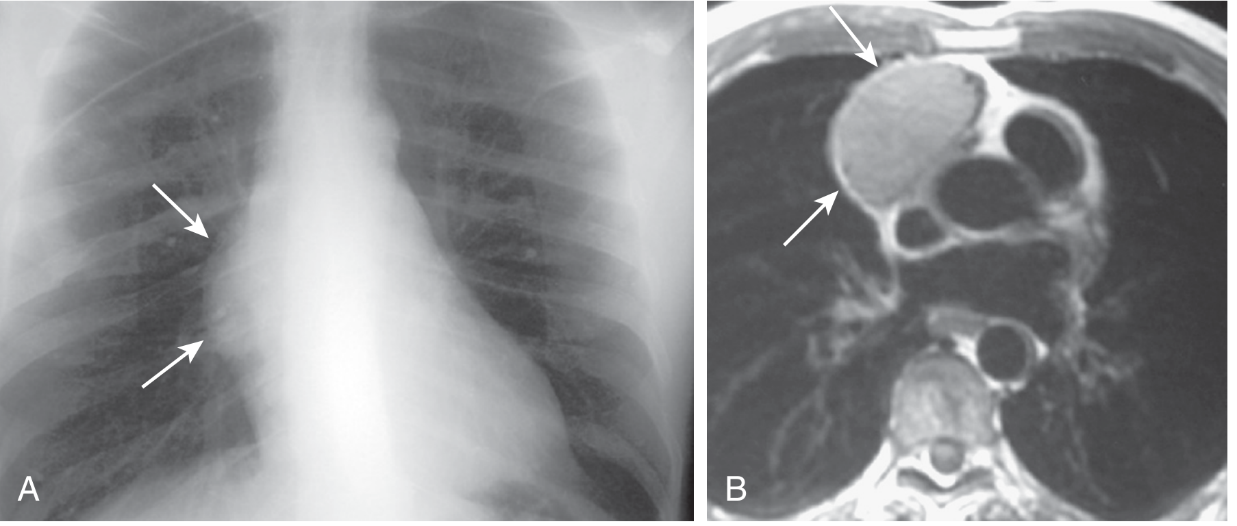
2. ANTERIOR MEDIASTINAL MASS (Thymic Involvement)
- PMBCL and nodular sclerosis HL classically present as a bulky anterior mediastinal mass involving the thymus (Murray & Nadel, line 4281)
- CT shows:
- Large soft-tissue anterior mediastinal mass (can be >10 cm)
- Heterogeneous - areas of haemorrhage or necrosis (hypodense areas on CT)
- Obliterates normal thymic tissue
- Lobulated margins - encases adjacent vessels
- Anterior mediastinum = "4 Ts": Thymoma, Teratoma, Thyroid lesion, Terrible lymphoma (HL/NHL)
Murray & Nadel: "These lymphomas present as a bulky anterior mediastinal mass involving the thymus, sometimes with haemorrhage or necrosis on chest CT."
3. SVC OBSTRUCTION
- SVC syndrome is a common presentation of PMBCL (less so for HL)
- CT shows:
- SVC compression or occlusion by anterior mediastinal mass
- Collateral venous channels (enlarged azygos, internal mammary, chest wall veins)
- Upper body oedema (clinical)
- Venous thrombosis within SVC (filling defect on contrast CT)
Murray & Nadel: "SVC syndrome is a common presentation of PMBCL, but less so for HL."
4. PULMONARY INVOLVEMENT
- More common in HL than NHL; occurs in advanced disease
- Direct extension from mediastinal disease into adjacent lung (most common mechanism)
- CT patterns of pulmonary lymphoma:
| Pattern | Description |
|---|---|
| Consolidation | Lobar/segmental; air bronchograms present; may mimic pneumonia |
| Nodules / masses | Multiple, well or poorly defined; no calcification (pre-treatment) |
| Peribronchovascular thickening | Along bronchovascular bundles from hilum |
| Interstitial pattern | Reticulonodular; may mimic ILD |
| Ground-glass opacity | Particularly in immunocompromised (consider infection as DDx) |
| Cavitation | Rare in HL; more common in NHL (large cell) |
| Direct extension | Mass extending from mediastinum with ill-defined pulmonary margin |
5. PLEURAL AND PERICARDIAL EFFUSION
- Result from compression of great vessels / lymphatic obstruction or direct pleural/pericardial involvement
- Murray & Nadel: "Compression of the great vessels may result in pleural and pericardial effusions"
- CT findings:
- Unilateral or bilateral pleural effusion (usually exudative)
- Pericardial effusion (cardiac tamponade in advanced disease)
- Pleural nodules / diffuse pleural thickening (direct pleural involvement by NHL)
- Pericardial thickening or nodularity
6. CHEST WALL INVOLVEMENT
- Extension of mediastinal or nodal disease through the chest wall
- CT shows: soft-tissue mass extending through intercostal spaces from mediastinal tumour
- More common in PMBCL and aggressive NHL subtypes
- Rib destruction (osteolytic lesions) - in advanced disease
7. SKELETAL INVOLVEMENT
- CT: lytic or mixed lytic-sclerotic rib/vertebral lesions (HL characteristically produces "ivory vertebra" - sclerotic vertebral body)
- Vertebral body involvement with paravertebral soft-tissue component
SPECIFIC LYMPHOMA TYPES - IMAGING FEATURES
Hodgkin Lymphoma (HL) - Most Exam-Relevant
| Feature | CT Finding |
|---|---|
| Most common site | Anterior + superior mediastinum |
| Nodal spread | Contiguous (neck → mediastinum → para-aortic) |
| Hilar nodes | Bilateral (characteristic) |
| Lung involvement | Direct extension; "flame-shaped" opacity from hilar region |
| Nodular sclerosis | Heterogeneous bulky mass; calcification common post-treatment |
| Thymus | Enlarged, heterogeneous anterior mediastinal mass |
Primary Mediastinal B-cell Lymphoma (PMBCL)
| Feature | CT Finding |
|---|---|
| Location | Bulky anterior mediastinal mass involving thymus |
| CT density | Heterogeneous; necrosis + haemorrhage within |
| Vascular | Compresses/invades SVC → SVC syndrome |
| Extrathoracic | Rare at first presentation; common in relapse |
| Post-treatment | Residual soft-tissue mass (fibrosis) - does NOT indicate active disease |
NHL - Pulmonary MALT Lymphoma
- MALT = Mucosa-Associated Lymphoid Tissue
- CT: consolidation with air bronchograms (most common); resembles organising pneumonia
- Nodules and masses; ground-glass halos
- Slow-growing; may be present for months before diagnosis
- Bilateral or unilateral; lower lobe predilection
ROLE OF PET-CT IN THORACIC LYMPHOMA
| Application | Details |
|---|---|
| Staging | Defines extent better than CT alone (detects nodes not enlarged on CT) |
| Response Assessment | Deauville 5-point scale - gold standard for interim and end-of-treatment response |
| Residual Mass | Distinguishes active tumour (FDG-avid) from fibrosis (FDG-negative) post-treatment |
| Biopsy Guidance | Identifies most metabolically active area (avoids sampling necrosis) |
| HL | Highly FDG-avid; PET-CT preferred for staging and response |
| NHL | FDG-avid in aggressive subtypes (DLBCL); variable in indolent subtypes |
Key exam point: Post-treatment residual anterior mediastinal mass on CT does NOT imply active disease - PET-CT distinguishes fibrosis (FDG-negative) from viable tumour (FDG-positive). This is the primary reason PET-CT has replaced CT for lymphoma response assessment.
CT FEATURES COMPARISON - HL vs NHL
| Feature | Hodgkin Lymphoma | Non-Hodgkin Lymphoma |
|---|---|---|
| Mediastinal involvement | >85% | 40-50% |
| Nodal spread | Contiguous | Non-contiguous (skip areas) |
| Anterior mediastinum | Common (thymus) | Common (PMBCL) |
| Hilar adenopathy | Bilateral | Unilateral more common |
| Pulmonary involvement | Direct extension from hilum | Nodules, consolidation |
| SVC syndrome | Less common | Common (PMBCL) |
| Pleural effusion | Secondary | Direct invasion possible |
| PET avidity | High | High (aggressive); Low (indolent) |
| Calcification | Post-treatment (common) | Rare |
CT FEATURES: POST-TREATMENT CHANGES
| Feature | Significance |
|---|---|
| Residual soft-tissue mass (no FDG uptake) | Fibrosis - benign; common after HL treatment |
| Calcification within nodes/mass | Post-radiation calcification - benign |
| FDG-avid residual mass | Active/recurrent disease - needs biopsy/management |
| "Thymic rebound" | Thymic enlargement after chemotherapy - mimics recurrence; FDG-negative or mildly positive |
HIGH-YIELD EXAM BULLETS
- Lymphoma = most common anterior + middle mediastinal mass in children; 10-20% of all mediastinal masses
- Bulky anterior mediastinal mass + thymic involvement + young female = PMBCL or nodular sclerosis HL
- CT: heterogeneous mass with necrosis / haemorrhage (hypodense areas) in bulky disease
- HL spreads contiguously (neck → mediastinum → para-aortic); NHL does not
- SVC syndrome = classic presentation of PMBCL (anterior mass compressing SVC)
- Pulmonary involvement in HL: direct extension from hilum; "flame-shaped" opacity
- MALT lymphoma: consolidation with air bronchograms (mimics pneumonia/organising pneumonia)
- Post-treatment residual mass + FDG-negative = fibrosis (not active disease) - key PET-CT application
- Thymic rebound post-chemotherapy mimics recurrence on CT - PET-CT differentiates
- Calcification in mediastinal nodes pre-treatment = against lymphoma (think granulomatous disease); post-treatment = common in HL
Described the calcium metabolism. Described imaging of hyperparathyroidism
CALCIUM METABOLISM AND IMAGING OF HYPERPARATHYROIDISM
RGUHS Radiology PG - 10 Marks
PART A: CALCIUM METABOLISM
Normal Calcium Distribution (Guyton & Hall)
| Compartment | % of Total Body Calcium |
|---|---|
| Bone | ~99% (reservoir) |
| Intracellular (organelles) | ~1% |
| Extracellular fluid | ~0.1% |
- Normal serum calcium: 9.4 mg/dL (2.4 mmol/L)
- Extracellular calcium exists in three forms:
- Protein-bound (mostly albumin): 41% - non-diffusible, non-ionised
- Complexed (citrate, phosphate): 9% - diffusible, non-ionised
- Ionised (free): 50% - physiologically active, diffusible
Clinically relevant: ionised calcium is what matters physiologically. Albumin level affects total calcium - correct total calcium for albumin.
THREE HORMONES REGULATING CALCIUM
1. Parathyroid Hormone (PTH)
| Target Organ | PTH Action | Result |
|---|---|---|
| Bone | Activates osteocytes (rapid) + osteoclast proliferation (slow) | ↑ Calcium + phosphate mobilisation from bone |
| Kidney (DCT) | ↑ Calcium reabsorption | ↑ Serum calcium |
| Kidney (PCT) | ↓ Phosphate reabsorption (phosphaturic effect) | ↓ Serum phosphate |
| Kidney | Activates 1-alpha hydroxylase | ↑ Active Vitamin D (1,25-dihydroxycholecalciferol) |
| Intestine | Indirect via Vitamin D | ↑ Calcium absorption |
- Rapid phase (minutes): Osteocytic osteolysis - activates existing bone cells
- Slow phase (days to weeks): Osteoclast proliferation → osteoclastic resorption
2. Vitamin D (1,25-Dihydroxycholecalciferol)
Skin (7-dehydrocholesterol + UV light → Vitamin D₃) → Liver (25-hydroxylation → 25-OH D₃) → Kidney (1-alpha hydroxylation → 1,25(OH)₂D₃ = active form)
- Intestine: ↑↑ Calcium and phosphate absorption (primary role)
- Bone: Supports bone mineralisation; in excess, can promote resorption
- Kidney: Mild calcium and phosphate reabsorption
- Feedback: High calcium → ↓ PTH → ↓ 1-alpha hydroxylase → ↓ active Vitamin D
3. Calcitonin
- Bone: ↓ Osteoclastic resorption → ↓ calcium release
- Kidney: ↑ Calcium and phosphate excretion
- Net effect: ↓ Ca²⁺, ↓ PO₄³⁻ (opposes PTH)
- More important in children; minor role in adults (Guyton & Hall, line 3704)
CALCIUM HOMEOSTASIS - REGULATORY LOOP
Low serum Ca²⁺
↓
Parathyroid CaSR senses ↓Ca²⁺
↓
↑ PTH secretion
↓
1. Bone: ↑ osteoclastic resorption → Ca²⁺ + PO₄ released
2. Kidney: ↑ Ca²⁺ reabsorption (DCT), ↑ PO₄ excretion (PCT)
3. Kidney: ↑ 1,25(OH)₂D₃ synthesis
4. Gut: ↑ Ca²⁺ absorption (via Vitamin D)
↓
↑ Serum Ca²⁺ → Negative feedback → ↓ PTH
- Exchangeable calcium salts on bone crystal surfaces (amorphous hydroxyapatite)
- Total ~5-10 g available for rapid buffering
- ~50% of excess extracellular Ca²⁺ removed within 70 minutes by this buffer
CALCIUM IN THE KIDNEY (Renal Handling)
- Filtered: ~59% of plasma calcium (ionised + complexed)
- Reabsorbed: 98% of filtered load
- PCT + loop of Henle + early DCT: ~90%
- Late DCT + collecting duct: ~10% (PTH-regulated)
- Excreted: ~200 mg/day (Guyton & Hall, line 3307)
PART B: HYPERPARATHYROIDISM - CLASSIFICATION
| Type | Cause | Calcium | Phosphate | PTH |
|---|---|---|---|---|
| Primary | Adenoma (85%), hyperplasia (15%), carcinoma (<1%) | ↑↑ | ↓ | ↑↑ |
| Secondary | Chronic renal failure, Vitamin D deficiency | ↓ or N | ↑ | ↑ (compensatory) |
| Tertiary | Autonomous PTH after prolonged secondary | ↑ | ↓ | ↑↑↑ |
- Primary hyperparathyroidism: 3-4x more common in women (Guyton & Hall, line 3729)
- Most common cause: solitary parathyroid adenoma (85%) (K.J. Lee, line 8618)
- Associated with: MEN 1 (Werner syndrome) and MEN 2a (K.J. Lee, line 8620)
- Classic mnemonic: "Bones, Stones, Groans, and Psychic Moans" (K.J. Lee, line 8622)
PART C: IMAGING OF HYPERPARATHYROIDISM
SKELETAL RADIOLOGY (Primary HPT Effects)
1. Subperiosteal Bone Resorption - PATHOGNOMONIC
- Most specific radiological sign of HPT
- Best seen at: radial aspect of middle phalanx of index/middle finger (classic site)
- Appears as: lace-like / irregular fraying of the subperiosteal cortex
- Other sites:
- Medial proximal tibia
- Medial femoral neck
- Humeral neck
- Lamina dura of teeth (loss = pathognomonic)
- Sacroiliac joints (subchondral resorption)
- Symphysis pubis
2. "Salt and Pepper" Skull
- Granular demineralisation of vault
- Fine mottled pattern from trabecular resorption
- Outer and inner tables lose definition
- On lateral skull XR: diffuse granular haziness replacing normal diploe
3. Brown Tumours (Osteitis Fibrosa Cystica)
- Guyton & Hall: "large punched-out cystic areas filled with osteoclasts in the form of giant cell osteoclast tumours" (line 3733)
- NOT true neoplasms - haemorrhagic foci of osteoclast activity with reactive fibrous tissue
- Imaging (X-ray/CT):
- Well-defined, expansile, lytic lesion in bone
- No sclerotic rim; cortex thin or absent
- Sites: mandible, ribs, pelvis, long bones (metaphysis)
- Heterogeneous on MRI (haemosiderin staining - mixed T1/T2 signal)
- May fracture through
- After treatment: brown tumours may calcify (become sclerotic)
4. Generalised Osteoporosis / Osteopenia
- Diffuse trabecular rarefaction on plain radiographs
- Loss of normal trabecular pattern
- Cortical thinning
- Vertebral body involvement → vertebral collapse (cod-fish vertebrae in severe cases)
5. Rugger-Jersey Spine (Secondary HPT - Renal Osteodystrophy)
- Alternating bands of dense bone (end plates) and lucent bone (vertebral body centre)
- Gives "rugger jersey" appearance on lateral lumbar spine X-ray
- Due to: ↑ bone turnover + secondary mineralisation defect
- More common in secondary HPT (renal osteodystrophy) than primary
6. Soft-tissue / Vascular Calcification
- Chondrocalcinosis (calcium pyrophosphate deposition in cartilage)
- Knee menisci, wrist (TFCC), symphysis pubis
- Vascular calcification (arteries)
- Soft-tissue calcification (periarticular, muscle)
- Nephrocalcinosis (medullary / cortical renal calcification)
7. Nephrocalcinosis and Nephrolithiasis
- Most common complication of mild HPT (Guyton & Hall, line 3747)
- Renal stones: calcium oxalate and calcium phosphate
- X-ray KUB: radiopaque stones (calcium)
- US: echogenic renal pyramids with shadowing (medullary nephrocalcinosis)
- CT (non-contrast): most sensitive for renal calculi
8. Pancreatitis
- Calcification in pancreatic parenchyma (chronic pancreatitis secondary to hypercalcaemia)
- CT: pancreatic calcification + duct dilatation
IMAGING FOR PARATHYROID GLAND LOCALISATION
1. Ultrasound (US) - First Line
- Sensitivity: 60-90% for single adenoma (K.J. Lee, line 8628)
- Real-time; no radiation; cheap; guides FNA if needed
- US appearance of parathyroid adenoma:
- Oval/bean-shaped hypoechoic nodule posterior to thyroid
- Well-defined margins
- Hypervascular on Doppler (feeding vessel = "polar artery sign")
- Separate from thyroid nodule
- Limitations: ectopic glands, retroesophageal, deep mediastinal, multigland disease, obese neck
2. Technetium-99m Sestamibi Scintigraphy (Tc-99m MIBI) - Most Important
- Sensitivity: 70-100% (K.J. Lee, line 8630)
- Sestamibi is concentrated in mitochondria-rich tissue (both thyroid + parathyroid initially)
- Principle (dual-phase):
- Early image (10-15 min): Both thyroid and parathyroid take up tracer
- Delayed image (2-3 hours): Sestamibi washes out of normal thyroid, retained in hypercellular parathyroid adenoma
- Abnormal gland = persistent uptake on delayed images
- SPECT/CT fusion: improved anatomical localisation
- Limitations (K.J. Lee, line 8632):
- Adenomas < 5 mm
- Multigland disease (4-gland hyperplasia)
- Co-existing thyroid pathology (goitre, nodules)
- Previous neck surgery
3. 4D-CT (Time-Resolved / Four-Dimensional CT)
- CT acquisition at 4 time-points (pre-contrast, arterial, venous, delayed)
- Parathyroid adenoma: early arterial enhancement, then rapid washout (kinetic pattern)
- Advantages: shows exact anatomical location (for surgical planning)
- Better than sestamibi for multigland disease and ectopic glands
- K.J. Lee (line 8634): "4D CT: time-lapsed imaging of the neck"
4. MRI
- Used for: ectopic / mediastinal parathyroid adenoma
- Signal: T1 isointense, T2 hyperintense (high water content)
- Post-gadolinium: enhancement
- No radiation; good soft-tissue contrast
- Less available; longer scan time
5. PET/CT - Emerging
- ¹¹C-choline and ¹⁸F-fluorocholine PET/CT
- Originally used for prostate cancer detection; now showing promise for parathyroid localisation
- Useful for: re-operative cases, negative sestamibi, multigland disease
- Radiotracer taken up by parathyroid adenoma due to high metabolic activity
6. Intraoperative PTH Monitoring
- Half-life of PTH: 3-5 minutes
- Criteria for success: >50% fall in PTH at 10 minutes post-excision confirms removal of offending gland
- K.J. Lee (line 8640): validates imaging and guides surgical decision-making
IMAGING COMPARISON TABLE
| Modality | Sensitivity | Best For | Limitations |
|---|---|---|---|
| Ultrasound | 60-90% | First-line; cervical adenomas | Ectopic, deep, obese |
| Tc-99m Sestamibi | 70-100% | Functional; single adenoma | <5 mm, multigland, thyroid disease |
| 4D CT | >85% | Precise anatomy; ectopic; re-op | Radiation; contrast |
| MRI | 70-80% | Mediastinal ectopic | Availability; time |
| Fluorocholine PET/CT | >90% (emerging) | Re-operative; failed sestamibi | Cost; availability |
SUMMARY TABLE: SKELETAL CHANGES IN HPT
| Sign | Location | Imaging |
|---|---|---|
| Subperiosteal resorption | Radial middle phalanx | X-ray - frayed cortex |
| Salt & pepper skull | Calvarium | Skull X-ray - granular |
| Brown tumours | Mandible, ribs, pelvis | X-ray/CT - expansile lytic |
| Loss of lamina dura | Teeth | Dental X-ray |
| Rugger jersey spine | Lumbar vertebrae | Lateral lumbar X-ray |
| Nephrocalcinosis | Renal medulla | US/CT |
| Chondrocalcinosis | Knee, wrist | X-ray |
| Osteoporosis | Generalised | X-ray/DXA |
HIGH-YIELD EXAM BULLETS
- Serum calcium forms: 41% protein-bound + 9% complexed + 50% ionised (active)
- PTH: ↑ bone resorption + ↑ renal Ca reabsorption + ↑ phosphaturia + ↑ Vit D activation → ↑Ca, ↓PO₄
- Vitamin D pathway: skin (UV) → liver (25-OH) → kidney (1-alpha-OH) → active 1,25(OH)₂D₃
- Primary HPT: adenoma 85%; women 3-4x more; MEN1 and MEN2a
- Pathognomonic radiology sign: subperiosteal resorption - radial aspect middle phalanx
- Osteitis fibrosa cystica = bone cysts (brown tumours) = giant cell osteoclast lesions = expansile lytic on X-ray
- Salt and pepper skull = granular demineralisation of calvarium in HPT
- Rugger jersey spine = secondary HPT / renal osteodystrophy (end-plate sclerosis)
- Sestamibi: delayed washout from adenoma = diagnostic; sensitivity 70-100%
- 4D CT: best for anatomy + ectopic + re-operative cases
- Nephrolithiasis = most common complication of mild primary HPT
- Intraoperative PTH: >50% fall at 10 minutes = successful gland removal
Explain about metabolic disorders of bone and how to arrive at diagnosis on plain radiography
METABOLIC DISORDERS OF BONE - PLAIN RADIOGRAPHIC DIAGNOSIS
RGUHS Radiology PG - 10 Marks
CLASSIFICATION OF METABOLIC BONE DISEASES
| Category | Disease | Primary Defect |
|---|---|---|
| Decreased bone mass | Osteoporosis | ↓ Bone quantity (normal mineralisation) |
| Defective mineralisation | Osteomalacia (adults) / Rickets (children) | Unmineralised osteoid excess |
| Increased bone turnover | Paget's Disease (Osteitis Deformans) | ↑ Osteoclastic + osteoblastic activity |
| HPT-related | Hyperparathyroidism / Renal Osteodystrophy | ↑ PTH-mediated bone resorption |
| Increased bone density | Osteopetrosis, Fluorosis | ↓ Osteoclastic function / fluoride deposition |
| Miscellaneous | Scurvy (Vit C), Acromegaly | Collagen defect / GH excess |
APPROACH TO METABOLIC BONE DISEASE ON PLAIN RADIOGRAPH
Systematic Assessment Framework
| Parameter | What to Assess |
|---|---|
| 1. Bone density | Increased / Decreased / Normal |
| 2. Cortex | Thinned / Thickened / Resorbed / Intact |
| 3. Trabeculae | Lost / Coarsened / Prominent / Disorganised |
| 4. Bone size/shape | Normal / Enlarged / Deformed |
| 5. Fractures | Insufficiency / Stress / Pathological / Pseudofractures |
| 6. Specific signs | Disease-specific patterns (see below) |
| 7. Distribution | Generalised vs focal; axial vs appendicular |
| 8. Soft tissues | Calcification present / absent |
1. OSTEOPOROSIS
Definition
Plain Radiograph Features
A. General / Axial Skeleton (Spine)
- Reduced bone density (osteopenia - radiolucency): radiographs detect only when >30-40% bone loss has occurred
- Cortical thinning - pencil-thin cortex of vertebral body end plates
- Vertical trabecular accentuation - horizontal trabeculae are resorbed first (load-bearing vertical ones remain prominent initially)
- "Empty box" vertebrae - prominent end plates + radiolucent centre
B. Vertebral Fractures (Most Important)
- Wedge fracture: anterior height loss > posterior height → kyphosis (dorsal spine)
- Biconcave / "Fish vertebra" / "Codfish vertebra": biconcave deformity of both end plates due to disc expansion into weakened vertebra (nuclear impression)
- Crush / Plana fracture: complete collapse of vertebral body (severe osteoporosis)
- "Picture frame" vertebra - dense end plates + lucent centre (can overlap with Paget's)
C. Long Bones
- Cortical thinning (cortical index: cortical thickness/total bone width <50% = abnormal)
- Ward's triangle: area of radiolucency in femoral neck (normal variant but exaggerated in osteoporosis)
- Singh Index (hip): grading of trabecular loss in femoral neck (Grade 1-6; Grade 1-3 = severe osteoporosis)
D. Peripheral
- Intracortical and endosteal tunnelling (scalloping of inner cortex)
- Sesamoid index: used in acromegaly differentiation
Differentiating Primary vs Secondary Osteoporosis on XR
- Uniform loss → Primary (postmenopausal / senile)
- Asymmetric / focal → Secondary (disuse, malignancy, drugs)
- Bone-within-bone appearance → Consider osteopetrosis, heavy metal poisoning
2. OSTEOMALACIA (Adults) / RICKETS (Children)
Definition
RICKETS (Children) - Radiograph Features
Active Rickets (Classic Signs)
- Widened growth plate - failure of provisional zone of calcification
- Cupping / Flaring / Fraying of metaphysis - most characteristic
- Cupping = concave metaphyseal margin (like a cup)
- Flaring = splaying / widening of metaphyseal margin
- Fraying = irregular, ill-defined metaphyseal zone
- Reduced bone density (osteopenia)
- Coarse trabeculation
- Delayed bone age (skeletal maturation delayed)
- Widened physeal plate (>2 mm is abnormal)
Sites Best Seen
- Distal radius / ulna (most commonly assessed site)
- Distal femur, proximal tibia
- Anterior rib ends (rachitic rosary - clinical)
- Knees (most florid changes)
Deformities (Chronic/Healing)
- Genu varum (bowing legs - vitamin D deficiency) or genu valgum
- Coxa vara (femoral neck angulation)
- Harrison's sulcus (chest: lateral rib indentation from diaphragm pull)
- Triradiate pelvis (trefoil deformity from acetabular softening)
- Craniotabes (skull softening)
- Protrusio acetabuli
Healing Rickets
- Dense band of provisional calcification reappears at metaphysis
- Fraying gradually resolves
- Deformities persist
OSTEOMALACIA (Adults) - Radiograph Features
- Generalised osteopenia (diffuse radiolucency)
- Looser Zones (Milkman fractures / Pseudofractures) - PATHOGNOMONIC
- Transverse radiolucent bands perpendicular to bone cortex
- Represent insufficiency fractures with unmineralised callus
- Bilateral and symmetrical (key distinguishing feature from true fractures)
- Sites: medial femoral neck, pubic rami, ribs, scapular axillary border, proximal ulna
- May progress to complete fracture
- Coarsened trabeculae - smudgy, ill-defined trabecular pattern
- Cortical thinning - endosteal resorption
- Bowing deformities - long bones bow under mechanical stress
- Triradiate pelvis - lateral compression of softened acetabula
- "Smudgy" bone density - indistinct bone margins
Key distinction: Osteomalacia = normal quantity of bone matrix but inadequate mineralisation. Osteoporosis = reduced quantity but normally mineralised matrix.
3. HYPERPARATHYROIDISM (covered in detail in previous answer)
Diagnostic Radiograph Features
- Subperiosteal resorption (radial middle phalanx) - PATHOGNOMONIC
- Salt and pepper skull
- Brown tumours (expansile lytic lesions)
- Loss of lamina dura of teeth
- Nephrocalcinosis / nephrolithiasis
- Chondrocalcinosis
Renal Osteodystrophy (Secondary HPT)
- Rugger jersey spine - dense end plates alternating with lucent body (horizontal striping on lateral lumbar XR)
- Subperiosteal resorption
- Soft-tissue calcification (metastatic)
- Osteopenia (osteomalacia component from Vit D deficiency)
- Mixed pattern on X-ray (osteosclerosis + osteopenia + resorption) = renal osteodystrophy
4. PAGET'S DISEASE (Osteitis Deformans)
Definition
Three Radiographic Phases
Phase 1 - Lytic / Active Phase
- Pure osteoclastic resorption
- Osteoporosis circumscripta - well-defined advancing lytic front in skull (flame-shaped lucency)
- "Blade of grass" / "Candle flame" sign in long bones - V-shaped advancing lytic front at the leading edge
Phase 2 - Mixed Phase (Most Common at Presentation)
- Combined lysis + sclerosis
- Coarsened, disorganised trabeculae - thickened, irregular trabecular pattern
- Cortical thickening (bone expanded)
- Bone enlargement (expanded, widened shaft)
- "Cotton wool" skull - mixed lytic and sclerotic areas giving patchy cotton wool appearance (classic)
- "Picture frame" vertebra - thickened cortical outline + enlarged vertebral body
- Pelvis: thickening of ilio-pectineal line (pelvic brim sign)
Phase 3 - Sclerotic / Inactive Phase
- Predominantly sclerotic/dense bone
- "Ivory vertebra" (single dense vertebral body - also seen in lymphoma, metastasis)
- Dense skull vault
Site-Specific Signs
| Site | Sign | Appearance |
|---|---|---|
| Skull | Cotton wool / Osteoporosis circumscripta | Mixed lytic-sclerotic patches |
| Long bones | Blade of grass sign; bowing | V-shaped lytic front; anterior tibial bowing |
| Vertebra | Picture frame vertebra / Ivory vertebra | Dense cortical outline + expanded body |
| Pelvis | Pelvic brim sign / Protrusio | Thickened ilio-pectineal line |
| Femur | Shepherd's crook deformity | Coxa vara with lateral bowing |
Complications Visible on X-ray
- Pathological fracture - "banana fracture" = transverse fracture in bowed tibia
- Sarcomatous change (Paget's sarcoma - osteosarcoma most common): aggressive lytic lesion, periosteal reaction, cortical destruction within pagetic bone; ~1% risk
- Nerve compression, basilar invagination
- High-output cardiac failure (not visible on XR)
5. SCURVY (Vitamin C Deficiency)
Mechanism
Radiograph Features (Children - Most Dramatic)
- Trümmerfeld zone - zone of destruction/rarefaction at metaphysis (adjacent to dense zone)
- Frankel's line - dense white line at zone of provisional calcification (pathognomonic)
- Trümmerfeld zone (corner sign/Pelkan spur) - marginal metaphyseal fractures → corner spurs
- Wimberger's ring sign - dense cortical ring around epiphysis (sclerotic rim of epiphysis)
- Pelican spur / Angulation deformity - periosteal haemorrhage → periosteal elevation and calcification
- Generalised osteopenia - "ground glass" bone density
- Subperiosteal haemorrhage → linear periosteal new bone formation after healing
6. OSTEOPETROSIS (Marble Bone Disease)
Mechanism
Radiograph Features
- Generalised sclerosis - markedly increased bone density throughout skeleton
- "Bone within bone" appearance (endobones) - ghost vertebrae / miniature bone within normal bone outline
- Sandwich vertebra - dense end plates with lucent body centre (similar to rugger jersey but in child)
- Erlenmeyer flask deformity - failure of normal metaphyseal modelling → flask-shaped widening of distal femur/proximal tibia
- Dense skull base - cranial nerve foramina narrowed → CN palsies
- Pathological fractures through dense but brittle bone
- "Club sandwich" / "Rugger jersey" spine
7. FLUOROSIS
Mechanism
Radiograph Features
- Generalised osteosclerosis (especially axial skeleton)
- Calcification of ligaments and tendons (interosseous membranes, spinal ligaments)
- Periosteal new bone formation - spiculated/exuberant
- "Chalky white" dense vertebrae
- "Bamboo spine" appearance (similar to ankylosing spondylitis but with denser bones)
- Osteophytes - at tendon/ligament insertions
PLAIN RADIOGRAPH DIAGNOSTIC ALGORITHM
Step 1: Is the Bone Dense or Lucent?
INCREASED DENSITY DECREASED DENSITY
| |
Osteosclerosis Osteopenia
| / \
• Osteopetrosis Normal Abnormal
• Fluorosis mineralisation mineralisation
• Paget (sclerotic) | |
• Renal OD OSTEOPOROSIS OSTEOMALACIA
• Metastases /RICKETS
(osteoblastic)
Step 2: Are There Specific Signs?
| Sign | Diagnosis |
|---|---|
| Subperiosteal resorption (middle phalanx) | Hyperparathyroidism |
| Looser zones / pseudofractures (bilateral symmetric) | Osteomalacia |
| Cupping + fraying + widened growth plate | Rickets |
| Cotton wool skull + bone enlargement | Paget's disease |
| Blade of grass sign in long bone | Paget's (lytic phase) |
| Rugger jersey spine | Renal osteodystrophy |
| Bone-within-bone / Erlenmeyer flask | Osteopetrosis |
| Wimberger ring + Frankel line + corner sign | Scurvy |
| Brown tumours (expansile lytic) | HPT / Osteitis fibrosa cystica |
| Fish vertebrae (biconcave) | Osteoporosis |
| Wedge / crush vertebra | Osteoporosis |
| Ligamentous calcification + sclerosis | Fluorosis |
Step 3: Check Distribution
| Distribution | Think |
|---|---|
| Spine predominantly | Osteoporosis, Paget's, fluorosis |
| Metaphyses | Rickets, scurvy, osteopetrosis |
| Radial middle phalanx | HPT (subperiosteal resorption) |
| Pelvis + long bones | Paget's, osteomalacia (Looser zones) |
| Skull + pelvis + tibia | Paget's (classic triad of sites) |
| Generalised symmetric | Osteoporosis, osteomalacia, osteopetrosis |
COMPARISON TABLE - KEY DISTINGUISHING FEATURES
| Feature | Osteoporosis | Osteomalacia | Rickets | Paget's | HPT |
|---|---|---|---|---|---|
| Density | ↓ | ↓↓ | ↓ | Mixed (↑↓) | ↓ |
| Cortex | Thinned | Thinned | Normal | Thickened | Resorbed |
| Trabeculae | Fewer, vertical | Smudgy | Irregular | Coarse, thick | Resorbed |
| Bone size | Normal | Normal | Deformed | Enlarged | Normal |
| Pathognomonic | Fish vertebra | Looser zones | Cupping + fraying | Cotton wool skull | Sub-periosteal resorption |
| Fractures | Insufficiency | Pseudofractures | Bowing | Banana/pathological | Pathological |
| Mineralisation | Normal | Defective | Defective | Normal | Normal |
| Soft-tissue Ca²⁺ | No | No | No | No | Yes (HPT) |
HIGH-YIELD EXAM BULLETS
- Osteoporosis: ↓ bone quantity, normal quality; fish vertebra + wedge fracture; Singh index for hip
- Osteomalacia: ↓ bone quality (unmineralised osteoid); Looser zones = bilateral symmetric pseudofractures (pathognomonic)
- Rickets: metaphyseal cupping + fraying + flaring + widened growth plate; distal radius most assessed
- Paget's: enlarged bone + coarsened trabeculae; blade of grass (lytic) → cotton wool skull (mixed) → ivory vertebra (sclerotic)
- Rugger jersey spine = renal osteodystrophy (secondary HPT)
- Subperiosteal resorption at radial middle phalanx = HPT pathognomonic
- Scurvy: Frankel's line + Wimberger ring + Trümmerfeld zone = children
- Osteopetrosis: bone-within-bone + Erlenmeyer flask + dense skull; osteoclast failure
- Fluorosis: dense bones + ligamentous calcification; endemic (high fluoride water)
- Looser zones are perpendicular to cortex, bilateral symmetric, at arteries (blood supply routes)
- Bone enlargement is unique to Paget's among metabolic bone diseases
- Radiograph detects osteoporosis only after >30-40% bone loss → DEXA scan is more sensitive