
Causes for black discolouration of chin and upper lip after medications in paediatric age group
Reading File
Now let me search the medical library and web simultaneously for this topic.
Finding Sources
Finding Sources
Searching the Web
black discolouration chin upper lip children after medication drug induced perioral hyperpigmentation paediatric causes
Now let me search the medical library with more targeted queries and also extract the most relevant web content:
Finding Sources
Finding Sources
Finding Sources
Reading Web Page
https://www.ncbi.nlm.nih.gov/books/NBK542253
Let me read the relevant textbook sections now:
Reading File
Reading File
Finding Sources
I have excellent data. Let me read more of that Dermatology 2-Volume Set table and the Andrews' section on drug-induced pigmentation:
Reading File
Reading File
Reading File
Searching PubMed
drug induced perioral hyperpigmentation children pediatric
I now have comprehensive information to provide a detailed, well-organized answer. Let me also fetch the relevant textbook image.
Loading Image
Searching Images
perioral hyperpigmentation black discolouration drug induced chin upper lip
I now have all the information needed for a comprehensive answer. Here is the full detailed response:
Black Discolouration of Chin and Upper Lip After Medications - Paediatric Age Group
This presentation is classified as perioral drug-induced hyperpigmentation (DIP) or medication-induced oral/perioral pigmentation. It results from several distinct mechanisms: direct deposition of drug or its metabolites in the dermis/epidermis, stimulation of melanocytes causing excess melanin synthesis, post-inflammatory pigmentary alteration, and chelation of hemosiderin to the medication. - Andrews' Diseases of the Skin, p. 149
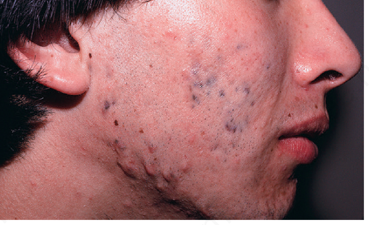
The image below shows minocycline-induced hyperpigmentation (type I - blue-black discolouration in areas of prior inflammation) as an example of how DIP can appear facially:
Most Relevant Drug Causes in Paediatrics
1. Minocycline (most common antibiotic cause)
- Produces blue-black to slate-gray pigmentation in three classic types:
- Type I: Blue-black at sites of prior inflammation (acne, scars) - can affect perioral/chin region
- Type II: Similar pigmentation on normal skin of anterior shins; perioral involvement possible
- Type III: Generalized muddy-brown, accentuated in sun-exposed areas including the face
- Also stains permanent teeth gray-green (most marked in midportion of tooth), oral mucosa, nail beds, sclera
- Risk increases with cumulative dose (up to 40% incidence after >1 year of therapy)
- Used in children for acne, MRSA coverage, and multibacillary leprosy (MDT)
- Histology: Pigment granules (iron + melanin) within macrophages in the dermis
- Andrews' Diseases of the Skin, p. 149
2. Antimalarials (chloroquine, hydroxychloroquine)
- Cause gray to blue-black pigmentation affecting the face including perioral region, extremities, ear cartilage, oral mucosa, and nails
- Affects up to 25-29% of patients on long-term therapy (>300 g cumulative dose)
- Pretibial pattern is most common; gingiva and hard palate may also be discoloured
- Mechanism: Dermal deposition of melanin-drug complexes and hemosiderin around capillaries
- May fade after discontinuation but rarely resolves completely
- Used in children for malaria, lupus, juvenile idiopathic arthritis
- Dermatology 2-Volume Set 5e, Table 67.5
3. Imatinib (tyrosine kinase inhibitor)
- Causes mucosal and perioral pigmentation; specifically documented in paediatric patients with childhood chronic myeloid leukaemia (CML)
- Reported in case series: dental hyperpigmentation in childhood CML
- Three case reports of mucosal pigmentation from imatinib including chin/lip region
- Mechanism: Stimulation of melanocytes via c-Kit pathway inhibition leading to paradoxical melanocyte activity
4. Chemotherapeutic agents
Used in paediatric oncology; several produce perioral/facial pigmentation:
| Drug | Pattern of Pigmentation | Mechanism |
|---|---|---|
| Busulfan | Generalised, resembles Addison disease | Increased basal melanin |
| 5-Fluorouracil (5-FU) | Perioral, palmar creases, pressure points, streaky | Increased epidermal melanin |
| Bleomycin | Flagellate/linear bands on trunk; also nail, pressure points | Increased melanin, no melanocyte increase |
| Cyclophosphamide | Diffuse, may affect perioral region | Stimulation of melanin synthesis |
| Doxorubicin | Diffuse hyperpigmentation | Melanocyte stimulation |
| Hydroxyurea | Melanonychia + skin diffuse darkening | Similar to zidovudine |
Dermatology 2-Volume Set 5e, Table 67.5
5. Clofazimine
- Used in paediatric multibacillary leprosy (MDT regimen)
- Causes pink to reddish-blue or brown pigmentation concentrated in lesions; highly disfiguring
- A major cause of non-compliance with MDT in children
- Mechanism: "Drug-induced lipofuscinosis" - PAS-positive brown granular pigment in foamy macrophages
- Andrews' Diseases of the Skin, p. 150
6. Phenothiazines (chlorpromazine, thioridazine)
- Slate-gray hyperpigmentation in sun-exposed areas including the face/perioral region after long-term use
- Used in children for behavioural disorders, psychosis, severe nausea
- Associated with corneal and lens opacities (ophthalmologic evaluation required)
- Histology: Golden-brown drug granules + increased dermal melanin in macrophages
- Andrews' Diseases of the Skin, p. 150
7. Tricyclic antidepressants (imipramine, clomipramine)
- Imipramine causes slate-gray to blue-gray hyperpigmentation in sun-exposed sites
- Imipramine-associated pigmentation can resolve within 1 year of stopping the drug
- Used in children for enuresis, depression, ADHD
- A 2022 case report and review (PMID 35364629) confirmed cutaneous and iris hyperpigmentation with imipramine
8. Zidovudine (AZT)
- Blue or brown hyperpigmentation, mainly in darkly pigmented individuals
- Primarily affects nails (melanonychia), but diffuse skin hyperpigmentation including face and lateral tongue also reported
- Dose-dependent; clears after discontinuation
- Used in HIV-positive children for prophylaxis and treatment
- Andrews' Diseases of the Skin, p. 150
9. Heavy metals
| Metal | Pattern |
|---|---|
| Bismuth | Generalized blue-gray discolouration of face, neck, dorsal hands; oral mucosa and gingiva involvement |
| Silver (argyria) | Diffuse slate-gray, especially sun-exposed areas including perioral |
| Lead | "Lead line" at gingival margin |
| Gold (chrysiasis) | Blue-gray in sun-exposed areas, periorbital |
| Arsenic | Bronze hyperpigmentation with "raindrop" pattern; palms, soles |
Dermatology 2-Volume Set 5e, Table 67.5
Pathophysiologic Mechanisms
| Mechanism | Example Drugs |
|---|---|
| Direct deposition of drug/metabolite in dermis | Minocycline (iron-melanin complexes), gold, silver, bismuth |
| Stimulation of melanocytes (increased melanin synthesis) | Antimalarials, chemotherapeutics, zidovudine |
| Post-inflammatory pigmentary alteration | Minocycline type III, bleomycin |
| Drug-induced lipofuscinosis | Clofazimine |
| Photosensitivity-mediated pigmentation | Amiodarone, phenothiazines, type III minocycline |
Key Differential Diagnosis in Paediatrics
When evaluating a child with perioral/chin hyperpigmentation after medications, also consider:
- Addison disease / adrenal insufficiency (endogenous ACTH-driven diffuse pigmentation - check electrolytes, cortisol)
- Peutz-Jeghers syndrome (perioral melanotic macules - genetic, associated with gastrointestinal polyps; rule out if family history)
- Laugier-Hunziker syndrome (acquired benign perioral and oral mucosal pigmentation)
- Incontinentia pigmenti (in neonates/infants)
- Post-inflammatory hyperpigmentation (from eczema, impetigo around the mouth)
- Fixed drug eruption (recurrent, same site each time a drug is taken)
Clinical Approach
- Detailed medication history - onset relative to start of drug, duration, cumulative dose
- Examine distribution - purely perioral (upper lip + chin) vs. oral mucosa vs. diffuse
- Wood's lamp - differentiates epidermal (enhanced) vs. dermal (not enhanced) pigment
- Skin biopsy if diagnosis uncertain - histopathology guides identification of pigment type
- Stop or substitute the offending drug if clinically feasible
- Rule out endocrinopathy (cortisol, ACTH, electrolytes) if diffuse pigmentation
- Ophthalmology referral if phenothiazine-associated (corneal/lens opacities)
Treatment options (if drug cannot be stopped): Topical hydroquinone, high-potency corticosteroids, tacrolimus, chemical peels, Q-switched or picosecond laser therapy for deeper dermal deposits. - StatPearls, Drug-Induced Pigmentation (NCBI NBK542253)
Summary for Paediatric Context: The most common causes of perioral black discolouration after medications in children are minocycline (acne/MRSA treatment), antimalarials (malaria/JIA), clofazimine (leprosy MDT), imatinib (childhood CML), and chemotherapeutic agents in oncology settings. The diagnosis is primarily clinical - correlating the timing of pigmentation onset with medication history. Most cases partially or fully resolve after stopping the offending drug, though dermal deposits (particularly minocycline and antimalarials) may persist for months to years.
This is a shared conversation. Sign in to Orris to start your own chat.