
Explain anoto my of testis
anatomy of testis diagram cross section
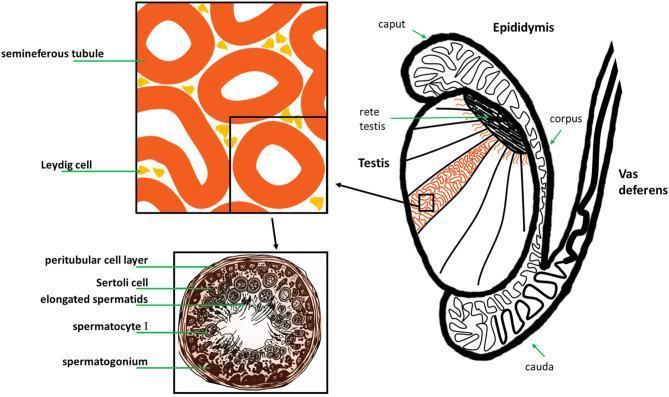
This medical illustration depicts the gross anatomy and histology of the human male reproductive system, specifically the testis and its associated ducts. The main diagram shows a sagittal-like section of the testis, highlighting internal lobules containing seminiferous tubules that converge at the rete testis. The epididymis is shown as a coiled ductal system attached to the posterior testis, divided into the caput (head), corpus (body), and cauda (tail), which then continues as the vas deferens. A magnified histological inset focuses on the interstitial space, showing Leydig cells clustered between cross-sections of seminiferous tubules. A second, high-magnification cross-section of a single seminiferous tubule illustrates the stages of spermatogenesis. Labeled structures include the peritubular cell layer (basal lamina), Sertoli cells for structural support, and germ cells in various stages of maturation: spermatogonia at the periphery, followed by spermatocytes I, and elongated spermatids positioned near the central lumen. This diagram serves as an educational tool for understanding the relationship between macroscopic anatomy and microscopic gametogenesis.
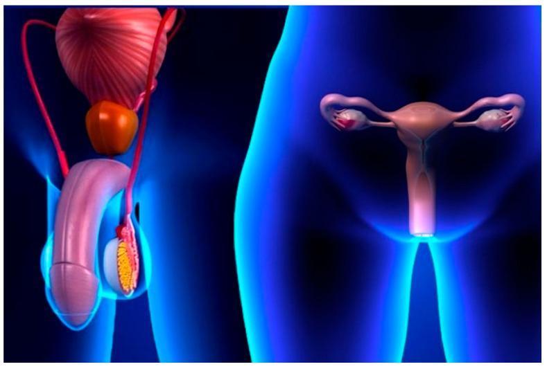
A 3D anatomical diagram illustrating the male and female reproductive systems side-by-side within stylized blue human silhouettes. On the left, the male anatomy includes the penis, a testis shown in cross-section within the scrotum, the epididymis, the vas deferens, the prostate gland, and the urinary bladder superiorly. On the right, the female anatomy depicts the uterus centrally, with the fallopian tubes extending laterally to the ovaries, and the vaginal canal positioned inferiorly. The visual highlights the pelvic location and relative spatial arrangement of primary and secondary sex organs. The image serves as an educational infographic for sexual health and reproductive anatomy, specifically in the context of public health strategies like the SAFE-S (Sexual behavior, Antibiotics, Facial cleanliness, Environmental improvement) strategy for controlling Chlamydia trachomatis and other sexually transmitted infections. The medical specialty is Urology and Gynecology, aimed at a general educational level.
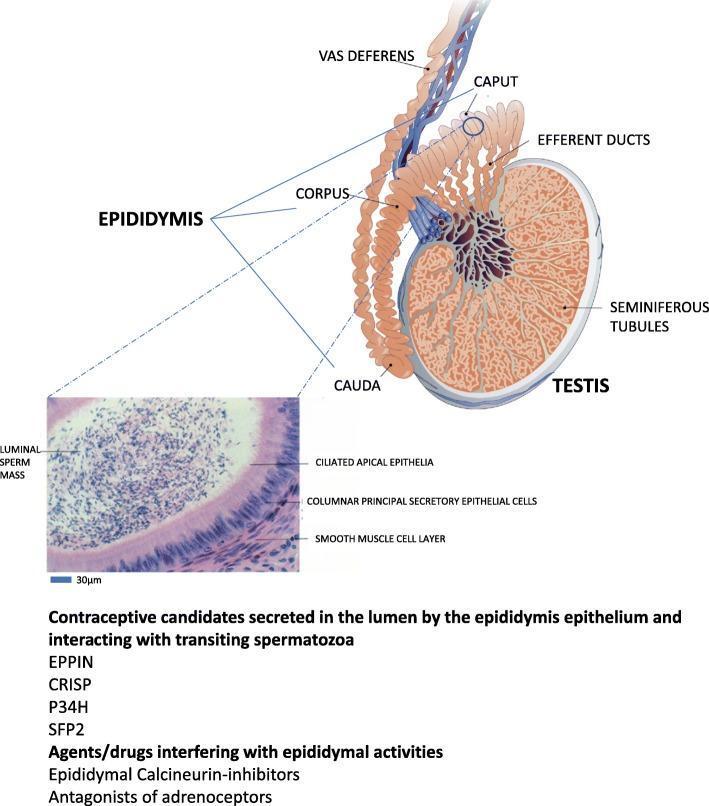
This educational graphic combines an anatomical diagram of the male reproductive system with a histological micrograph, illustrating the mammalian epididymis. The primary diagram shows the testis with internal seminiferous tubules connecting via efferent ducts to the epididymis, which is divided into three distinct segments: the caput (head), corpus (body), and cauda (tail). The cauda eventually transitions into the vas deferens. An inset micrograph, labeled with a 30μm scale bar, provides a high-magnification cross-section of an epididymal tubule. This histological view identifies four key components: the central luminal sperm mass, the ciliated apical epithelia, the underlying columnar principal secretory epithelial cells, and an external smooth muscle cell layer responsible for peristalsis. The image serves as a conceptual framework for understanding post-testicular sperm maturation and identifying potential contraceptive targets, such as EPPIN, CRISP, P34H, and SFP2, or pharmacological agents like calcineurin-inhibitors and adrenoceptor antagonists that interfere with epididymal function.
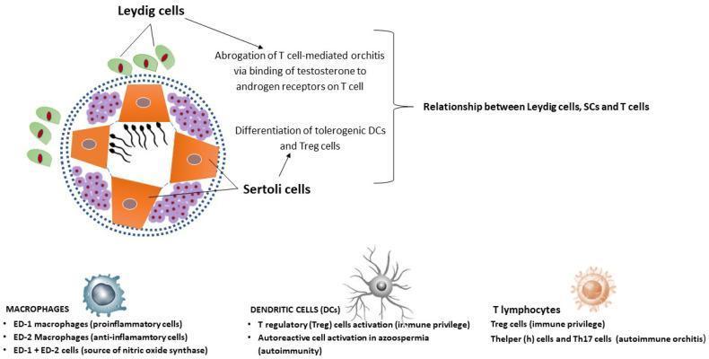
This medical illustration depicts the pathophysiology and immune microenvironment of the human testis, focusing on the maintenance of immune privilege. The central diagram shows a cross-section of a seminiferous tubule featuring orange Sertoli cells (SCs) supporting developing spermatozoa. These are surrounded by purple immune cell clusters within the interstitial space. Exterior to the tubule, green Leydig cells are shown, noting their role in abrogating T cell-mediated orchitis via testosterone binding to androgen receptors. The diagram highlights the 'Relationship between Leydig cells, SCs, and T cells' in promoting the differentiation of tolerogenic dendritic cells (DCs) and T regulatory (Treg) cells. Accompanying text categorizes testicular immune cells: Macrophages (proinflammatory ED-1, anti-inflammatory ED-2, and NOS-producing types); Dendritic Cells (involved in Treg activation and autoimmunity in azoospermia); and T lymphocytes (Treg cells for immune privilege vs. Th and Th17 cells in autoimmune orchitis). The illustration serves as an educational tool for understanding the balance between immune tolerance and autoimmune pathology in male reproductive health.
Anatomy of the Testis
1. Overview and Location
- Costanzo Physiology 7th Ed, p. 1144
2. Gross Anatomy and Coverings
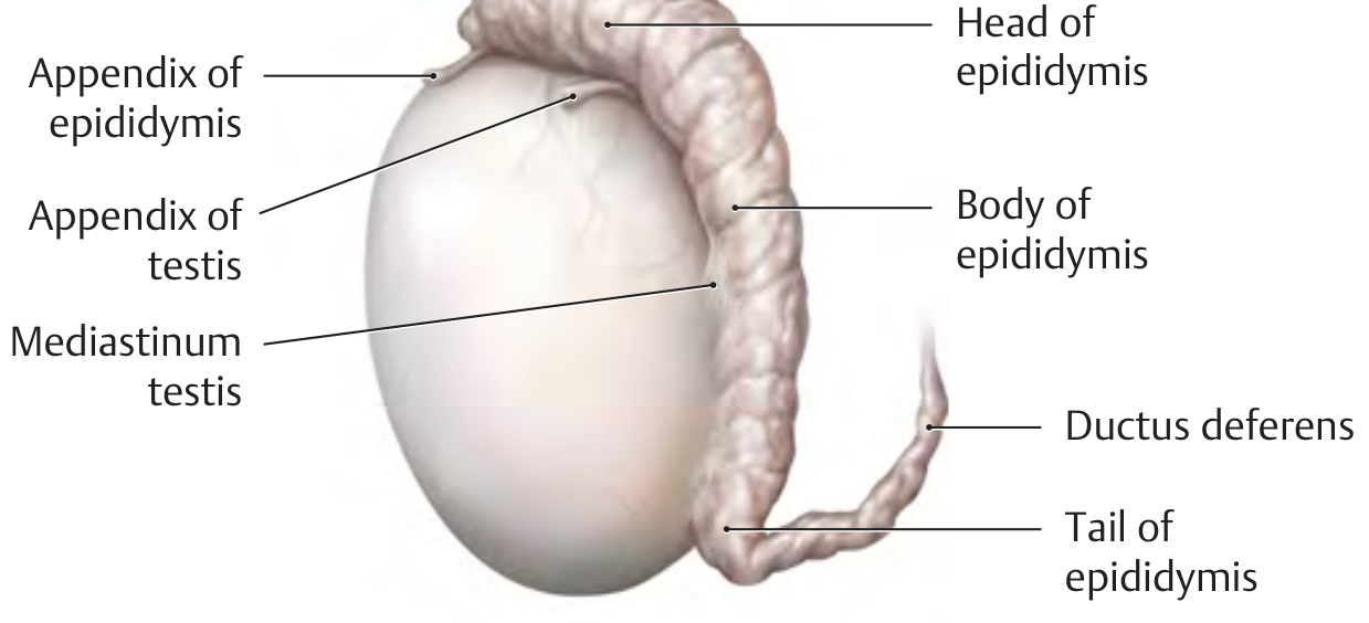
Coverings (from outermost to innermost):
| Layer | Description |
|---|---|
| Tunica vaginalis | Double-layered peritoneal sac; visceral layer covers the testis, parietal layer lines the internal spermatic fascia; a slit-like serous cavity between them contains a small amount of fluid |
| Tunica albuginea | Tough fibrous capsule enclosing the testicular tissue; gives it a rubbery consistency |
| Tunica vasculosa | Inner vascular layer |
- THIEME Atlas of General Anatomy and Musculoskeletal System, p. 247
Mediastinum Testis
3. Internal Structure
Lobules
- THIEME Atlas, p. 247; Histology: A Text and Atlas, p. 2162
Seminiferous Tubules
- Make up ~80% of testis volume in adults
- Convoluted closed loops, 120-300 µm in diameter, ~600 m in total length
- Arranged in lobules, surrounded by a connective tissue lamina propria containing peritubular myoid cells
- Empty into the rete testis at the mediastinum
A. Sertoli Cells ("Nurse Cells")
-
Tall columnar cells resting on the basal lamina; extend to the lumen
-
Form tight junctions with each other, creating the blood-testis barrier (BTB)
-
The BTB divides the tubule into:
- Basal compartment: contains diploid spermatogonia (stem cells)
- Luminal (adluminal) compartment: contains spermatocytes and spermatids
-
Functions of Sertoli cells:
- Provide nutrients to developing sperm (isolated from the bloodstream)
- Form the blood-testis barrier (selective permeability)
- Secrete aqueous fluid to transport sperm toward the epididymis
- Secrete androgen-binding protein (ABP) to maintain high local testosterone
- Secrete inhibin B (feedback inhibitor of FSH)
-
Costanzo Physiology, p. 1148
B. Spermatogenic Cells
- Spermatogonia (type A and B) - stem cells at the periphery
- Primary spermatocytes - largest cells; undergoing meiosis I
- Secondary spermatocytes - brief stage; undergo meiosis II
- Spermatids - haploid; undergo spermiogenesis
- Spermatozoa - fully formed sperm released into the lumen
Interstitial (Leydig) Cells
-
Found in the connective tissue between seminiferous tubules, making up ~20% of testis
-
Large polygonal cells with central nuclei and abundant smooth ER
-
Produce 95% of adult male testosterone (and other androgens: DHEA, androstenedione)
-
Testosterone has local (paracrine) effects supporting spermatogenesis and endocrine effects on other organs
-
Stimulated by LH from the anterior pituitary
-
Guyton & Hall Medical Physiology; Smith & Tanagho's General Urology
4. Ductal System (Sperm Pathway)
| Structure | Description |
|---|---|
| Seminiferous tubules | Site of spermatogenesis |
| Straight tubules (tubuli recti) | Short straight segments leading to the rete |
| Rete testis | Network of channels in the mediastinum |
| Efferent ductules (10-15) | Connect rete testis to the head of the epididymis |
| Epididymis | Head (caput), body (corpus), tail (cauda) - runs along posterior testis |
| Ductus (vas) deferens | Continuous from epididymis tail; passes through inguinal canal |
| Ejaculatory duct | Short segment opening into prostatic urethra |
- Harrison's Principles of Internal Medicine 22E, p. (403-2); THIEME Atlas, p. 247
5. Blood Supply and Lymphatics
- Testicular artery (from abdominal aorta at L2) - descends in the spermatic cord
- Pampiniform venous plexus - wraps around the testicular artery; acts as a countercurrent heat exchanger to cool arterial blood
- Venous drainage: right testicular vein drains directly into the IVC; left testicular vein drains into the left renal vein (clinically important in varicocele)
- Lymphatics drain to the para-aortic lymph nodes (NOT inguinal - clinically important in testicular tumors)
6. Spermatogenesis (Summary)
- Spermatogonial phase: Type A spermatogonia self-renew; Type B spermatogonia divide mitotically to form primary spermatocytes
- Spermatocyte phase: Meiosis I (lasts ~22 days) → secondary spermatocytes; then Meiosis II (hours) → haploid spermatids
- Spermiogenesis: Spermatids remodel into mature spermatozoa - DNA condensation, acrosome cap formation, flagellum development, cytoplasm reduction
- Histology: A Text and Atlas, p. 2163; Harrison's 22E
7. Development
- Intermediate mesoderm (urogenital ridges)
- Mesodermal epithelium (primary sex cords)
- Primordial germ cells (migrate from the yolk sac)
- Histology: A Text and Atlas, p. 2162
Quick Reference Summary
| Feature | Detail |
|---|---|
| Size | ~5 cm × 3 cm, 18 mL volume |
| Temperature | 35-36°C (1-2°C below core) |
| Seminiferous tubules | 80% of volume, ~600 m total length |
| Lobules | ~250-370, each with 1-4 tubules |
| Sperm production | ~128 million/day |
| Spermatogenesis duration | ~74 days |
| Testosterone source | Leydig cells (interstitial, 20% of testis) |
| Lymph drainage | Para-aortic nodes |
| Venous asymmetry | Right → IVC; Left → left renal vein |