
Embryology of ear
Reading File
Now let me search the medical library for ear embryology content in parallel.
Finding Sources
Finding Sources
Excellent - found dedicated sections. Let me read all of them in parallel.
Reading File
Reading File
Reading File
Good content. Let me get the more detailed KJ Lee section (which was truncated) and the Scott-Brown block 5 content on inner ear development.
Reading File
Reading File
Let me get the rest of the KJ Lee embryology section (middle ear, inner ear, ossicles) and the Scott-Brown table content:
Reading File
Reading File
Excellent! I have enough comprehensive content. Let me also fetch the auricle diagram image for the answer:
Loading Image
Reading File
I now have comprehensive information from three authoritative sources. Let me compile the full answer.
Embryology of the Ear
The ear develops from three distinct embryological sources - ectoderm, mesoderm/neural crest, and endoderm - producing the external, middle, and inner ear through coordinated but partly independent processes.
1. External Ear (Auricle and External Auditory Canal)
Auricle
The auricle develops from the first and second pharyngeal arches (branchial arches) starting at week 6.
Six mesenchymal condensations - the Hillocks of His - arise around the margins of the first pharyngeal groove:
| Hillock | Origin | Structure Formed |
|---|---|---|
| 1 | 1st arch | Tragus |
| 2 | 1st arch | Helical crus |
| 3 | 1st arch | Helix |
| 4 | 2nd arch | Antihelix |
| 5 | 2nd arch | Antitragus |
| 6 | 2nd arch | Lobule and lower helix |
The concha is formed from three areas of the first groove (ectoderm):
- Concha cavum - from the middle part of the first groove
- Concha cymba - from the upper part of the first groove
- Intertragal incisure - from the lowest part of the first groove
Timeline:
- Week 6: Hillocks appear
- Week 7: Cartilage formation begins
- Week 12: Auricle formed by hillock fusion
- Week 20: Adult shape reached (adult size not until age 9)
The developing pinna is initially located in the neck and ascends to its normal position by week 20.
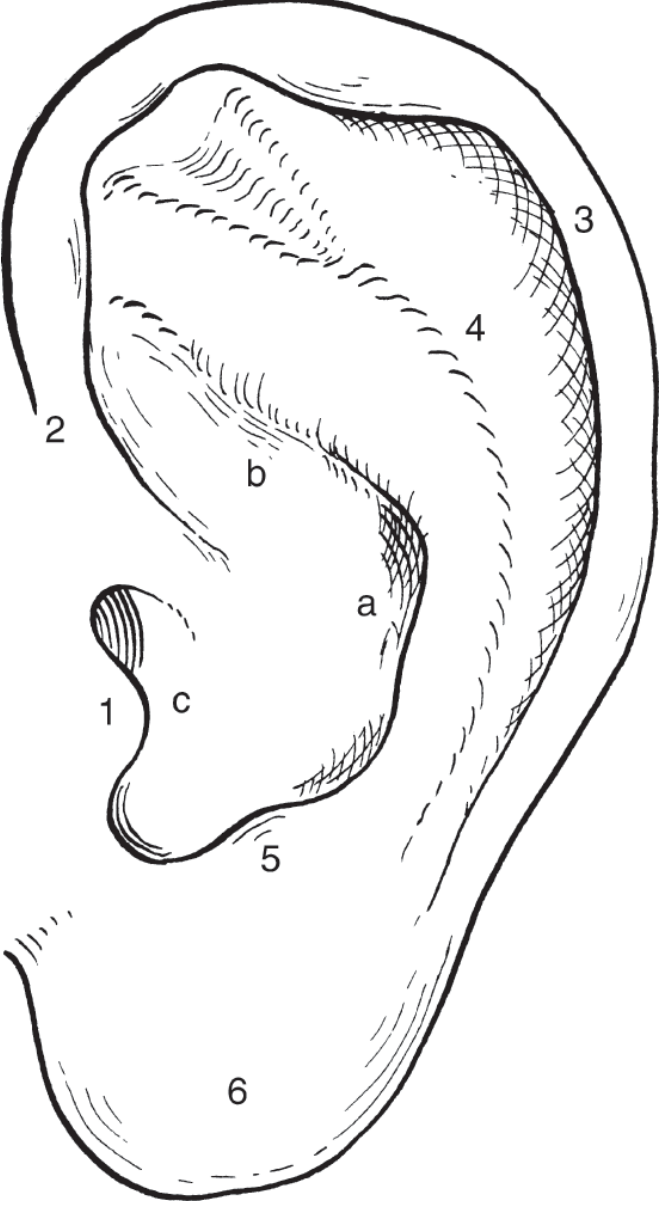
Embryology of the auricle - K.J. Lee's Essential Otolaryngology, Fig. 13-23
External Auditory Canal (EAC)
- Week 8: Surface ectoderm at the upper end of the first pharyngeal groove thickens, forming a solid epithelial core (the meatal plate) that grows inward toward the middle ear. Simultaneously, the concha cavum deepens to form the outer one-third of the EAC.
- Week 21: The meatal plate begins to resorb ("hollow out") to form a channel.
- Week 28: Canalization is complete, forming the inner two-thirds of the EAC. The innermost ectoderm remains as the squamous layer of the tympanic membrane.
- Birth: EAC is not yet ossified and not adult-sized.
- Age 3: Ossification of the tympanic ring and bony canal complete.
- Age 9: Adult size reached.
2. Middle Ear, Eustachian Tube, and Tympanic Membrane
Eustachian Tube and Middle Ear Space
These develop from the first pharyngeal pouch (endoderm):
- Week 3: The first pharyngeal pouch lies laterally on either side of the developing tongue. As the third arch enlarges, it compresses the space between the second arch and the pharynx (the first pouch), forming the Eustachian tube. The lateral "outpocketing" expands to become the middle ear space.
- Week 10: Pneumatization begins.
- Week 23: The antrum appears.
- Weeks 12-28: Four primary mucosal sacs develop, each becoming a specific anatomic region:
| Mucosal Sac | Middle Ear Region |
|---|---|
| Saccus anticus | Anterior pouch of von Troltsch |
| Saccus medius | Epitympanum and petrous area |
| Saccus superior | Posterior pouch of von Troltsch, part of mastoid, inferior incudal space |
| Saccus posterior | Round and oval window niches, sinus tympani |
- The middle ear remains filled with mucoid connective tissue until birth, after which subepithelial resorption and pneumatization continue.
- Eustachian tube length: ~17 mm at birth, growing to ~35 mm in adulthood.
- Because of proximity to 1st, 2nd, and 3rd arches, cranial nerves V, VII, and IX are all found in the middle ear.
Tympanic Membrane
Appears at week 28; derived from all three germ layers:
| Germ Layer | Layer of TM |
|---|---|
| Ectoderm (1st pharyngeal groove) | Squamous (outer) layer |
| Mesoderm | Fibrous (middle) layer |
| Endoderm (1st pharyngeal pouch) | Mucosal (inner) layer |
Auditory Ossicles
Malleus and Incus (from the 1st pharyngeal arch - Meckel's cartilage, and 2nd arch - Reichert's cartilage):
- Week 6: Malleus and incus appear as a single mass.
- Week 8: They separate; the malleoincudal joint forms.
- Week 16: Ossicles reach adult size; ossification begins (first at the long process of the incus).
- Week 17: Ossification spreads to the neck and manubrium of the malleus.
- At birth: Malleus and incus are of adult size and shape. Part of the manubrium remains cartilaginous throughout life.
Specific arch contributions:
- Head and neck of malleus - Meckel's cartilage (1st arch)
- Anterior process of malleus - Process of Folius (membranous bone/mesenchyme)
- Manubrium of malleus - Reichert's cartilage (2nd arch)
- Body and short process of incus - Meckel's cartilage (1st arch)
- Long process of incus - Reichert's cartilage (2nd arch)
Stapes (from 2nd pharyngeal arch - Reichert's cartilage, plus otic mesenchyme):
- Week 4.5: Mesenchymal blastema forms; the facial nerve (CN VII) divides it into stapes, interhyale, and laterohyale.
- Week 7: Stapes ring forms around the stapedial artery. The lamina stapedialis (otic mesenchyme) becomes the footplate and annular ligament.
- Week 8.5: Incudostapedial joint forms.
- Week 10: Stapes changes from ring to stirrup shape.
- Week 19: Ossification begins at the obturator surface of the base.
- Week 28: Ossification complete, except for the vestibular surface of the footplate - which remains cartilaginous throughout life.
- The interhyale → stapedius muscle and tendon; the laterohyale → posterior wall of middle ear.
3. Inner Ear
The inner ear develops from surface ectoderm (the otic placode) - it is the first part of the ear to develop and also the earliest to reach adult size.
Timeline:
- Week 3 (day 22-23): Ectodermal thickening lateral to the developing hindbrain forms the otic placode.
- Week 4: The otic placode invaginates, forms the otic pit, then pinches off to form the otocyst (otic vesicle), surrounded by mesoderm. The common macula first appears at this stage.
- Week 5: The otic vesicle divides into:
- Dorsal utricular part → utricle, semicircular ducts, endolymphatic duct and sac
- Ventral saccular part → saccule and cochlear duct (cochlear duct → spiral organ/organ of Corti)
- Week 6: Semicircular canals begin to form as three flattened pouches growing at right angles from the utricle. The superior canal is the first fully formed (by week 6).
- Week 7: Basal turn of the cochlea begins to form.
- Week 8: Utricle and semicircular canals are fully formed (pars superior complete). The otic capsule begins as mesenchymal precartilage condensation.
- Week 11: Vestibular end organs (cristae and maculae) complete, with sensory and supporting cells.
- Week 12: Cochlea completes its 2.5 turns.
- Week 15: Membranous labyrinth (without the organ of Corti) is complete.
- Week 15: 14 ossification centers visible in otic capsule.
- Week 20: Stria vascularis and tectorial membrane development complete.
- Week 23: Bony labyrinth (otic capsule) ossification complete; membranous and bony labyrinths are adult size from this point. Inner and outer ridge cells differentiate: inner ridge → spiral limbus; outer ridge → hair cells, pillar cells, Hensen cells, Deiters cells.
- Week 25: Full adult outline of membranous labyrinth is present.
- Week 26: Organ of Corti achieves its full structural differentiation.
- The endolymphatic sac is the first structure to appear in the labyrinth and the last to stop growing (continues until adulthood).
- The fissula ante fenestram may remain cartilaginous throughout life.
Associated neural crest cells around the otocyst become the:
- Spiral (cochlear) ganglion
- Scarpa's (vestibular) ganglion
- Geniculate ganglion (facial nerve)
4. Summary Table
| Structure | Embryological Origin |
|---|---|
| Auricle | Hillocks of His (1st + 2nd pharyngeal arches) |
| External auditory canal (outer 1/3) | Ectoderm of 1st pharyngeal groove |
| External auditory canal (inner 2/3) | Meatal plate (1st pharyngeal groove ectoderm) |
| Tympanic membrane | Ectoderm + mesoderm + endoderm |
| Middle ear space | 1st pharyngeal pouch (endoderm) |
| Eustachian tube | 1st pharyngeal pouch (endoderm) |
| Malleus head/neck + incus body/short process | Meckel's cartilage (1st arch) |
| Malleus manubrium + incus long process | Reichert's cartilage (2nd arch) |
| Stapes | 2nd arch + otic mesenchyme |
| Inner ear (membranous labyrinth) | Otic vesicle (surface ectoderm) |
| Bony labyrinth (otic capsule) | Periotic mesoderm |
5. Clinical Correlates
- Congenital aural atresia: EAC atresia results from failure of meatal plate canalization (weeks 21-28), while the auricle may be normally formed - since auricle and EAC develop from different processes.
- Low-set ears / preauricular pits / accessory auricles: Minor anomalies of auricular hillocks; severe malformation is associated with trisomy 13 and trisomy 18.
- Congenital deafness: Most commonly from autosomal recessive inheritance. Rubella virus infection near the end of the embryonic period (especially weeks 6-12) causes damage to the developing spiral organ, resulting in sensorineural deafness.
- Ossicular anomalies: First and second arch abnormalities cause conductive deafness; the stapes footplate (otic mesenchyme origin) may be spared when ossicular chain anomalies arise from arch defects alone.
- Foramen of Huschke: A persistent defect in the anterior bony canal wall (incomplete ossification), which in infants may allow spread of infection between the middle ear and parotid region.
Sources: K.J. Lee's Essential Otolaryngology (9th ed.), Ch. 13 | Scott-Brown's Otorhinolaryngology Head & Neck Surgery Vol. 2, Ch. 46 | The Developing Human: Clinically Oriented Embryology (Moore & Persaud)
This is a shared conversation. Sign in to Orris to start your own chat.