
Glen cath procedure
Glenn procedure bidirectional cavopulmonary shunt congenital heart surgery
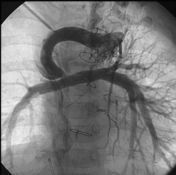
This diagnostic angiogram of the thoracic vasculature illustrates complex post-surgical anatomy in a pediatric patient following palliative cardiac surgery. The central feature is a bidirectional cavopulmonary shunt (Glenn procedure) showing a focal stenosis at the anastomosis site between the superior vena cava and the pulmonary artery conduit. The image demonstrates a prominent, arching aorta and relatively hypoplastic pulmonary arteries. Significant pathology is noted by the presence of numerous, tortuous, and enlarged collateral vessels originating from both internal mammary arteries, which provide compensatory blood flow to the lungs. Several surgical artifacts are visible, including metallic sternotomy wires in the midline and vascular clips along the larger vessels. A metallic coil is also identifiable, indicating a previous interventional coil embolization of collateral vessels. This image serves as a clinical example of postoperative complications in congenital heart disease management, specifically highlighting stenotic shunts and the development of aortopulmonary collaterals.
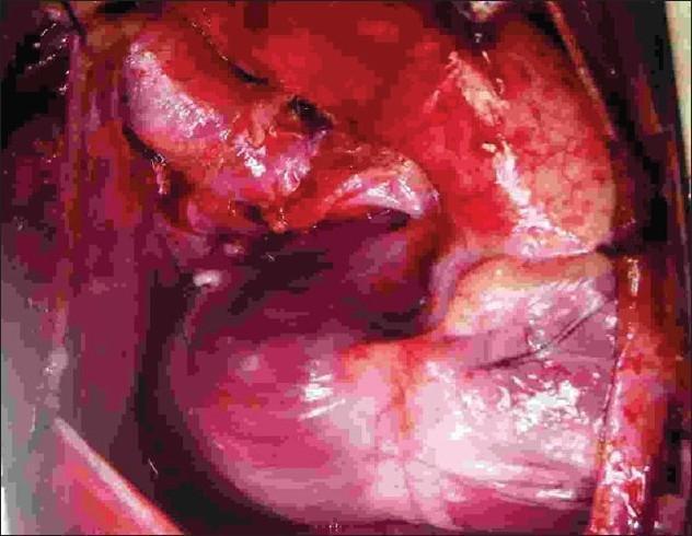
This clinical photograph displays an intraoperative view of a completed pediatric cardiac surgical procedure via a midline sternotomy. The image primarily demonstrates a Bidirectional Glenn (BDG) shunt, characterized by an end-to-side anastomosis between the superior vena cava (SVC) and the superior aspect of the right pulmonary artery (RPA). Visible in the surgical field are the smooth, moist textures of vascular tissues with clear suture lines indicating the anastomotic site. The SVC has been transected from its cardiac end and repositioned to provide direct venous drainage into the pulmonary circulation. Additionally, the field reflects the state following the division of a previously placed right Blalock-Taussig (BT) shunt. The surgical area is well-visualized with anatomical landmarks including the SVC-RPA junction and the surrounding pericardial space. This visual serves as an educational reference for staged palliative surgery in patients with single ventricle physiology or complex congenital heart disease, illustrating the transition from systemic-to-pulmonary shunting to a superior cavopulmonary connection.
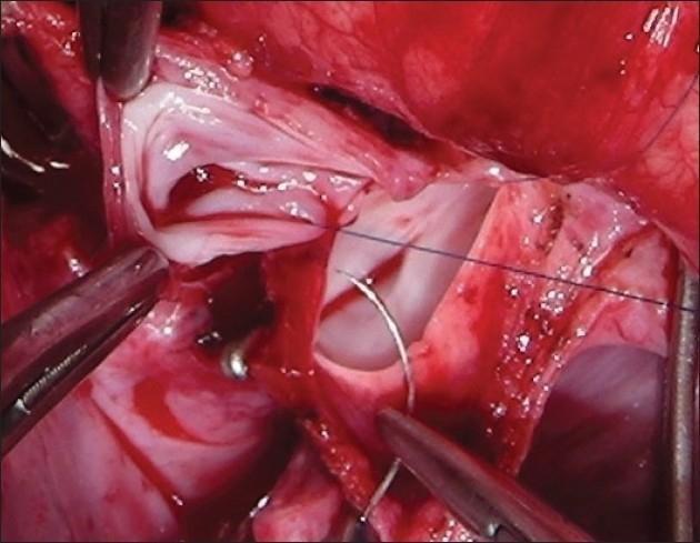
This intraoperative clinical photograph captures a Bidirectional Glenn (BDG) shunt procedure, specifically demonstrating an end-to-side anastomosis between the superior vena cava (SVC) and the right pulmonary artery (PA). The surgical field shows the dissected vessels held in a bloodless state. Multiple surgical instruments are visible, including vascular forceps and clamps used to manipulate and stabilize the vessel walls. A curved surgical needle and fine 6-0 polypropylene stay sutures are actively positioned at the anastomosis site to secure the junction. The interior lumen of the vascular structures is visible, showing the smooth endothelial surface and the thickness of the vessel walls. This image illustrates a critical step in pediatric cardiac surgery for congenital heart defects, emphasizing the precise suturing technique required to establish reliable pulmonary blood flow from the systemic venous circulation.
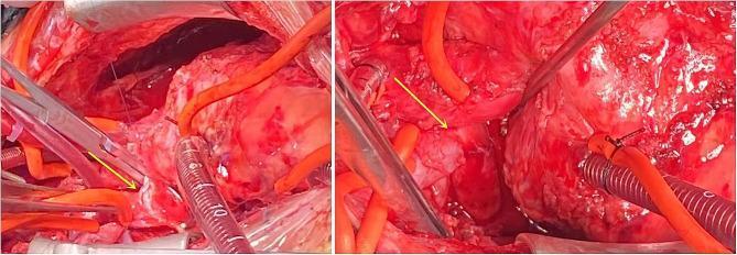
Two intraoperative clinical photographs demonstrate a bidirectional Glenn procedure (superior cavopulmonary anastomosis) in a pediatric surgical field. The images show the end-to-side anastomosis where the superior vena cava (SVC) has been surgically joined to the right pulmonary artery (RPA). Surgical instruments, including metal forceps and needle holders, are positioned around the vascular structures. Fine prolene sutures are visible at the anastomosis site, indicated by yellow arrows. The operative field includes various specialized equipment: a transparent measuring suction tip with metric markings (millimeter scale) for sizing, and multiple small-diameter orange silastic cannulas/perfusion lines. Corrugated bypass tubing and metallic retractors are also present at the periphery. This procedure aims to redirect deoxygenated blood from the upper body directly to the lungs, bypassing the right heart in the setting of right ventricular hypoplasia and severe tricuspid stenosis. The visual focus is on the vascular reconstruction and the precise surgical technique required for congenital heart disease correction.
Glenn Procedure (Bidirectional Cavopulmonary Connection - BCPC)
What Is It?
The Three-Stage Fontan Pathway
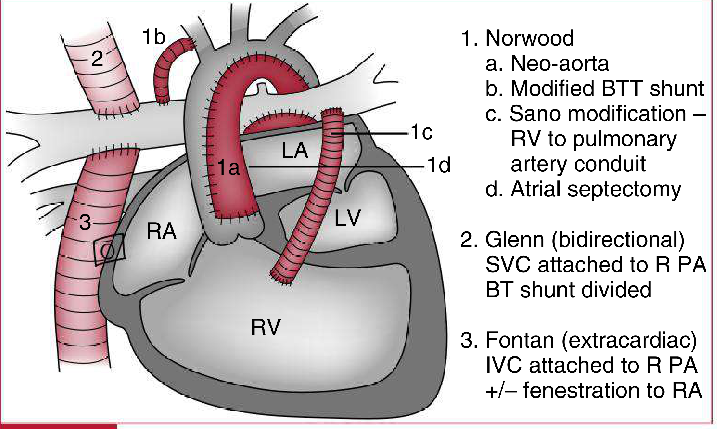
| Stage | Procedure | Age | Goal | Expected SpO2 |
|---|---|---|---|---|
| Stage 1 | Norwood / Hybrid | Neonatal | Create unobstructed systemic outflow; provide pulmonary blood flow via BT shunt or Sano conduit | 75-85% |
| Stage 2 | Bidirectional Glenn (BCPC) | 3-9 months | SVC-to-PA anastomosis; reduces volume overload of single ventricle | 80-85% |
| Stage 3 | Fontan (TCPC) | 18-24 months | IVC connected to PA; full cavopulmonary separation | >92% |
- Harriet Lane Handbook, 23rd ed., Table 7.12
- Mulholland and Greenfield's Surgery, 7th ed.
Indications
- Hypoplastic Left Heart Syndrome (HLHS) - the most common indication
- Tricuspid atresia
- Pulmonary atresia with intact ventricular septum
- Other single-ventricle defects (any functionally univentricular heart)
- Also used as a "1.5-ventricle repair" when the right ventricle is too hypoplastic to fully support pulmonary circulation but not quite single-ventricle physiology
The Procedure: Step by Step
- Cardiopulmonary bypass is established (can sometimes be done off-pump)
- The SVC is divided from the right atrium
- An end-to-side anastomosis is created between the cut end of the SVC and the superior aspect of the right pulmonary artery - this allows blood to flow into both right and left pulmonary arteries (hence "bidirectional")
- The SVC-RA stump is oversewn
- The prior palliative shunt (BT shunt or Sano conduit) is divided and removed
- Suturing uses fine prolene (typically 6-0) at the anastomotic site

Hemodynamics After Glenn
- Upper body venous blood (SVC) flows passively to the lungs - there is no right ventricular pump driving it
- This reduces the volume load on the single functioning ventricle (previously, the entire cardiac output was being shunted to the lungs AND the body - a huge volume overload)
- Lower body blood (IVC) still returns to the heart and is ejected to the body, causing mild residual cyanosis (SpO2 80-85%)
- The IVC blood does not yet reach the lungs - that is accomplished at Stage 3 (Fontan)
Pre-operative Assessment (Imaging Goals Before Glenn)
- Branch PA anatomy - any stenosis or distortion from the prior shunt
- Aortic arch obstruction or residual coarctation (in HLHS)
- Systemic ventricular volume and function
- AV valve regurgitation
- Adequacy of atrial communication
- Bilateral SVCs (must be identified - bilateral SVCs require bilateral Glenn connections)
- Systemic-pulmonary collateral vessels
- Pulmonary vascular resistance must be low enough (PVR <2-3 Wood units/m²) to allow passive flow
Post-operative Monitoring (Pre-Fontan Assessment)
| Assessment | Why |
|---|---|
| Branch PA anatomy and SVC-PA connection patency | Stenosis will cause Fontan failure |
| Systemic venous anatomy (SVC, IVC, hepatic veins) | Plan the IVC connection |
| Ventricular function and AV valve | Must be normal for Fontan |
| Veno-venous collaterals (SVC to IVC) | Common after Glenn; steal pulmonary flow |
| Pulmonary arterial pressure (<20 mmHg) | Key Fontan prerequisite |
| Pulmonary vein stenosis | Can cause the circulation to fail |
| At GA: measure SVC/IJV pressure directly | Proxy for PA pressure |
Complications
- Anastomotic stenosis at the SVC-PA junction (can cause SVC syndrome)
- Aortopulmonary collaterals - compensatory vessels that form and cause volume overload (may require coil embolization)
- Veno-venous collaterals - divert blood from lungs and worsen cyanosis
- Pleural effusions / chylothorax
- Arrhythmias
- Ventricular dysfunction
Glenn vs. Hemi-Fontan
- Glenn: SVC divided and anastomosed end-to-side to RPA; SVC-RA junction oversewn
- Hemi-Fontan: SVC-PA connection made while keeping a patch across the SVC-RA junction (easier conversion to Fontan later); some evidence suggests slightly improved fluid dynamics with hemi-Fontan