
Cycloplegicdrugs mech of action and clinical significance
cycloplegic drugs mechanism of action eye pupil dilation
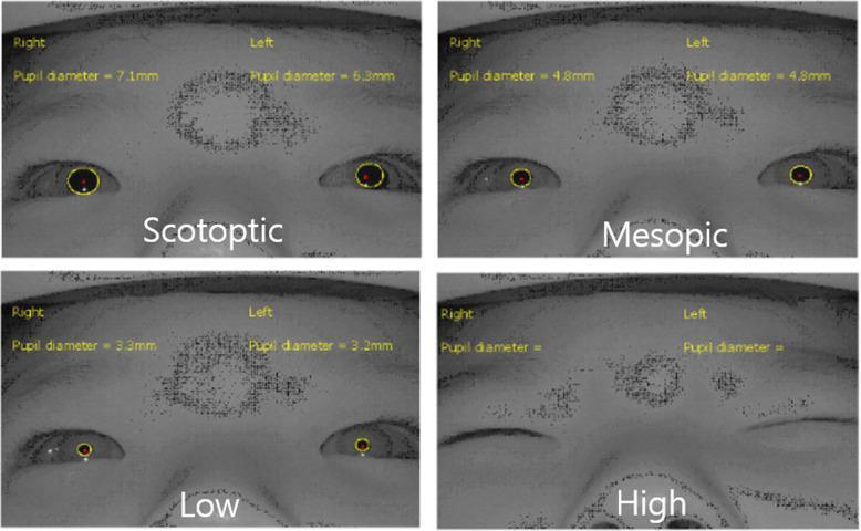
This clinical comparison chart presents four near-infrared snapshots of a patient's eyes across different light intensities (Scotopic, Mesopic, Low, and High) to assess pupil dynamics and reflexive eye closure. The scotopic phase shows maximal dilation with pupil diameters of 7.1mm (right) and 6.3mm (left). The mesopic and low-intensity phases demonstrate progressive pupillary constriction (4.8mm and ~3.2mm respectively) with maintained eyelid aperture. In the high-intensity phase (100 cd/m2), total reflexive eye closure (TEC) is observed, obscuring the pupils entirely. Each image features automated pupillometry tracking with yellow contours outlining the pupils and red central markers. The visual material is representative of neuro-ophthalmological monitoring of photosensitivity and the pupillary light reflex. The images were captured using a vision monitoring system specifically designed for real-time assessment of eyelid position and pupil diameter in varying luminance environments.
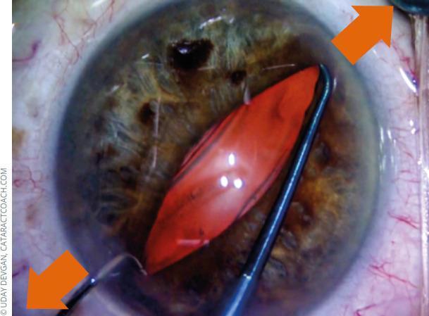
This clinical photograph captures an intraoperative ophthalmologic procedure, specifically a mechanical pupil stretching technique used during cataract surgery or in cases of small pupil management (miosis). The image shows the anterior segment of a human eye with two surgical hooks positioned approximately 180 degrees apart. These instruments are engaged at the pupillary margin, applying outward radial tension toward the limbus to expand the pupil, which appears elongated and oval-shaped. The iris exhibits a mixed brownish-green pigmentation. Within the center of the dilated pupil, an orange-red reflex or structure is visible, likely representing the lens nucleus or a red reflex from the fundus. The surrounding sclera and bulbar conjunctiva show mild hyperemia with visible fine vasculature. Orange arrows are superimposed on the image to indicate the directional force of the mechanical dilation. This visual demonstrates a critical surgical step in facilitating access to the lens when pharmacological dilation is insufficient.
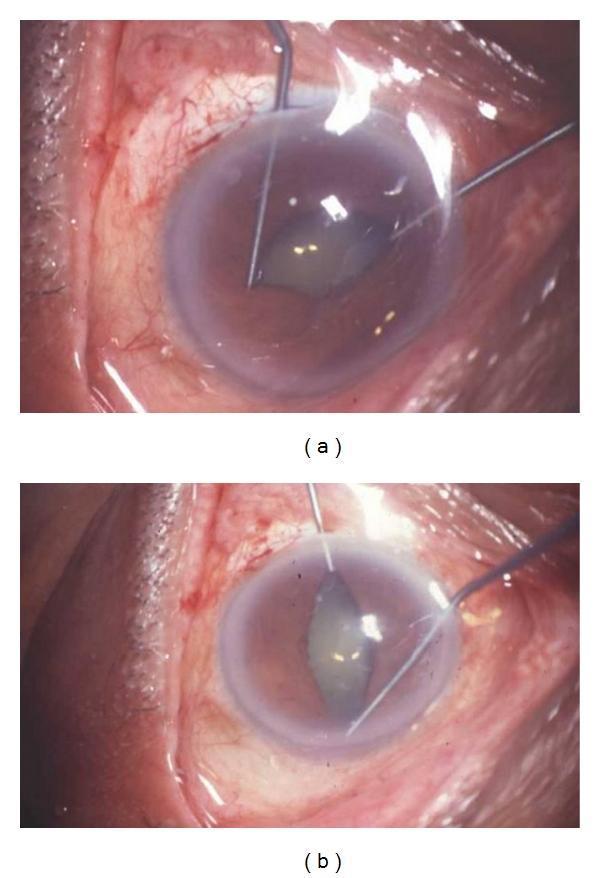
Two intraoperative clinical photographs (a and b) demonstrate the use of Kuglen hooks for mechanical pupillary dilation during ophthalmic surgery. The images show a close-up of a human eye with two metallic, angled Kuglen hooks inserted into the anterior chamber. The hooks are positioned at opposite margins of the iris, applying outward traction to stretch the pupil. In frame (a), the traction is applied horizontally, resulting in an elongated, oval-shaped pupil. In frame (b), the hooks are used in a perpendicular orientation to further expand the pupillary aperture. The pupil appears irregular and non-circular due to mechanical stretching. The surrounding sclera and conjunctiva exhibit prominent perilimbal injection and vascularization, consistent with inflammation often seen in uveitic eyes undergoing cataract surgery. The cornea appears slightly hazy, and the ocular surface is moist with visible light reflections. These maneuvers are typically employed to manage small pupils or lyse posterior synechiae to ensure adequate visualization of the lens and capsule during phacoemulsification.
Cycloplegic Drugs: Mechanism of Action and Clinical Significance
Mechanism of Action
1. Cycloplegia (paralysis of the ciliary muscle)
- The ciliary muscle (which controls accommodation/lens shape) is innervated by parasympathetic fibers via M3 receptors.
- Blocking these receptors causes paralysis of the ciliary muscle, eliminating the ability to accommodate (focus on near objects).
- This is the defining cycloplegic effect.
- Cycloplegia requires higher concentrations or more prolonged application than simple mydriasis alone.
2. Mydriasis (pupil dilation)
- The iris sphincter (pupillary constrictor) muscle is also cholinergically innervated (M3 receptors).
- Blocking these receptors causes the sphincter to relax, allowing the sympathetically innervated dilator pupillae to dominate, producing mydriasis.
- Cycloplegia and mydriasis always coexist; you cannot achieve cycloplegia without concurrent mydriasis.
Per Goodman & Gilman's: "Cycloplegia requires higher concentrations or more prolonged application of antagonist and thus cannot be achieved without concomitant mydriasis."
Individual Cycloplegic Agents
| Agent | Concentration | Onset | Duration |
|---|---|---|---|
| Tropicamide | 0.5%, 1% | 20-30 min | 3-6 hours |
| Cyclopentolate | 0.5%, 1%, 2% | 20-45 min | ~24 hours |
| Homatropine | 2%, 5% | 20-90 min | 2-3 days |
| Scopolamine | 0.25% | 20-45 min | 4-7 days |
| Atropine | 0.5%, 1%, 2% | 30-40 min | 1-2 weeks |
Clinical Significance
1. Cycloplegic Refraction (Most Important Use)
- Used to measure refractive errors accurately, especially in children.
- Children have strong accommodative tone that can mask the true refractive error.
- Cycloplegia eliminates the accommodation reflex, preventing the child from involuntarily over-focusing during testing, giving a true measure of hyperopia, myopia, or astigmatism.
- Cyclopentolate is the most commonly used agent for cycloplegic refraction in children due to moderate duration.
- Atropine is reserved for high hyperopia in young children due to its longer and more complete cycloplegia.
- Per Lippincott: "Topical atropine...permits the measurement of refractive errors without interference by the accommodative capacity of the eye." Shorter-acting agents (cyclopentolate, tropicamide) have largely replaced atropine due to prolonged mydriasis (7-14 days with atropine vs. 6-24 hours with others).
2. Fundus Examination / Dilated Fundoscopy
- Mydriasis allows clear visualization of the optic disc and peripheral retina.
- Routine adult dilation: Phenylephrine 2.5% + Tropicamide 1% (repeat in 15-30 min if needed).
- Children: Phenylephrine 2.5% + Tropicamide 1% + Cyclopentolate 1-2%.
- Preterm infants/neonates: Lower concentrations to minimize systemic absorption toxicity.
3. Uveitis / Iridocyclitis (Therapeutic Use)
- Cycloplegics are used to rest the ciliary muscle and reduce pain from ciliary spasm.
- They prevent posterior synechiae (adhesions between iris and lens) by keeping the pupil dilated.
- Atropine or homatropine are preferred for their longer duration in this context.
4. Myopia Control (Emerging/Established Use)
- Low-dose atropine 0.01% is used for myopia progression control in children, taking advantage of the drug's action on ocular growth pathways (possibly via non-accommodative mechanisms involving retinal/scleral M receptors).
5. Pre-operative and Post-operative Use
- Used before and after eye surgery to maintain mydriasis, prevent iris spasm, and reduce inflammation.
6. Keratitis
- Cycloplegics are used therapeutically in keratitis to relieve photophobia and ciliary spasm.
Important Contraindications and Precautions
- Angle-closure glaucoma - Dilation pushes the iris root against the trabecular meshwork, obstructing aqueous outflow and precipitating an acute attack. Dilating drops are contraindicated in most types of angle-closure glaucoma and in eyes with severely narrow anterior chamber angles.
- Darkly pigmented eyes - More iris pigment absorbs the drug; dilation tends to be less effective at the same concentration.
- Pediatric toxicity - Children are particularly sensitive to systemic absorption; high fever, restlessness, delirium, and tachycardia can occur (atropine toxicity).
- Elderly - Risk of confusion, disorientation, urinary retention.
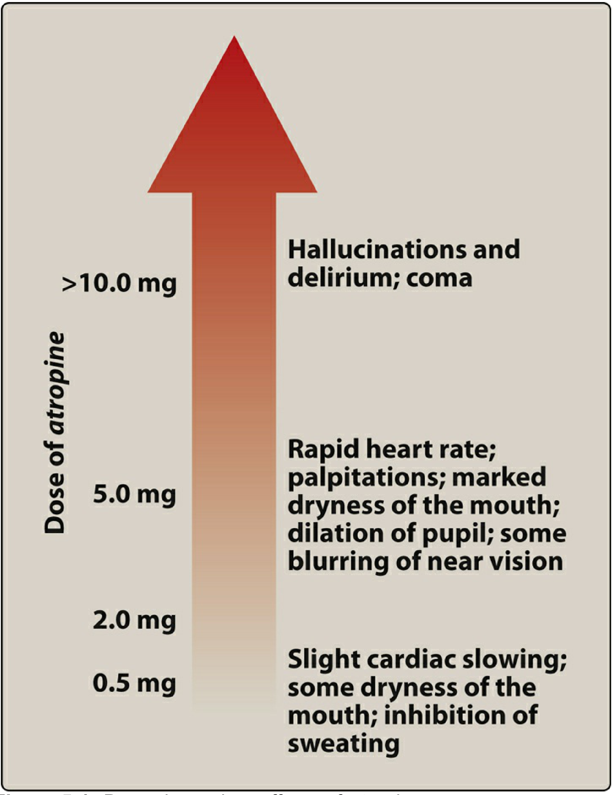
Atropine Dose-Dependent Systemic Effects
Summary Table
| Feature | Detail |
|---|---|
| Drug class | Muscarinic (M3) receptor antagonists |
| Target structures | Ciliary muscle + iris sphincter |
| Primary ocular effects | Cycloplegia (no accommodation) + Mydriasis |
| Key clinical uses | Cycloplegic refraction, fundoscopy, uveitis, keratitis, myopia control |
| Most short-acting | Tropicamide (3-6 h) |
| Most long-acting | Atropine (1-2 weeks) |
| Workhorse for refraction | Cyclopentolate |
| Main contraindication | Angle-closure glaucoma |