
Pentoxyphylline
I'll look up Pentoxifylline from the medical library right away.
Searching sources...
Reading File
Now searching the medical library and PubMed simultaneously:
Finding Sources
Searching PubMed
pentoxifylline clinical uses mechanisms
Good - found relevant sections across multiple textbooks. Now reading the most informative ones:
Reading File
Reading File
Finding Sources
Now reading the GI/liver and pharmacology sections:
Reading File
Finding Sources
Searching PubMed
pentoxifylline
Excellent. I now have comprehensive information to provide a thorough answer. Let me compile everything.
Pentoxifylline (PTX)
Drug Class: Xanthine derivative (methylxanthine) - nonspecific phosphodiesterase (PDE) inhibitor
Mechanism of Action
Pentoxifylline has a multi-faceted mechanism:
- Hemorheological effects: Reduces blood viscosity by increasing RBC deformability and flexibility, enabling them to pass through narrowed microvascular channels more easily
- Anti-platelet: Reduces platelet aggregation and platelet-activating factor (PAF) synthesis and inhibits its action on neutrophils
- Anti-inflammatory: Downregulates TNF-alpha production and release; inhibits the TGF-beta1-mediated pathway of inflammation
- Anti-fibrotic: Prevents deposition of collagen type I; inhibits fibroblast proliferation; attenuates TGF-beta-mediated elastogenesis
- Fibrinolytic: Increases fibrinolytic activity
- Vasodilatory (peripheral): Can cause mild hypotension through peripheral vasodilation
The net result is improved blood flow through narrowed or diseased vessels and reduced tissue inflammation/fibrosis.
FDA-Approved Indications
1. Intermittent Claudication (IC) - Peripheral Arterial Disease
- Approved in 1984 - the first oral drug approved for this condition in the US
- Cilostazol and pentoxifylline are the only two FDA-approved drugs for IC symptom relief in the US
- Dose: 400 mg three times daily (max 1,800 mg/day)
- Produces ~30% improvement in maximal walking distance (vs. ~54% for cilostazol)
- The Society for Vascular Surgery guideline recommends pentoxifylline (400 mg TID) as an alternative for patients with contraindications to cilostazol (e.g., heart failure, since cilostazol is contraindicated in any level of CHF)
- Current Surgical Therapy, 14e
Off-Label / Investigational Uses
2. Peyronie's Disease (PD)
- Inhibits tunica albuginea-derived fibroblast proliferation in vitro
- Reduces collagen I, alpha-smooth muscle actin, and plaque size in animal models (by up to 95%)
- In a randomized double-blind, placebo-controlled trial (n=228), pentoxifylline significantly reduced disease progression (11% vs. 42% in placebo), improved penile curvature, plaque volume, and IIEF score
- The AUA 2015 PD guideline acknowledges uncertain efficacy and states replication is needed before formal recommendation
- Campbell-Walsh-Wein Urology
3. Alcoholic (Alcohol-Associated) Hepatitis
- Rationale: Anti-TNF and anti-inflammatory properties; also reduces hepatorenal syndrome risk
- STOPAH Trial (large RCT): No significant survival advantage over placebo at 28 days or 1 year (OR 1.07; 95% CI 0.77-1.49; P = 0.69)
- Some hepatologists still use it in selected patients given its good safety profile and as an alternative when glucocorticoids are contraindicated
- Combined with IL-1 receptor antagonist and zinc in some experimental protocols
- Sleisenger & Fordtran's GI and Liver Disease - PTX is now considered of unlikely survival benefit by most guidelines
4. Chronic Venous Leg Ulcers
- Improves microcirculation and reduces lipodermatosclerosis
- Multiple RCTs and reviews support benefit as an adjunct to compression therapy
- Dermatology 5e (Fitzpatrick)
5. Leishmaniasis
- Used as an adjunct (with antimony IV or other leishmanicidal agents) for certain species:
- Leishmania aethiopica - combined with liposomal amphotericin B or miltefosine
- L. braziliensis and other New World species - adjunct to antimony
- Fitzpatrick's Dermatology
6. Osteoradionecrosis of the Jaw (ORN)
- Often used as PENTOCLO protocol (pentoxifylline + tocopherol ± clodronate)
- Improves tissue oxygenation in radiation-damaged bone
- Recent 2024 systematic review (PMID: 38964469) examined evidence for ORN and medication-related osteonecrosis
7. Radiation Proctitis (Chronic)
- Oral pentoxifylline studied for chronic radiation-induced proctitis (PMID: 37608385)
8. Inner Ear Diseases
- A 2025 systematic review (PMID: 40332573) evaluated pentoxifylline in inner ear conditions (e.g., sudden sensorineural hearing loss), citing its microcirculatory and anti-inflammatory effects
9. Endometriosis / Infertility
- A systematic review of 4 trials (n=334) showed no significant effect on endometriosis-related infertility outcomes
- Not recommended for this indication
- Berek & Novak's Gynecology
Dosing Summary
| Indication | Dose |
|---|---|
| Intermittent claudication | 400 mg TID (max 1,800 mg/day) with meals |
| Alcoholic hepatitis | 400 mg TID x 28 days (historical; now not recommended) |
| Peyronie's disease (off-label) | 400 mg TID |
| Venous ulcers (off-label) | 400-800 mg TID |
Formulation: Extended-release (ER) tablets 400 mg. Take with food to reduce GI side effects.
Adverse Effects
| System | Effects |
|---|---|
| GI | Nausea, vomiting, dyspepsia, anorexia (most common) |
| CNS | Headache, dizziness, drowsiness |
| Cardiovascular | Peripheral vasodilation -> hypotension; exacerbation of hypertension (paradoxically, due to some reports) |
| General | Malaise, flushing |
Monitor blood pressure - pentoxifylline is a peripheral vasodilator and can induce hypotension.
Contraindications / Precautions
- Prior intolerance to other methylxanthines (caffeine, theophylline, theobromine)
- Recent cerebral or retinal hemorrhage
- Use with caution in patients on anticoagulants or antiplatelet agents (additive effects)
- Reduce dose in severe renal impairment
Key Clinical Evidence Updates (2023-2026)
- Alcoholic hepatitis (2026): A recent meta-analysis (PMID: 40518060) in Clinical Gastroenterology and Hepatology (March 2026) further confirms the limited role of pentoxifylline in severe alcohol-associated hepatitis - glucocorticoids remain the preferred pharmacological strategy despite modest benefit.
- Osteoradionecrosis (2024): Systematic review (PMID: 38964469) supports use of pentoxifylline + tocopherol (PENTOCLO) for ORN management.
- Inner ear diseases (2025): New systematic review (PMID: 40332573) evaluates emerging role in sudden hearing loss and other inner ear conditions.
Summary - Key Points for Exams/Clinics
- MOA: Nonspecific PDE inhibitor + hemorheological + anti-inflammatory + anti-fibrotic + anti-TNF
- FDA approved for: Intermittent claudication (PAD) - alternative to cilostazol when CHF is present
- Alcoholic hepatitis: Was used historically; STOPAH trial showed NO survival benefit - now largely abandoned
- Peyronie's disease: Anti-fibrotic rationale strong; clinical evidence promising but guideline recommendation pending replication
- Side effects: Mainly GI (nausea, dyspepsia); monitor BP
- Dose: 400 mg TID standard
Acrocyanosis
Acrocyanosis is a persistent, painless bluish discoloration of the hands and feet (and sometimes face) caused by vasospasm of small cutaneous vessels, leading to increased oxygen extraction in the periphery. It is typically benign, symmetric, and worsens with cold. Let me pull detailed textbook content on this.
Searching sources...
Finding Sources
Searching PubMed
acrocyanosis
Good results. Reading the main textbook sections now:
Reading File
Reading File
Now fetching the clinical image and reading the treatment section:
Loading Image
Reading File
I now have all the information needed for a thorough, well-illustrated answer.
Acrocyanosis
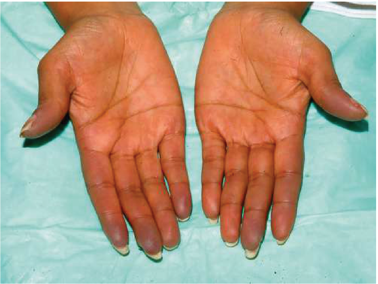
Persistent, painless, symmetrical bluish discoloration of both hands in acrocyanosis. - Goldman-Cecil Medicine
Definition
Acrocyanosis is a persistent, painless, symmetrical bluish (cyanotic) discoloration of the hands, feet, or both. It occurs in primary and secondary forms. The word derives from Greek: akron (extremity) + kyanos (blue).
- Goldman-Cecil Medicine
Pathophysiology
Acrocyanosis was originally thought to result from vasospasm of small cutaneous arteries and arterioles, causing reduced blood flow and consequent oxygen desaturation in the venules (increased O2 extraction -> deoxyhaemoglobin -> blue color).
More recent data suggest that low perfusion pressures and sluggish capillary flow result in capillary constriction. Either way, the end result is stagnant deoxygenated blood pooling in the microvasculature of the skin.
Epidemiology & Predisposing Factors
| Feature | Detail |
|---|---|
| Most common in | Young women, 2nd-4th decades of life |
| Climate | More common in cooler temperatures |
| Familial predisposition | Reported |
| Prevalence in anorexia nervosa | ~20-40% |
| Prevalence in cancer patients | Up to 25% |
Primary vs Secondary Acrocyanosis
Primary Acrocyanosis
- Benign, idiopathic condition
- Underlying cause unknown; a role for estrogen is suggested (explains female predominance)
- Symmetric, painless, no tissue loss
- No ulceration or gangrene
Secondary Acrocyanosis
- Associated with an underlying systemic condition
- May be asymmetrical
- Can be associated with pain, ulceration, tissue loss, or gangrene
- Requires investigation and treatment of the underlying cause
Secondary causes include:
| Category | Examples |
|---|---|
| Connective tissue disorders | Ehlers-Danlos syndrome |
| Hematological | Cold agglutinins, cryoglobulins, cryofibrinogens, antiphospholipid antibodies |
| Malignancy | Any cancer (up to 25% of all cancer patients) |
| Hypoxemia | Cardiopulmonary disease |
| Eating disorders | Anorexia nervosa, chronic starvation |
| Neurological | Spinal cord injury |
| Toxic | Arsenic poisoning |
| Drugs | Vasopressors, tricyclic antidepressants |
| Injection drug use | "Puffy hand syndrome" (injecting into hands/fingers) |
| Myeloproliferative disorders | Should be excluded in new-onset acrocyanosis in elderly patients |
Clinical Features
- Distribution: Hands and feet primarily; also forearms, nose, ears, and nipples
- Color: Bluish/cyanotic of varying shades, symmetric
- Persistent (not episodic/paroxysmal - key distinction from Raynaud's)
- Aggravated by: Cold exposure, emotional stress, dependent limb position
- Relieved by: Elevation of the limb, warming
- Associated: Clamminess and hyperhidrosis of hands and feet
- No pain, no ulceration, no tissue loss (in primary form)
Differential Diagnosis
| Condition | Key Distinguishing Features |
|---|---|
| Raynaud's phenomenon | Episodic (not persistent); classic triphasic color change (white -> blue -> red); triggered by cold/stress; may have ulceration |
| Peripheral cyanosis | Due to systemic hypoxia; cyanosis also present on mucous membranes; ABG shows low PaO2 |
| Pernio (chilblains) | Painful, erythematous-to-violaceous lesions; inflammatory; blisters possible; cold + damp exposure; may occur on a background of acrocyanosis |
| Erythromelalgia | Warm, red, burning extremities - the opposite of acrocyanosis; worsened by heat |
Key differentiating point from peripheral cyanosis: In acrocyanosis, mucous membranes are normal (no central hypoxia), and ABG is normal.
Neonatal / Newborn Note
In neonates, acrocyanosis of hands and feet in the first hours of life is physiological and normal - it reflects the transitional circulation and resolves quickly. It should be distinguished from central cyanosis, which is always pathological.
Diagnosis
Diagnosis is clinical, based on history and physical examination.
Workup (to exclude secondary causes):
- Complete blood count
- Metabolic profile
- Antiphospholipid antibodies
- Cold agglutinins
- Cryofibrinogens
- Cryoglobulins
- Testing for connective tissue disorders (ANA, anti-Scl-70, etc.)
- If elderly with new onset: exclude myeloproliferative disorder
Remittent Necrotizing Acrocyanosis (Severe Form)
A severe variant characterized by:
- Enhanced susceptibility to cooling and pain
- Ulceration and gangrene of fingers
- Arteriolar occlusion by thrombi or intimal proliferation
- Cold allodynia and cold hyperalgesia may be present
- Fitzpatrick's Dermatology
Treatment
Primary Acrocyanosis
Treatment is largely supportive - the prognosis is excellent.
| Measure | Notes |
|---|---|
| Reassurance | Most important - primary form is benign |
| Avoid cold and damp | Main lifestyle measure |
| Wear gloves | Practical protection |
| Avoid tobacco | Smoking worsens vasospasm |
| Keep skin warm | Supportive |
Pharmacological (if needed):
- Alpha-adrenergic blockers: Prazosin 3 mg/day - may be trialed
- Calcium channel blockers: Amlodipine or nifedipine - generally well tolerated
- These are the same agents used for Raynaud's, applied here when symptoms are bothersome
Secondary Acrocyanosis
- Treat the underlying cause
- Prognosis depends entirely on the underlying condition
Prognosis
- Primary: Excellent. Benign, chronic but harmless condition.
- Secondary: Depends on the underlying disorder.
Key Exam Points
- Persistent symmetric painless acrocyanosis = primary (benign)
- Asymmetric + pain + ulcers = secondary - investigate
- Distinguish from Raynaud's: Raynaud's is episodic with triphasic color change; acrocyanosis is persistent with no triphasic pattern
- Distinguish from central cyanosis: mucous membranes are normal in acrocyanosis
- Strongly associated with anorexia nervosa (20-40%)
- Role of estrogen suspected - explains female predominance
- New onset in elderly: always exclude myeloproliferative disorder
- Treatment: reassurance + cold avoidance; prazosin or CCBs if needed
- In neonates: physiological in first hours of life
Compare acrocyanosis vs Raynaud phenomenon - clinical differentiation
Searching sources...
Finding Sources
Finding Sources
Good. Reading the Raynaud sections from Goldman-Cecil, Fitzpatrick's, and Robbins in parallel:
Reading File
Reading File
Reading File
Now reading the primary vs secondary Raynaud features table and the nailfold capillaroscopy/investigation section:
Reading File
Reading the treatment section and fetching the Raynaud image:
Reading File
Loading Image
All the data needed. Here is the full comparison:
Acrocyanosis vs Raynaud Phenomenon - Clinical Differentiation
Side-by-Side Visual Comparison
| Acrocyanosis | Raynaud Phenomenon | |
|---|---|---|
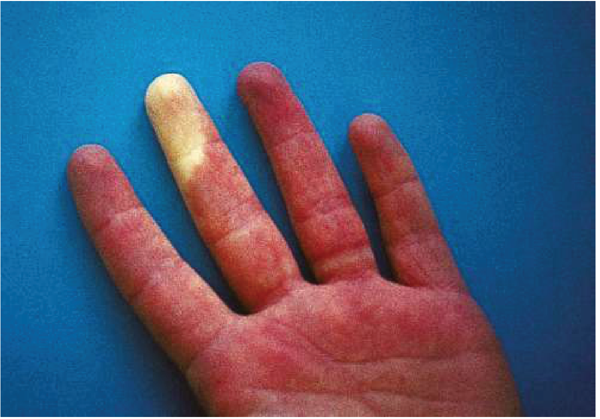
| Persistent, symmetric, diffuse blue-violet discoloration of both hands | One finger white (ischemic), rest red/hyperemic - mid-attack |
 |
Core Pathophysiology Contrast
| Feature | Acrocyanosis | Raynaud Phenomenon |
|---|---|---|
| Mechanism | Persistent arteriolar/capillary constriction with low flow and stagnant deoxygenation | Episodic, exaggerated vasoconstrictive response to cold or emotional stimuli |
| Flow state | Sluggish but continuous - slow oxygen extraction | Sudden shut-off then sudden reopening |
| Vessel level | Small cutaneous arterioles and capillaries | Digital arteries and arterioles |
| Oxygen state | Chronic mild deoxygenation (venular pooling) | Acute anoxia then reactive hyperemia |
Master Comparison Table
| Clinical Feature | Acrocyanosis | Raynaud Phenomenon |
|---|---|---|
| Nature of discoloration | Persistent, constant, never fully resolves | Episodic, paroxysmal, attack-based |
| Color change | Fixed blue-violet (one shade) - no triphasic change | Classic triphasic: White (pallor) → Blue (cyanosis) → Red (rubor/hyperemia) |
| Symmetry | Always symmetric (primary form) | Bilateral or unilateral (secondary form can be asymmetric) |
| Pain | Absent (primary form is painless) | Present during attacks - paresthesias, burning, numbness |
| Trigger | Cold worsens it; present even at rest | Triggered by cold exposure or emotional stress |
| Reversibility | Does not fully reverse with rewarming | Fully reverses on rewarming - digits return to normal |
| Distribution | Hands, feet, forearms, nose, ears, nipples - diffuse | Digits (fingers > toes); thumb often spared; middle and ring fingers most common |
| Sweating | Hyperhidrosis and clamminess present | No sweating during attacks |
| Ulceration/gangrene | Absent in primary form | Present in secondary Raynaud (esp. scleroderma) |
| Tissue changes | Less sclerotic | Atrophy of skin, subcutaneous tissue, muscles in chronic cases |
| Elevation of limb | Improves color | No significant effect during an attack |
| Dependency of limb | Worsens color | Not a major modifier |
| Mucous membranes | Normal (no central hypoxia) | Normal |
| Primary sex predilection | Young women (2nd-4th decade) | Young women (affects 3-15% of young women) |
| Associated conditions | Anorexia nervosa (20-40%), malignancy, Ehlers-Danlos, spinal cord injury, drug users | Scleroderma (>90% of SSc patients), SLE, RA, Sjögren's syndrome, hypothyroidism, drugs (beta-blockers, ergots), vibration exposure |
| Family history | Reported | Strong (30-50% have first-degree relative) |
| Role of estrogen | Suggested (female predominance) | Associated |
The Triphasic Color Change of Raynaud's - Explained
COLD / STRESS TRIGGER
        ↓
  WHITE (Pallor)
  - Arterial vasoconstriction → no blood entering digits
  - "Dead white" appearance
        ↓
  BLUE (Cyanosis)
  - Post-capillary venular constriction persists
  - Deoxygenation of trapped blood
  - Blue/purple hue
        ↓
  RED (Rubor/Hyperemia)
  - Rewarming → vasodilation
  - Rush of oxygenated blood
  - Bright red, burning sensation
Note: All three phases are not always seen - pallor alone may be the only finding. In darker skin types: pallor → purple → pink/dull red.
- Robbins & Kumar Basic Pathology
Key Distinguishing Points (Quick Memory Aid)
| Point | Acrocyanosis | Raynaud |
|---|---|---|
| "Always there" | Yes - persistent | No - comes and goes |
| Color sequence | Monotone blue | Tri-color (W→B→R) |
| Pain | No | Yes (during attack) |
| Goes away with warmth | Partly improves, does not fully resolve | Completely resolves |
| Sweaty hands | Yes | No |
| Clue disease | Anorexia nervosa | Scleroderma |
Investigations - How They Differ
For Both - Baseline Workup
- CBC, metabolic profile
- Cold agglutinins, cryoglobulins, cryofibrinogens
- Antiphospholipid antibodies
- Connective tissue screen (ANA, anti-Scl-70, anti-centromere)
Raynaud-Specific
| Test | Purpose |
|---|---|
| Nailfold capillaroscopy | Most useful - abnormal nailfold capillaries (dilated, distorted loops, "giant" capillaries, avascular areas) indicate secondary Raynaud / early scleroderma; normal in primary Raynaud and acrocyanosis |
| Photoplethysmography / pulse volume recordings | Quantify degree of vasospasm |
| Laser Doppler flux | Blood flow measurement |
| Duplex ultrasonography | Structural vessel disease |
| MR/contrast angiography | Severe cases with ischemia |
| ESR, CRP, TSH, SPEP, urinalysis | Screen for secondary causes |
| Specific autoantibodies (anti-Scl-70, anti-centromere) | If ANA positive or rheumatologic features |
Treatment Comparison
| Approach | Acrocyanosis | Raynaud Phenomenon |
|---|---|---|
| First-line | Reassurance + cold avoidance | Cold avoidance + keep core warm + biofeedback |
| Pharmacological - 1st | CCBs (amlodipine/nifedipine) if needed; prazosin 3 mg/day | Calcium channel blockers (nifedipine 10-20 mg q6h; amlodipine 2.5-10 mg/day) - Grade A evidence |
| Pharmacological - 2nd | - | Topical nitroglycerin (effective for both primary and secondary) |
| Pharmacological - 3rd | - | PDE-5 inhibitors (sildenafil 20-50 mg TID, tadalafil 5-20 mg/day) |
| Pharmacological - 4th | - | Bosentan (endothelin-1 blocker) for digital ulcers in secondary |
| Pharmacological - 5th | - | Iloprost (prostacyclin analogue, IV) for severe ischemia |
| Surgical | Not needed | Sympathectomy, spinal cord stimulation for refractory severe cases |
| Treat underlying cause | Yes (secondary form) | Yes (secondary form - essential) |
Secondary Raynaud - Causes to Know
| Category | Examples |
|---|---|
| Rheumatologic (most common) | Scleroderma (>90% of SSc), SLE, RA, Sjögren's, MCTD |
| Obstructive arterial | Atherosclerosis, Buerger's disease, arterial embolism |
| Occupational | Hypothenar hammer syndrome, hand-arm vibration syndrome, frostbite |
| Endocrine | Hypothyroidism |
| Hematologic | Polycythemia vera, myeloma, cryoglobulinemia, cold agglutinins |
| Drugs | Beta-blockers, ergots, amphetamines, cocaine, clonidine, OCP, cyclosporine, antineoplastics |
| Infections | Hepatitis B |
| Structural | Thoracic outlet syndrome, subclavian artery stenosis, AV fistula |
| Toxic | Lead, arsenic poisoning |
Every patient with Raynaud phenomenon should be evaluated for secondary causes, since it may be the first manifestation of scleroderma or other connective tissue disease. - Robbins & Kumar Basic Pathology
Prognosis Contrast
| Acrocyanosis | Raynaud Phenomenon | |
|---|---|---|
| Primary form | Excellent - benign, cosmetic concern only | Excellent - chronic but benign; atrophy of skin/subcutaneous tissue possible; ulceration/gangrene rare |
| Secondary form | Depends on underlying cause | Depends on underlying cause - scleroderma-associated Raynaud can lead to digital ulcers, critical ischemia, and amputation |
This is a shared conversation. Sign in to Orris to start your own chat.