
Surgical Anatomy of sigmoid colon, carcinoma of sigmoid colon full details and management
NCI colon cancer treatment PDQ sigmoid colon surgery staging management 2025
colon cancer surgery management
-
Surgical anatomy of the sigmoid colon
- Extent, peritoneal relations, mesocolon, sigmoid mesentery
- Arterial supply, venous and lymphatic drainage
- Autonomic nerves, ureter, gonadal vessels, hypogastric nerves, and other operative danger points
- Applied anatomy for sigmoid colectomy and high/low ligation of the inferior mesenteric artery
-
Carcinoma of the sigmoid colon
- Risk factors, pathology, molecular testing, routes of spread
- Clinical presentation and complications: obstruction, perforation, bleeding, fistula
- Differential diagnosis
-
Diagnosis and staging
- Colonoscopy and biopsy, CEA, CT chest-abdomen-pelvis
- TNM staging, assessment of resectability, mismatch-repair/MSI testing
- Evaluation of obstruction, synchronous tumors, and metastases
-
Management
- Principles and steps of elective oncologic sigmoid colectomy
- Laparoscopic versus open surgery, lymph-node harvest, margins, anastomosis and stoma decisions
- Stage-specific adjuvant chemotherapy
- Management of acute obstruction or perforation
- Locally advanced, metastatic, recurrent, and palliative disease
- Follow-up surveillance and prognosis
Explain the surgical anatomy with a labeled diagram
labeled surgical anatomy diagram of sigmoid colon sigmoid mesocolon inferior mesenteric artery left ureter hypogastric nerves
sigmoid colectomy anatomy
surgical anatomy sigmoid colon inferior mesenteric artery ureter sigmoid mesocolon authoritative
A precise labeled surgical anatomy diagram of the sigmoid colon for medical students, anterior intraoperative view. Show descending colon entering the S-shaped intraperitoneal sigmoid colon and rectosigmoid junction at S3. Show sigmoid mesocolon as an inverted V attached to posterior abdominal wall, with apex at the left common iliac artery bifurcation. Label inferior mesenteric artery, left colic artery, two to four sigmoid arteries, superior rectal artery, marginal artery of Drummond, inferior mesenteric vein, left ureter running retroperitoneally beneath the mesocolon, left gonadal vessels, psoas major, left common iliac vessels, hypogastric nerves, and Toldt's white line. Include a small inset showing safe surgical dissection plane between mesocolic fascia and retroperitoneum, emphasizing ureter and gonadal vessels preservation. Clearly label all structures.
Surgical anatomy of the sigmoid colon
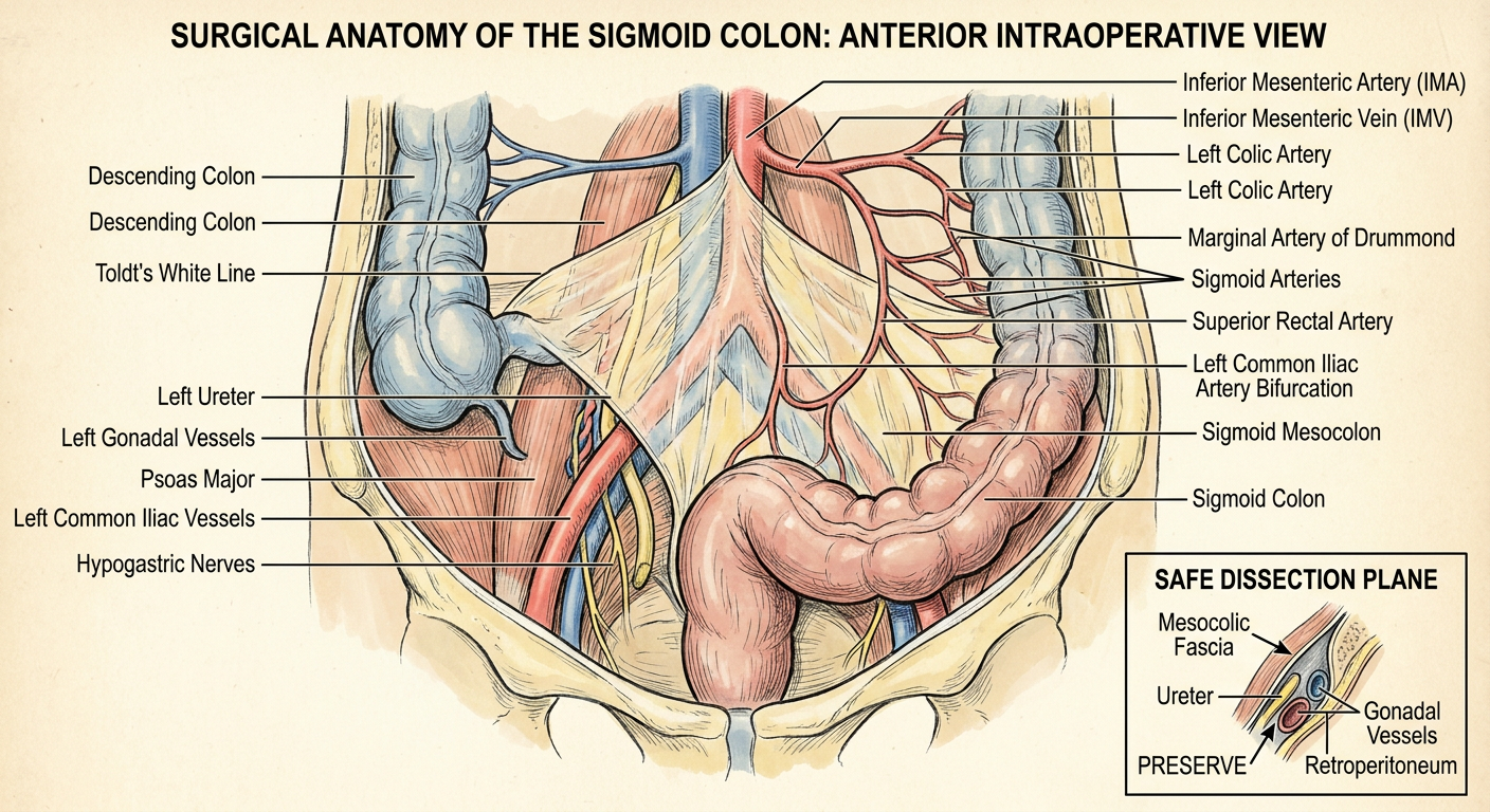
1. Situation, extent, and external features
- The sigmoid colon is the mobile, S-shaped terminal part of the colon.
- It continues from the descending colon at the pelvic brim and ends at the rectosigmoid junction, approximately at the S3 vertebral level.
- Surgically, the most reliable anatomical landmark for the rectosigmoid junction is the confluence of the three taeniae coli. The rectum has no taeniae coli, appendices epiploicae, or haustra. Fischer's Mastery of Surgery, p. 767.
- Typical length is variable, often about 25 to 40 cm. A long, redundant sigmoid and long mesocolon predispose to sigmoid volvulus.
- Unlike the descending colon, it is fully peritonealized and therefore mobile, except at the root of its mesocolon. Maingot's Abdominal Operations, p. 767.
2. Peritoneum and sigmoid mesocolon
Root of the sigmoid mesocolon
- Apex: near the bifurcation of the left common iliac artery into external and internal iliac arteries.
- Left limb: runs upward along the medial border of the left psoas major.
- Right limb: runs downwards into the pelvis to approximately S3.
Intersigmoid recess
3. Important posterior relations and danger structures
| Structure | Relation to sigmoid mesocolon | Surgical relevance |
|---|---|---|
| Left ureter | Retroperitoneal, deep to the sigmoid mesocolon; crosses the pelvic brim near the iliac vessels | Must be identified and protected before dividing the mesocolon or vascular pedicle |
| Left gonadal vessels | Usually lie with or close to the ureter on the posterior abdominal wall | Can be injured during mobilization |
| Left psoas major | Posterior to the left limb of the mesocolic root | Useful orientation landmark |
| Left common iliac artery and vein | Related to the apex of the mesocolic root | The ureter crosses anteriorly near this region |
| Hypogastric nerves / superior hypogastric plexus | Deep and medial at the root of the mesocolon and pelvic inlet | Preserve to avoid postoperative urinary and sexual dysfunction |
| Sacral promontory | Posterior pelvic landmark near the distal dissection | Helps orient the rectosigmoid and superior rectal pedicle |
Operative principle
4. Arterial supply
IMA branches relevant to sigmoid colectomy
-
Left colic artery
- Usually the first branch of the IMA.
- Ascending branch supplies descending colon.
- Descending branch communicates with the upper sigmoid arterial supply.
-
Sigmoid arteries
- Usually 2 to 4 branches.
- Run inferolaterally within the sigmoid mesocolon.
- Supply the lower descending colon and sigmoid colon.
-
Superior rectal artery
- Terminal continuation of IMA.
- Supplies the upper rectum and rectosigmoid region.
- Descends over the left common iliac vessels into the pelvis.
-
Marginal artery of Drummond
- A continuous arterial arcade close to the mesenteric border of the colon.
- Connects branches of the superior and inferior mesenteric systems.
- Its preservation helps maintain perfusion of the proximal colon and anastomosis.
Watershed area
5. Venous drainage
- Sigmoid veins accompany the sigmoid arteries.
- They drain into the inferior mesenteric vein (IMV).
- The IMV typically ascends on the left side, passes behind the pancreas, and usually drains into the splenic vein, hence ultimately into the portal venous system.
6. Lymphatic drainage
- Epicolic nodes: on bowel wall and appendices epiploicae
- Paracolic nodes: along marginal vessels
- Intermediate nodes: along sigmoid arteries and left colic artery
- Principal/apical nodes: around the IMA origin
7. Nerve supply
Sympathetic
- Mainly from lumbar splanchnic nerves through the inferior mesenteric plexus and superior hypogastric plexus.
- Sympathetic fibers travel along the IMA and its branches.
Parasympathetic
- From pelvic splanchnic nerves, S2-S4, reaching the distal colon via the inferior hypogastric plexuses.
Surgical relevance
- Bladder dysfunction or urinary retention
- Retrograde ejaculation in men
- Erectile dysfunction
- Sexual dysfunction in women
8. Applied anatomy in oncologic sigmoid colectomy
A. Lateral-to-medial approach
- Incise the white line of Toldt, the lateral peritoneal reflection of the descending/sigmoid colon.
- Mobilize the sigmoid and descending colon medially.
- Enter the avascular retroperitoneal plane.
- Identify and preserve the left ureter and gonadal vessels.
- Mobilize sufficiently to achieve a tension-free colorectal anastomosis.
B. Medial-to-lateral approach
- Elevate the sigmoid mesocolon.
- Identify the IMA and define the retroperitoneal plane.
- Protect the left ureter, gonadal vessels, and hypogastric nerves.
- Control the vascular pedicle according to oncologic and perfusion requirements.
- Divide mesocolon and mobilize lateral attachments.
C. Vascular ligation options
-
High tie: IMA divided close to its origin from the aorta.
- Provides apical nodal clearance and added colonic length.
- May threaten autonomic nerves or compromise perfusion if collateral circulation is limited.
-
Low tie: preservation of the left colic artery with division distal to it.
- May preserve perfusion to the proximal limb.
- Still requires appropriate lymph-node clearance around the IMA when indicated.
Key operative safety checklist
- Left ureter seen and protected
- Gonadal vessels retained in the retroperitoneum
- Hypogastric nerves protected
- Correct plane maintained outside the mesocolic fascia
- Adequate proximal vascular supply for the anastomosis
- Sufficient colon mobilized to create a tension-free colorectal anastomosis
Carcinoma sigmoid colon full details
Which section should I explain first in full detail?
-
Surgical anatomy of the sigmoid colon
- Extent, peritoneal relations, mesocolon, sigmoid mesentery
- Arterial supply, venous and lymphatic drainage
- Autonomic nerves, ureter, gonadal vessels, hypogastric nerves, and other operative danger points
- Applied anatomy for sigmoid colectomy and high/low ligation of the inferior mesenteric artery
-
Carcinoma of the sigmoid colon
- Risk factors, pathology, molecular testing, routes of spread
- Clinical presentation and complications: obstruction, perforation, bleeding, fistula
- Differential diagnosis
-
Diagnosis and staging
- Colonoscopy and biopsy, CEA, CT chest-abdomen-pelvis
- TNM staging, assessment of resectability, mismatch-repair/MSI testing
- Evaluation of obstruction, synchronous tumors, and metastases
-
Management
- Principles and steps of elective oncologic sigmoid colectomy
- Laparoscopic versus open surgery, lymph-node harvest, margins, anastomosis and stoma decisions
- Stage-specific adjuvant chemotherapy
- Management of acute obstruction or perforation
- Locally advanced, metastatic, recurrent, and palliative disease
- Follow-up surveillance and prognosis
1. Situation, extent, and external features
- The sigmoid colon is the mobile, S-shaped terminal part of the colon.
- It continues from the descending colon at the pelvic brim and ends at the rectosigmoid junction, approximately at the S3 vertebral level.
- Surgically, the most reliable anatomical landmark for the rectosigmoid junction is the confluence of the three taeniae coli. The rectum has no taeniae coli, appendices epiploicae, or haustra. Fischer's Mastery of Surgery, p. 767.
- Typical length is variable, often about 25 to 40 cm. A long, redundant sigmoid and long mesocolon predispose to sigmoid volvulus.
- Unlike the descending colon, it is fully peritonealized and therefore mobile, except at the root of its mesocolon. Maingot's Abdominal Operations, p. 767.
2. Peritoneum and sigmoid mesocolon
Root of the sigmoid mesocolon
- Apex: near the bifurcation of the left common iliac artery into external and internal iliac arteries.
- Left limb: runs upward along the medial border of the left psoas major.
- Right limb: runs downwards into the pelvis to approximately S3.
Intersigmoid recess
3. Important posterior relations and danger structures
| Structure | Relation to sigmoid mesocolon | Surgical relevance |
|---|---|---|
| Left ureter | Retroperitoneal, deep to the sigmoid mesocolon; crosses the pelvic brim near the iliac vessels | Must be identified and protected before dividing the mesocolon or vascular pedicle |
| Left gonadal vessels | Usually lie with or close to the ureter on the posterior abdominal wall | Can be injured during mobilization |
| Left psoas major | Posterior to the left limb of the mesocolic root | Useful orientation landmark |
| Left common iliac artery and vein | Related to the apex of the mesocolic root | The ureter crosses anteriorly near this region |
| Hypogastric nerves / superior hypogastric plexus | Deep and medial at the root of the mesocolon and pelvic inlet | Preserve to avoid postoperative urinary and sexual dysfunction |
| Sacral promontory | Posterior pelvic landmark near the distal dissection | Helps orient the rectosigmoid and superior rectal pedicle |
Operative principle
4. Arterial supply
IMA branches relevant to sigmoid colectomy
-
Left colic artery
- Usually the first branch of the IMA.
- Ascending branch supplies descending colon.
- Descending branch communicates with the upper sigmoid arterial supply.
-
Sigmoid arteries
- Usually 2 to 4 branches.
- Run inferolaterally within the sigmoid mesocolon.
- Supply the lower descending colon and sigmoid colon.
-
Superior rectal artery
- Terminal continuation of IMA.
- Supplies the upper rectum and rectosigmoid region.
- Descends over the left common iliac vessels into the pelvis.
-
Marginal artery of Drummond
- A continuous arterial arcade close to the mesenteric border of the colon.
- Connects branches of the superior and inferior mesenteric systems.
- Its preservation helps maintain perfusion of the proximal colon and anastomosis.
Watershed area
5. Venous drainage
- Sigmoid veins accompany the sigmoid arteries.
- They drain into the inferior mesenteric vein (IMV).
- The IMV typically ascends on the left side, passes behind the pancreas, and usually drains into the splenic vein, hence ultimately into the portal venous system.
6. Lymphatic drainage
- Epicolic nodes: on bowel wall and appendices epiploicae
- Paracolic nodes: along marginal vessels
- Intermediate nodes: along sigmoid arteries and left colic artery
- Principal/apical nodes: around the IMA origin
7. Nerve supply
Sympathetic
- Mainly from lumbar splanchnic nerves through the inferior mesenteric plexus and superior hypogastric plexus.
- Sympathetic fibers travel along the IMA and its branches.
Parasympathetic
- From pelvic splanchnic nerves, S2-S4, reaching the distal colon via the inferior hypogastric plexuses.
Surgical relevance
- Bladder dysfunction or urinary retention
- Retrograde ejaculation in men
- Erectile dysfunction
- Sexual dysfunction in women
8. Applied anatomy in oncologic sigmoid colectomy
A. Lateral-to-medial approach
- Incise the white line of Toldt, the lateral peritoneal reflection of the descending/sigmoid colon.
- Mobilize the sigmoid and descending colon medially.
- Enter the avascular retroperitoneal plane.
- Identify and preserve the left ureter and gonadal vessels.
- Mobilize sufficiently to achieve a tension-free colorectal anastomosis.
B. Medial-to-lateral approach
- Elevate the sigmoid mesocolon.
- Identify the IMA and define the retroperitoneal plane.
- Protect the left ureter, gonadal vessels, and hypogastric nerves.
- Control the vascular pedicle according to oncologic and perfusion requirements.
- Divide mesocolon and mobilize lateral attachments.
C. Vascular ligation options
-
High tie: IMA divided close to its origin from the aorta.
- Provides apical nodal clearance and added colonic length.
- May threaten autonomic nerves or compromise perfusion if collateral circulation is limited.
-
Low tie: preservation of the left colic artery with division distal to it.
- May preserve perfusion to the proximal limb.
- Still requires appropriate lymph-node clearance around the IMA when indicated.
Key operative safety checklist
- Left ureter seen and protected
- Gonadal vessels retained in the retroperitoneum
- Hypogastric nerves protected
- Correct plane maintained outside the mesocolic fascia
- Adequate proximal vascular supply for the anastomosis
- Sufficient colon mobilized to create a tension-free colorectal anastomosis
-
Definition and epidemiology
- A sigmoid-colon adenocarcinoma is a left-sided colon cancer.
- It must be distinguished from rectal cancer, because rectal cancers may require pelvic MRI, neoadjuvant treatment, and different operative planning.
-
Etiology and risk factors
- Age, adenomatous polyps, family history, Lynch syndrome, familial adenomatous polyposis
- Inflammatory bowel disease, obesity, smoking, alcohol, diet, diabetes, prior colorectal cancer
-
Pathology and molecular biology
- Usual histology: adenocarcinoma
- Grading, mucinous/signet-ring variants, lymphovascular and perineural invasion
- MMR/MSI, RAS, BRAF, HER2, and NTRK testing where relevant
-
Clinical features
- Altered bowel habit, progressive constipation, colicky lower-abdominal pain, bleeding, iron-deficiency anemia, weight loss
- Left-sided lesions commonly cause annular narrowing and large-bowel obstruction
- Complications: obstruction, perforation, abscess, fistula, metastases
-
Diagnosis and preoperative work-up
- Colonoscopy with biopsy and examination of the entire colon
- CEA, CBC, renal/liver tests
- CT chest-abdomen-pelvis for staging
- Completion colonoscopy if an obstructing lesion prevents full initial examination
-
TNM staging and prognosis
- T stage by depth of bowel-wall invasion
- N stage by regional lymph nodes
- M stage by liver, lung, peritoneal, or distant spread
-
Operative management
- Elective oncologic sigmoid colectomy with regional lymphadenectomy and colorectal anastomosis when safe
- Laparoscopic or open approach
- Margins, vascular ligation, mesocolic excision, lymph-node assessment, and stoma decisions
-
Stage-wise adjuvant and systemic therapy
- Surgery alone for most stage I disease
- Selective adjuvant chemotherapy for high-risk stage II
- Oxaliplatin-based chemotherapy for most stage III disease
- Systemic, biologic, immunotherapy, metastasis-directed, and palliative options for stage IV disease
-
Emergency presentation
- Resuscitation and staging where feasible
- Stenting, diverting stoma, primary resection/anastomosis, or Hartmann procedure depending on obstruction/perforation, physiological status, and local expertise
-
Surveillance
- Clinical review, CEA, CT in appropriate stages, and colonoscopic surveillance.
Overview
Create a quick-reference PDF on sigmoid carcinoma
colon cancer adjuvant chemotherapy management
NCI Colon Cancer Treatment PDQ stage II III IV treatment surveillance 2025
~/
mkdir -p "/tmp/workspace/sigmoid-carcinoma-pdf/src" "/tmp/workspace/sigmoid-carcinoma-pdf/output" "/tmp/workspace/sigmoid-carcinoma-pdf/assets" && curl -L --fail --silent --show-error "https://cdn.orris.care/image-gen/12cd4cd640584118a7dbb41964c1669d.png" -o "/tmp/workspace/sigmoid-carcinoma-pdf/assets/sigmoid_anatomy.png" && python -c "from PIL import Image; p='/tmp/workspace/sigmoid-carcinoma-pdf/assets/sigmoid_anatomy.png'; im=Image.open(p); print(im.size)"
~/sigmoid-carcinoma-pdf
~/sigmoid-carcinoma-pdf/src/create_pdf.py
from reportlab.lib import colors
from reportlab.lib.enums import TA_CENTER, TA_LEFT
from reportlab.lib.pagesizes import A4
from reportlab.lib.styles import getSampleStyleSheet, ParagraphStyle
from reportlab.lib.units import cm
from reportlab.platypus import (SimpleDocTemplate, Paragraph, Spacer, Table, TableStyle,
                                PageBreak, Image, KeepTogether)
from reportlab.pdfbase.pdfmetrics import stringWidth
from reportlab.pdfbase.ttfonts import TTFont
from reportlab.pdfbase import pdfmetrics
from reportlab.lib.colors import HexColor
from reportlab.pdfgen import canvas
from reportlab.lib.utils import ImageReader
from pathlib import Path

ROOT = Path('/tmp/workspace/sigmoid-carcinoma-pdf')
OUT = ROOT / 'output' / 'Sigmoid_Carcinoma_Quick_Reference.pdf'
ASSET = ROOT / 'assets' / 'sigmoid_anatomy.png'

NAVY = HexColor('#12304A')
TEAL = HexColor('#087E8B')
PALE = HexColor('#EAF5F5')
GOLD = HexColor('#C98C1A')
LIGHTGOLD = HexColor('#FFF6E3')
INK = HexColor('#1D2935')
MUTED = HexColor('#52616B')
RULE = HexColor('#C9D5DB')
RED = HexColor('#A73636')

styles = getSampleStyleSheet()
styles.add(ParagraphStyle(name='TitleX', parent=styles['Title'], fontName='Helvetica-Bold', fontSize=23, leading=27, textColor=NAVY, spaceAfter=5))
styles.add(ParagraphStyle(name='SubTitleX', parent=styles['Normal'], fontName='Helvetica', fontSize=10.2, leading=14, textColor=MUTED, spaceAfter=12))
styles.add(ParagraphStyle(name='H1X', parent=styles['Heading1'], fontName='Helvetica-Bold', fontSize=14.5, leading=18, textColor=NAVY, spaceBefore=9, spaceAfter=6))
styles.add(ParagraphStyle(name='H2X', parent=styles['Heading2'], fontName='Helvetica-Bold', fontSize=10.8, leading=13, textColor=TEAL, spaceBefore=6, spaceAfter=3))
styles.add(ParagraphStyle(name='BodyX', parent=styles['BodyText'], fontName='Helvetica', fontSize=8.7, leading=11.7, textColor=INK, spaceAfter=4))
styles.add(ParagraphStyle(name='SmallX', parent=styles['BodyText'], fontName='Helvetica', fontSize=7.2, leading=9.4, textColor=MUTED, spaceAfter=2))
styles.add(ParagraphStyle(name='BoxX', parent=styles['BodyText'], fontName='Helvetica', fontSize=8.5, leading=11.1, textColor=INK))
styles.add(ParagraphStyle(name='TableX', parent=styles['BodyText'], fontName='Helvetica', fontSize=7.6, leading=9.3, textColor=INK))
styles.add(ParagraphStyle(name='TableHeadX', parent=styles['BodyText'], fontName='Helvetica-Bold', fontSize=7.5, leading=9.2, textColor=colors.white))
styles.add(ParagraphStyle(name='FootX', parent=styles['BodyText'], fontName='Helvetica', fontSize=6.7, leading=8.5, textColor=MUTED))

def P(text, style='BodyX'):
    return Paragraph(text, styles[style])

def bullets(items):
    return [P('• ' + x) for x in items]

def panel(title, items, accent=TEAL):
    content = [P(f'<b>{title}</b>', 'H2X')] + [P('• ' + x, 'BoxX') for x in items]
    t = Table([[content]], colWidths=[8.0*cm])
    t.setStyle(TableStyle([
        ('BACKGROUND',(0,0),(-1,-1),PALE if accent==TEAL else LIGHTGOLD),
        ('BOX',(0,0),(-1,-1),0.6,accent),
        ('LINEBEFORE',(0,0),(0,-1),3,accent),
        ('LEFTPADDING',(0,0),(-1,-1),9),('RIGHTPADDING',(0,0),(-1,-1),7),
        ('TOPPADDING',(0,0),(-1,-1),5),('BOTTOMPADDING',(0,0),(-1,-1),5),
    ]))
    return t

def table(data, widths, header=True):
    converted=[]
    for r,row in enumerate(data):
        converted.append([P(cell, 'TableHeadX' if header and r==0 else 'TableX') for cell in row])
    t=Table(converted,colWidths=widths,repeatRows=1 if header else 0,hAlign='LEFT')
    cmds=[('VALIGN',(0,0),(-1,-1),'TOP'),('GRID',(0,0),(-1,-1),0.28,RULE),
          ('LEFTPADDING',(0,0),(-1,-1),5),('RIGHTPADDING',(0,0),(-1,-1),5),
          ('TOPPADDING',(0,0),(-1,-1),4),('BOTTOMPADDING',(0,0),(-1,-1),4)]
    if header:
        cmds += [('BACKGROUND',(0,0),(-1,0),NAVY),('TEXTCOLOR',(0,0),(-1,0),colors.white)]
    for r in range(1 if header else 0,len(data)):
        if r%2==0: cmds.append(('BACKGROUND',(0,r),(-1,r),HexColor('#F6F9FA')))
    t.setStyle(TableStyle(cmds))
    return t

class NumberedCanvas(canvas.Canvas):
    def __init__(self,*args,**kwargs):
        canvas.Canvas.__init__(self,*args,**kwargs)
        self.pages=[]
    def showPage(self):
        self.pages.append(dict(self.__dict__))
        self._startPage()
    def save(self):
        page_count=len(self.pages)
        for state in self.pages:
            self.__dict__.update(state)
            self.setStrokeColor(RULE); self.setLineWidth(.4)
            self.line(1.35*cm,1.32*cm,19.65*cm,1.32*cm)
            self.setFont('Helvetica',6.8); self.setFillColor(MUTED)
            self.drawString(1.35*cm,.85*cm,'SIGMOID CARCINOMA | QUICK REFERENCE | Educational use only')
            self.drawRightString(19.65*cm,.85*cm,f'Page {self._pageNumber} of {page_count}')
            canvas.Canvas.showPage(self)
        canvas.Canvas.save(self)

doc=SimpleDocTemplate(str(OUT),pagesize=A4,rightMargin=1.35*cm,leftMargin=1.35*cm,topMargin=1.2*cm,bottomMargin=1.65*cm)
story=[]
# Cover / page 1
story += [P('SIGMOID CARCINOMA', 'TitleX'), P('Clinical and surgical quick-reference | Adult colonic adenocarcinoma', 'SubTitleX')]
meta=Table([[P('<b>Scope</b><br/>Diagnosis, staging, surgery, adjuvant treatment, emergencies, follow-up.', 'BoxX'),P('<b>Key distinction</b><br/>A sigmoid lesion is managed as <b>colon cancer</b>. Confirm it is not rectal cancer, because staging and neoadjuvant planning differ.', 'BoxX')]],colWidths=[8.7*cm,8.7*cm])
meta.setStyle(TableStyle([('BACKGROUND',(0,0),(0,0),PALE),('BACKGROUND',(1,0),(1,0),LIGHTGOLD),('BOX',(0,0),(-1,-1),.4,RULE),('INNERGRID',(0,0),(-1,-1),.4,RULE),('VALIGN',(0,0),(-1,-1),'TOP'),('LEFTPADDING',(0,0),(-1,-1),8),('RIGHTPADDING',(0,0),(-1,-1),8),('TOPPADDING',(0,0),(-1,-1),7),('BOTTOMPADDING',(0,0),(-1,-1),7)]))
story += [meta, Spacer(1,9), P('ANATOMY THAT CHANGES THE OPERATION','H1X')]
im=Image(str(ASSET),width=17.3*cm,height=9.42*cm)
story += [im, P('Operative landmarks: IMA branches, sigmoid mesocolon, left ureter, gonadal vessels, hypogastric nerves, and the plane of mesocolic dissection.', 'SmallX'), Spacer(1,5)]
left=panel('Surgical anatomy', ['Sigmoid colon is intraperitoneal and suspended by an inverted V-shaped sigmoid mesocolon.', 'IMA branches: left colic, 2-4 sigmoid arteries, then superior rectal artery.', 'During mobilization: identify and preserve left ureter, gonadal vessels, and hypogastric nerves.', 'Oncologic specimen follows arterial lymphatic drainage.'], TEAL)
right=panel('Curative operation', ['Oncologic sigmoid colectomy: bowel margins plus intact mesocolon and regional nodes.', 'Primary colorectal anastomosis only if well perfused and tension-free.', 'Laparoscopic approach is appropriate where expertise and patient factors allow.', 'Pathology should assess margins, nodal yield, T/N stage, grade, LVI/PNI, tumor deposits, and MMR status.'], GOLD)
story += [Table([[left,right]],colWidths=[8.6*cm,8.6*cm],style=[('VALIGN',(0,0),(-1,-1),'TOP'),('LEFTPADDING',(0,0),(-1,-1),0),('RIGHTPADDING',(0,0),(-1,-1),0)]), Spacer(1,5)]
story += [P('<b>Urgent red flags:</b> complete obstruction, perforation/peritonitis, sepsis, uncontrolled bleeding, or severe acute colonic dilatation require urgent surgical assessment.', 'BoxX')]
story.append(PageBreak())

# page 2
story += [P('1. PRESENTATION, DIAGNOSIS, AND STAGING','H1X')]
story += [P('<b>Common presentation:</b> progressive change in bowel habit, constipation or colicky left-lower-quadrant pain, occult/overt rectal bleeding, iron-deficiency anemia, fatigue, weight loss, or acute large-bowel obstruction. A left-sided lesion often produces a constricting annular lesion.', 'BodyX')]
story += [table([
['Domain','Quick-reference actions'],
['Confirm diagnosis','Full colonoscopy with biopsy where feasible. Document lesion site, circumferential extent, and obstruction. If colonoscopy cannot traverse an obstruction, complete colonic evaluation after decompression/resection.'],
['Baseline tests','CBC, renal and liver profile, electrolytes, albumin/nutritional assessment, and <b>CEA</b> before treatment. CEA is useful for trend monitoring, not for diagnosis alone.'],
['Staging imaging','Contrast CT of chest, abdomen, and pelvis. Consider liver MRI for indeterminate or potentially resectable liver lesions.'],
['Tumor biology','Perform universal <b>MMR IHC or MSI</b> testing. In metastatic/unresectable disease, test RAS and BRAF; assess HER2 amplification and NTRK fusion where treatment selection may be affected.'],
['MDT review','Review fitness, obstruction/perforation, resectability, imaging, pathology, genetic risk, and systemic-treatment plan.']
],[3.2*cm,14.1*cm])]
story += [Spacer(1,6), P('TNM AT A GLANCE','H2X')]
story += [table([
['Category','Meaning'],
['T1 / T2','Submucosa / muscularis propria invasion'],
['T3','Through muscularis propria into pericolic tissue'],
['T4a / T4b','Penetrates visceral peritoneum / directly invades or adheres to other organs or structures'],
['N1 / N2','1-3 regional nodes or tumor deposits in N1c / 4 or more regional nodes'],
['M1','Distant metastatic disease, commonly liver, lung, peritoneum, or distant nodes']
],[3.2*cm,14.1*cm])]
story += [Spacer(1,5), panel('Do not confuse sigmoid and rectal cancer', ['The surgical rectosigmoid landmark is confluence of taeniae coli. If a lesion is in the rectum, pelvic MRI and neoadjuvant therapy considerations differ.', 'A low lesion should be accurately localized before treating it as sigmoid cancer.'], GOLD)]
story.append(PageBreak())

# page 3
story += [P('2. MANAGEMENT PATHWAY','H1X')]
story += [table([
['Clinical scenario','Usual management principle'],
['Stage I (T1-2 N0)','Oncologic resection is standard. Selected low-risk T1 lesions may be cured endoscopically only when pathology confirms favorable features and complete excision.'],
['Stage II (T3-4 N0)','Surgery. Discuss adjuvant fluoropyrimidine-based therapy if high risk: T4, obstruction/perforation, inadequate nodal assessment, lymphovascular/perineural invasion, poor differentiation, positive/close margin, tumor budding, or other adverse features. dMMR/MSI-H modifies expected benefit from fluoropyrimidine alone.'],
['Stage III (any T, N+)','Surgery followed by adjuvant oxaliplatin-fluoropyrimidine therapy for most fit patients, commonly CAPOX or FOLFOX. Regimen and duration are individualized by recurrence risk, toxicity, comorbidity, and patient preference.'],
['Resectable synchronous metastases','Multidisciplinary plan. Resection/ablation may be appropriate for selected liver or lung metastases, with sequencing of systemic therapy and colon/metastasis surgery individualized.'],
['Unresectable/metastatic disease','Systemic therapy guided by performance status, molecular profile, tumor sidedness, resectability goals, and symptoms. Include fluoropyrimidine/oxaliplatin/irinotecan backbones, biologic agents, and immunotherapy for MSI-H/dMMR tumors as appropriate.'],
['Locally advanced T4','Aim for R0 <b>en bloc</b> resection of involved structures, never peel tumor off an adherent organ. Consider preoperative systemic therapy in selected cases via MDT.']
],[4.1*cm,13.2*cm])]
story += [Spacer(1,7), P('ELECTIVE ONCOLOGIC SIGMOID COLECTOMY','H2X')]
story += [Table([[panel('Core steps', ['Survey abdomen and liver; obtain cytology/biopsy if unexpected disease will alter plan.', 'Mobilize sigmoid and left colon in the mesocolic plane. Identify left ureter and gonadal vessels before mesenteric division.', 'Control relevant sigmoid/IMA vascular pedicle and remove associated mesocolic lymphovascular drainage.', 'Resect bowel with oncologically appropriate margins; assess perfusion and construct tension-free colorectal anastomosis.', 'Consider diversion or end colostomy when anastomosis is unsafe.'], TEAL),panel('Pathology checklist', ['Tumor type and grade', 'Depth and adjacent-organ/peritoneal involvement', 'Margins and radial/mesenteric margin where relevant', 'Number of nodes examined and positive', 'Tumor deposits, LVI, PNI, budding', 'MMR/MSI status'], GOLD)]],colWidths=[8.6*cm,8.6*cm],style=[('VALIGN',(0,0),(-1,-1),'TOP'),('LEFTPADDING',(0,0),(-1,-1),0),('RIGHTPADDING',(0,0),(-1,-1),0)])]
story.append(PageBreak())

# page 4
story += [P('3. EMERGENCIES, FOLLOW-UP, AND EXAM PEARLS','H1X')]
story += [P('MALIGNANT LARGE-BOWEL OBSTRUCTION','H2X')]
story += [table([
['Situation','Practical approach'],
['Stable, potentially curable left-sided obstruction','Resuscitate, correct fluid/electrolyte disturbances, stage where possible, and involve colorectal/endoscopy teams early. In selected patients at expert centers, self-expanding metal stent as a bridge to elective surgery may be considered.'],
['Perforation, peritonitis, sepsis, ischemia, or unstable patient','Emergency surgery. Source control and survival take priority. Options include resection with end colostomy (Hartmann procedure), diversion, or selected primary anastomosis depending on contamination, perfusion, physiology, and expertise.'],
['Unresectable obstruction','Endoscopic stent or diverting stoma can provide palliation; integrate systemic therapy and goals-of-care planning.']
],[4.2*cm,13.1*cm])]
story += [Spacer(1,7), P('POSTOPERATIVE SURVEILLANCE','H2X')]
story += [P('Use a structured program tailored to pathologic stage, recurrence risk, fitness for further therapy, and national guidance. Typical curative-intent surveillance includes clinical review and serial CEA, periodic CT chest-abdomen-pelvis for higher-risk stage II/III disease, and colonoscopy at approximately 1 year after surgery. If the colon was not completely examined preoperatively because of obstruction, complete colonoscopy should occur earlier after surgery. Subsequent colonoscopy interval is determined by findings and guideline pathway.', 'BodyX')]
story += [Spacer(1,6), P('HIGH-YIELD EXAM POINTS','H2X')]
story += [table([
['Point','Why it matters'],
['Hematogenous spread','Portal drainage makes the <b>liver</b> the most common site; lung and peritoneum are also frequent metastatic sites.'],
['Lymphatic spread','Follows epicolic → paracolic → intermediate → principal nodes along sigmoid/IMA vessels.'],
['Obstructing left-sided tumor','Assess competence of ileocecal valve and proximal colonic dilation. Do not undertake an unsafe primary anastomosis in a septic, unstable, or grossly contaminated setting.'],
['Anastomotic safety','Adequate mobilization, no tension, good arterial inflow, healthy bowel ends, and physiologically suitable patient.'],
['Genetics','dMMR/MSI-H suggests possible Lynch syndrome and predicts sensitivity to immune checkpoint blockade in advanced disease; arrange appropriate hereditary-cancer evaluation when indicated.']
],[4.1*cm,13.2*cm])]
story += [Spacer(1,7), panel('Safety statement', ['This is an educational quick-reference, not a substitute for multidisciplinary cancer care, local protocols, operative judgment, pathology review, or medical-oncology consultation.', 'For a patient with suspected obstruction, perforation, sepsis, acute abdomen, or significant bleeding, seek urgent in-person surgical assessment.'], RED)]
story += [Spacer(1,8), P('<b>Selected sources</b>', 'H2X'), P('• Sabiston Textbook of Surgery, 21st ed., colon cancer chapter.  • Bailey & Love’s Short Practice of Surgery, 28th ed., colorectal cancer and staging.  • Mulholland and Greenfield’s Surgery, 7th ed., adjuvant therapy.  • National Cancer Institute, Colon Cancer Treatment (PDQ), updated 12 Feb 2025.  • SEOM-GEMCAD-TTD clinical guideline for adjuvant treatment of colon cancer (2024), PMID: 38914755.', 'FootX')]

doc.build(story, canvasmaker=NumberedCanvas)
print(OUT)
python "/tmp/workspace/sigmoid-carcinoma-pdf/src/create_pdf.py" && python -c "from pypdf import PdfReader; p='/tmp/workspace/sigmoid-carcinoma-pdf/output/Sigmoid_Carcinoma_Quick_Reference.pdf'; r=PdfReader(p); print('pages=',len(r.pages)); print('page1 chars=',len(r.pages[0].extract_text())); print('total chars=',sum(len(x.extract_text() or '') for x in r.pages))"
python -c "import fitz; p='/tmp/workspace/sigmoid-carcinoma-pdf/output/Sigmoid_Carcinoma_Quick_Reference.pdf'; d=fitz.open(p); pg=d[0]; pix=pg.get_pixmap(matrix=fitz.Matrix(1.2,1.2),alpha=False); pix.save('/tmp/workspace/sigmoid-carcinoma-pdf/output/page1_preview.png'); print(d.page_count, pg.rect)"