Hypoxia
Reading File
search_medical_text
hypoxia types causes pathophysiology classification
search_medical_text
hypoxia clinical features diagnosis management treatment
search_medical_image
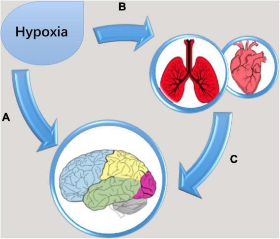
hypoxia oxygen delivery pathophysiology diagram
GradeResponse
GradeResponse
GradeResponse
Hypoxia
Hypoxia is a state of inadequate oxygen supply to tissues relative to metabolic demand. It is distinct from hypoxemia (low arterial PO₂), though hypoxemia is one of its most common causes.
Classification & Causes
Hypoxia is classically divided into four types (Harrison's, p. 272):
| Type | Mechanism | Common Causes |
|---|---|---|
| Hypoxic hypoxia (hypoxemia) | Low PaO₂ due to reduced alveolar O₂ | High altitude, hypoventilation, V/Q mismatch, diffusion impairment, right-to-left shunt |
| Anemic hypoxia | Reduced O₂-carrying capacity | Anemia, CO poisoning, methemoglobinemia |
| Stagnant (circulatory) hypoxia | Impaired O₂ delivery due to low blood flow | Heart failure, shock, peripheral vascular disease |
| Histotoxic hypoxia | Cells cannot utilize O₂ | Cyanide poisoning, H₂S toxicity |
Pathophysiology
Oxygen delivery (DO₂) = Cardiac Output × Arterial O₂ Content
Arterial O₂ content = (Hb × 1.34 × SaO₂) + (PaO₂ × 0.003)
When DO₂ falls below tissue oxygen consumption (VO₂), cells shift from aerobic to anaerobic metabolism, producing lactate and leading to cellular dysfunction and, ultimately, death.
CNS Effects (Harrison's, p. 1140)
The CNS — especially higher cortical centers — is highly sensitive to hypoxia:
- Mild/acute: Impaired judgment, motor incoordination (resembling alcohol intoxication)
- High altitude: Headache (cerebral vasodilation), nausea, dizziness, insomnia, fatigue, somnolence
- HAPE (High-Altitude Pulmonary Edema): Pulmonary arterial/venous constriction → capillary leakage → worsening hypoxia (a vicious cycle)
- HACE (High-Altitude Cerebral Edema): Severe headache, papilledema, coma (rare)
- Severe: Brainstem regulatory failure → respiratory arrest → death
Adaptation to Hypoxia
The body mounts several compensatory responses:
| Timeframe | Response |
|---|---|
| Immediate | Hyperventilation (hypoxic ventilatory response via carotid bodies), tachycardia, increased cardiac output |
| Hours–days | Cerebral & coronary vasodilation; pulmonary vasoconstriction (HPV — Euler-Liljestrand reflex) |
| Days–weeks | Erythropoietin (EPO) release → polycythemia; increased 2,3-DPG (rightward shift of O₂-Hb curve) |
| Molecular | HIF-1α (Hypoxia-Inducible Factor) activation → upregulates VEGF, glycolytic enzymes, EPO |
Clinical Features
| System | Manifestations |
|---|---|
| Respiratory | Dyspnea, tachypnea, use of accessory muscles |
| Cardiovascular | Tachycardia, hypertension (early), hypotension/bradycardia (late/severe) |
| CNS | Agitation, confusion, coma, seizures |
| Skin/Mucosa | Central cyanosis (SaO₂ < ~85%), mottling |
| Metabolic | Lactic acidosis, elevated lactate |
Diagnosis
- Pulse oximetry (SpO₂): Quick, non-invasive; normal ≥ 95% at sea level. Note: unreliable in CO poisoning (falsely normal).
- Arterial Blood Gas (ABG): Gold standard — measures PaO₂, PaCO₂, pH, SaO₂, and allows calculation of A-a gradient.
- A-a gradient = PAO₂ − PaO₂ (normal < 10–15 mmHg in young adults)
| A-a Gradient | Likely Mechanism |
|---|---|
| Normal | Hypoventilation, low inspired FiO₂ |
| Elevated | V/Q mismatch, diffusion impairment, right-to-left shunt |
- Lactate: Elevated (>2 mmol/L) indicates inadequate tissue O₂ utilization
- CBC, COHb, MetHb to exclude anemic/toxic causes
- Imaging (CXR, CT) to identify pulmonary etiology
Management
Treatment is directed at the underlying cause plus supportive oxygenation:
- Supplemental O₂: Titrate to SpO₂ 94–98% (88–92% in hypercapnic COPD)
- Airway management: Non-rebreather mask → high-flow nasal cannula (HFNC) → NIV → mechanical ventilation as severity escalates
- Treat cause:
- Pneumonia → antibiotics
- PE → anticoagulation/thrombolysis
- Heart failure → diuresis, afterload reduction
- CO poisoning → 100% O₂ or hyperbaric O₂
- Cyanide poisoning → hydroxocobalamin
- HAPE: Descent, supplemental O₂, nifedipine or PDE-5 inhibitors, portable hyperbaric chambers
- Positioning: Prone positioning improves V/Q matching in ARDS
Hypoventilation as a Cause (Harrison's, p. 2202)
Hypoventilation elevates PaCO₂ and reduces PaO₂ by the alveolar gas equation. Causes include:
- Central hypoventilation syndrome
- Obesity hypoventilation syndrome
- Neuromuscular disease, sedative drug overdose
Treatment targets the primary cause; NIV (BiPAP) is a mainstay for chronic hypoventilatory hypoxia.
Key distinction: Hypoxia ≠ hypoxemia. Hypoxemia always causes hypoxia (if severe enough), but hypoxia can occur with normal PaO₂ (e.g., anemia, cyanide poisoning). Clinical assessment must integrate SpO₂, hemoglobin, cardiac output, and tissue perfusion together.
This is a shared conversation. Sign in to Orris to start your own chat.