
Differences between aspiration pnempnia and bronchitis pnemonia on xray
Reading File
Searching sources...
This is a visual/radiology question. Let me search the medical library and image database in parallel.
Finding Sources
Finding Sources
Searching Images
aspiration pneumonia chest xray
Searching Images
bronchopneumonia chest xray
Good results from the textbook. Now let me read the relevant sections and fetch the textbook images.
Reading File
Reading File
Excellent - rich content found. Now let me fetch the key images and read the aspiration section in Murray & Nadel.
Loading Image
Loading Image
Reading File
Let me fetch one more key image - the aspiration bronchopneumonia progression image and the pneumococcal bronchopneumonia image:
Loading Image
Finding Sources
I now have very rich, authoritative content from multiple textbooks. Let me compile the full answer.
Aspiration Pneumonia vs. Bronchopneumonia on X-ray (and CT)
These two conditions overlap significantly in appearance - aspiration pneumonia is actually a type of bronchopneumonia on imaging - but they have key distinguishing features in distribution, pattern, and clinical context.
1. Bronchopneumonia (Bronchial/Lobular Pneumonia) - General Pattern
Pathophysiology: Infection begins in the bronchi/bronchioles and spreads outward, producing peribronchiolar inflammation that stays lobular before progressing to segmental consolidation.
X-ray Findings:
- Patchy, multifocal airspace opacities with poorly defined margins
- Bilateral and widespread distribution - often lower lobe predominant but not restricted to dependent zones
- No air bronchogram typically (distinguishes it from lobar pneumonia)
- Peribronchial thickening and bronchovascular markings
- Can be unilateral or bilateral, involving multiple segments
CT Findings (HRCT):
- Centrilobular ill-defined nodules and branching linear ("tree-in-bud") opacities
- Multifocal lobular areas of consolidation
- Airspace nodules
Common organisms: S. aureus, H. influenzae, P. aeruginosa, anaerobes
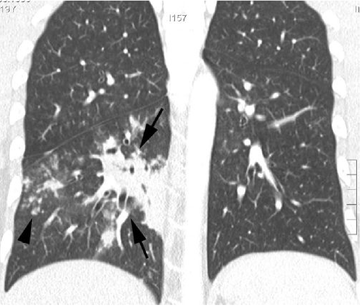
Fig: Bronchopneumonia (H. influenzae) - CT shows focal right lower lobe consolidation with air bronchogram, poorly defined margins, small nodules and tree-in-bud opacities.
2. Aspiration Pneumonia - Distinguishing Features
Pathophysiology: Material (oropharyngeal secretions, food, gastric contents) is inhaled and deposits in gravity-dependent lung zones. The pattern is that of bronchopneumonia, but the location is the key clue.
X-ray Findings:
| Feature | Detail |
|---|---|
| Distribution | Gravity-dependent segments - right lower lobe (most common), superior segment of lower lobes, posterior segments of upper lobes |
| Laterality | Commonly right-sided (right main bronchus is more vertical) |
| Pattern | Patchy peribronchial consolidation/nodular foci - similar to bronchopneumonia |
| Volume loss | Often present (posterior displacement of fissures) |
| Speed of onset | Can appear rapidly - new opacities within days of witnessed aspiration |
| Pleural effusion | May be present (small) |
| Tracheal shift | Toward affected side if significant consolidation/atelectasis |
CT Findings:
- Peribronchial consolidation in dependent lung regions
- Extensive lower lobe, dependent consolidation with volume loss
- Small centrilobular nodules (bronchiolitis component)
- May progress to cavitation/lung abscess (common complication)
- If oral contrast was given: contrast material visible directly in affected bronchi/parenchyma (pathognomonic)
3. Side-by-Side Comparison Table
| Feature | Bronchopneumonia | Aspiration Pneumonia |
|---|---|---|
| Distribution | Multifocal, bilateral, lower lobe predominant | Dependent segments (RLL, posterior UL, superior LL) |
| Laterality | Bilateral or unilateral | Often right-sided |
| Pattern | Patchy peribronchial opacities | Peribronchial + dependent consolidation |
| Air bronchogram | Usually absent | Variable |
| Volume loss | Uncommon early | Common (posterior fissure displacement) |
| Progression | Gradual | Rapid (days after witnessed aspiration) |
| Cavitation/abscess | Less common | More frequent complication |
| Clinical context | Any age, infection-driven | Impaired swallowing, altered consciousness, intubation |
| Tree-in-bud CT | Present | Present (bronchiolitis component) |
4. Key Practical Points
-
Aspiration pneumonia IS a form of bronchopneumonia - the patterns overlap, and the textbooks explicitly state they may be identical in appearance. The distinction rests mainly on location (dependent zones) and clinical history (dysphagia, altered consciousness, witnessed aspiration).
-
Right lower lobe predominance in aspiration is because the right main bronchus is more vertically oriented, especially in an upright or semi-recumbent patient.
-
Rapid development is a hallmark - a clear CXR at admission can show multifocal lower lobe opacity within days.
-
Watch for complications of aspiration that bronchopneumonia less commonly shows early: lung abscess (air-fluid level in a thick-walled cavity), foul-smelling sputum, anaerobic organisms.
Aspiration Pneumonia - Textbook Images
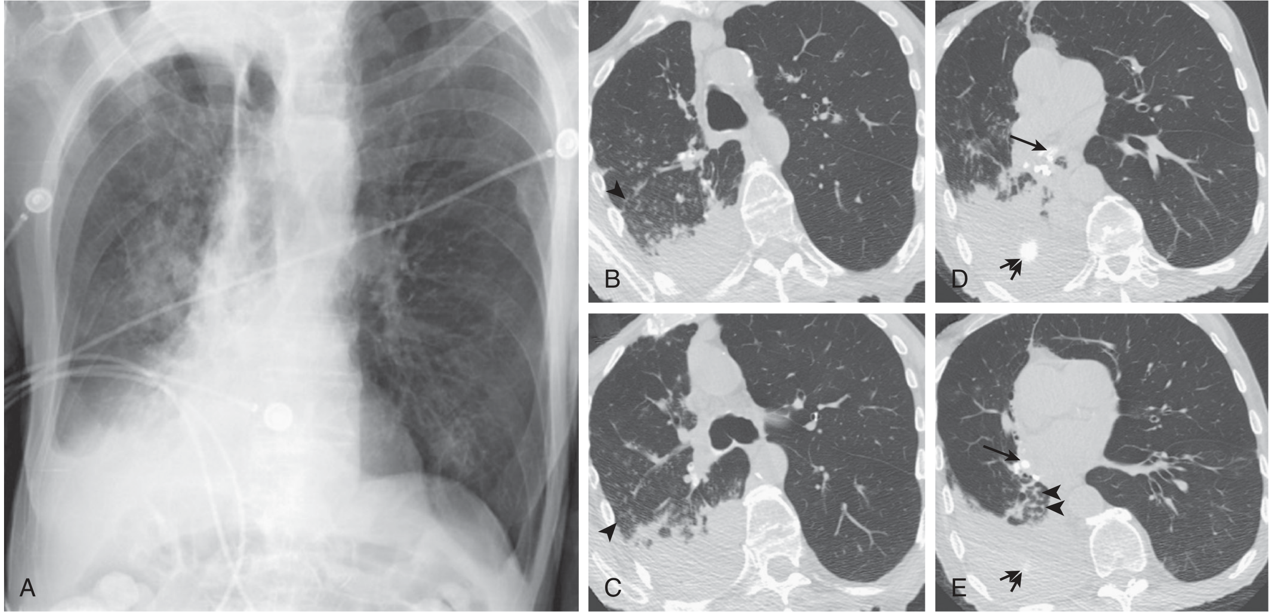
Aspiration Pneumonia (eFigure 46.4, Murray & Nadel): Right lower lobe consolidation with volume loss, tracheal shift toward the right. CT shows right lower lobe consolidation with centrilobular nodules consistent with bronchopneumonia + bronchiolitis in a dependent distribution.
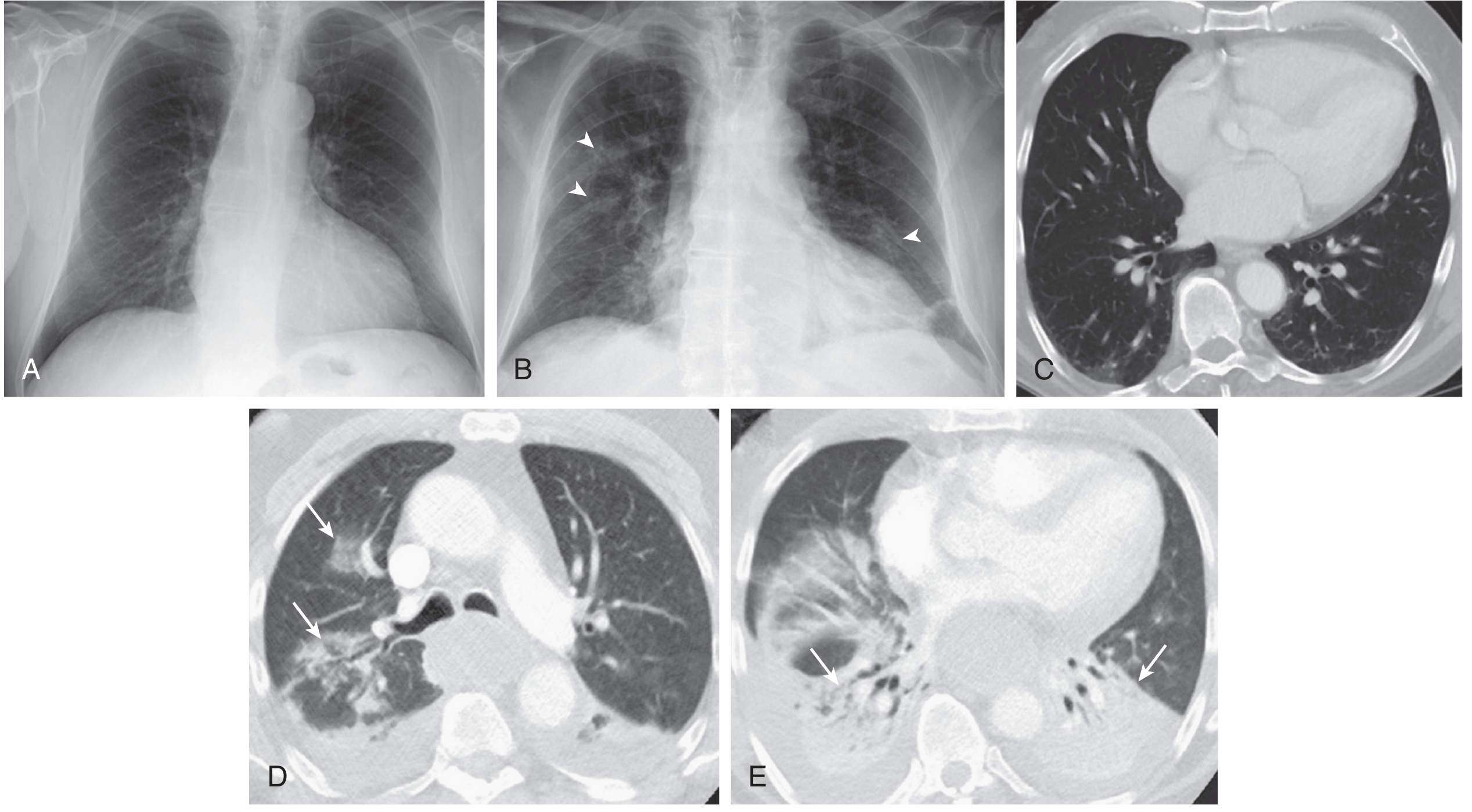
Aspiration Bronchopneumonia progression (eFigure 46.35, Murray & Nadel): (A) Clear CXR at admission. (B) Multifocal peribronchial nodular foci (arrowheads) after witnessed aspiration. (D-E) CT shows peribronchial consolidation and extensive dependent lower lobe consolidation with volume loss.
Sources: Grainger & Allison's Diagnostic Radiology, p. 123 | Murray & Nadel's Textbook of Respiratory Medicine, pp. 1036-1050 | Goldman-Cecil Medicine | Fishman's Pulmonary Diseases
This is a shared conversation. Sign in to Orris to start your own chat.